
Abbas Dhami
Specialist Diagnostic Radiographer
You have had scans. You have seen your GP. Yet the hip pain continues. This is a frustration shared by thousands of patients across the UK. The answer often lies not in what was scanned, but in how — because most routine scans are taken lying down and cannot show what your body does when standing under real load.
EOS imaging for hip pain offers a fundamentally different approach. It scans your full skeleton while you stand, revealing how posture, pelvic tilt, and whole-body alignment contribute to your pain. This blog explains what EOS shows, why it matters, and when it can make a real difference to your diagnosis.
Many patients spend months or even years searching for answers without clear results. In many of these cases, the missing piece is not another scan, but a better way of seeing how the body works under real conditions. This is where EOS imaging becomes valuable, as it focuses on function, alignment, and real-life movement rather than just static images.
What Is EOS Imaging for Hip Assessment?
Unlike routine scans, EOS assesses the hip under real load. That makes it far more useful for understanding how posture, balance, and movement affect pain.
Standing, weight-bearing hip imaging explained
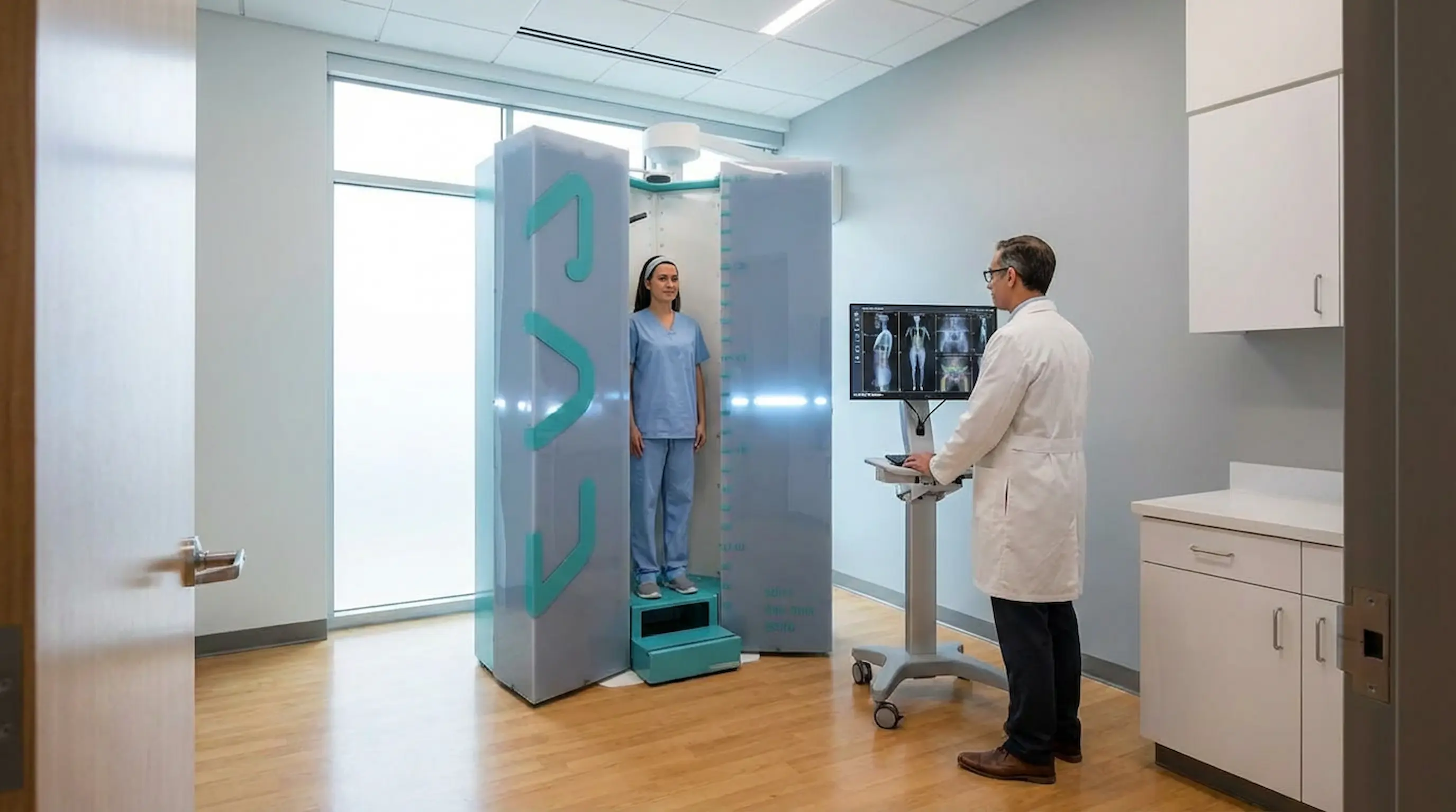
EOS captures simultaneous front and side images of your full skeleton in a single 15-second scan — taken while you stand. This weight-bearing position is critical. It shows the hip joint under real load, not at rest, which is when most hip pain actually occurs.
Low-dose EOS imaging for safer hip scans
EOS uses up to 90% less radiation than a standard CT scan, thanks to Nobel Prize-winning detector technology originally developed at CERN. It is safe for adults, suitable for repeat use, and produces sharper skeletal detail than many conventional alternatives.
Why full-body alignment matters in hip diagnosis
The hip does not function in isolation. EOS scans the entire skeleton — from the skull to the feet — so doctors can assess how your spine, pelvis, and hip joints work together. This full-body context is what separates EOS from every other imaging tool.
EOS Imaging vs. Standard X-Ray vs. MRI — Quick Comparison
This comparison shows how EOS imaging differs from standard X-rays and MRI when assessing alignment, weight-bearing function, and full-body skeletal detail.
| Feature | Standard X-Ray | MRI Scan | EOS Imaging |
|---|---|---|---|
| Taken standing | No | No | Yes |
| Full-body view | No | No | Yes |
| 3D reconstruction | No | No | Yes |
| Radiation level | Moderate | None | 90% less than CT |
| Alignment data | Limited | None | Full |
| Weight-bearing | No | No | Yes |
| Soft tissue detail | No | Yes | No |
Why Standard Hip Imaging May Miss the Real Cause
Many causes of hip pain do not become clear on routine imaging alone. In many cases, the real issue is linked to how the joint works under load, not just how it looks at rest.
Limits of a standard standing hip X-ray
A routine hip X-ray gives a single flat view, usually lying down. It shows the bone shape but not how the joint behaves under body weight. Small alignment problems, pelvic shifts, and postural imbalances are completely invisible in this position.
How posture, pelvis, and spine affect hip pain
Hip pain is often driven by what is happening above and around the joint — a tilted pelvis, a rotated spine, or a leg length difference. Routine imaging rarely captures these contributing factors, which is why so many patients receive a normal result despite ongoing pain.
How EOS hip alignment scans reveal hidden issues
Because EOS scans the whole body in a standing position, it can identify the chain of imbalances from spine to hip in one image. This makes it uniquely effective at finding the real cause of hip pain that standard scans consistently overlook.
Hip Pain, Posture, and Pelvic Alignment
Hip pain is often shaped by the way the body stands and carries weight. Small postural changes can place uneven stress through the hips over time.
How posture directly affects hip pain
The way you hold your body — whether you lean to one side, carry one shoulder lower, or overarch your lower back — directly affects how load passes through the hip joints. Over time, these postural habits create uneven pressure that leads to pain, stiffness, and joint wear.
Pelvic tilt and its impact on hip loading
Pelvic tilt hip pain is more common than many people realise. When the pelvis tilts forward, backward, or sideways, the hip joint moves with it — changing the angle at which forces pass through the socket. EOS identifies pelvic tilt accurately in the standing position, where it actually matters.
Why full-body hip posture assessment is important
A hip posture assessment through EOS looks beyond the joint itself. It shows how your feet, knees, pelvis, and spine all influence hip mechanics — giving clinicians a complete picture rather than just a view of the painful spot.
Spinopelvic Alignment in Hip Pain
The hip does not move independently of the rest of the body. Changes in spinal posture and pelvic angle can directly affect how force travels through the joint.
What spinopelvic alignment means for the hip
Spinopelvic alignment describes the angular relationship between your lumbar spine and pelvis. When this relationship is off even slightly, it changes the resting position of the hip joint and alters how forces travel through it during movement.
How spine–pelvis–hip connection affects movement
The spine, pelvis, and hip function as one kinetic chain. A stiff or curved lower spine forces the pelvis to compensate, which in turn shifts load unevenly onto one hip. EOS is the only routine imaging system that measures this entire chain in a single standing scan.
Impact of imbalance on hip pain and treatment
Identifying spinopelvic imbalance changes treatment decisions significantly. Physiotherapy, orthotics, and surgical planning can all be tailored more precisely once the full alignment picture is known. Without it, treatment often addresses symptoms rather than causes.
Leg Length Difference and Hip Pain
Even a small difference in leg length can affect the way the pelvis sits and how force moves through the hips. Over time, this imbalance can lead to pain, altered walking patterns, and strain on surrounding joints.
How leg length discrepancy causes hip pain
Even a few millimetres of difference between leg lengths can tilt the pelvis and overload one hip joint. Over time this leads to pain on one side, altered gait, and compensatory changes in the lower back. Leg length discrepancy hip pain is frequently underdiagnosed because it is invisible on lying-down scans.
True vs functional leg length differences
A true leg length difference is structural — one bone is physically shorter. A functional difference is caused by pelvic tilt or muscle imbalance. Both require different treatment approaches, and EOS can clearly distinguish between the two by measuring the skeleton in a weight-bearing position.
Role of weight-bearing EOS hip imaging
Weight-bearing hip imaging gives a real-world measurement of leg length. When you stand, any difference immediately affects pelvic position and hip loading. EOS captures this precisely, providing millimetre-accurate data that cannot be reliably obtained from a lying-down scan.
EOS Imaging for Hip Dysplasia
Why EOS is better for hip dysplasia assessment
Hip dysplasia occurs when the hip socket does not fully cover the ball of the joint. In adults, this often develops into chronic pain and early arthritis. EOS imaging hip dysplasia assessment is superior to routine X-ray because it measures socket coverage in the standing, loaded position which is where the instability actually manifests.
Benefits in adult hip dysplasia imaging
Adult hip dysplasia imaging with EOS provides the exact acetabular angle measurements needed for treatment planning. It shows how the hip behaves under body weight and whether alignment factors such as pelvic tilt are making the dysplasia worse.
Key hip alignment factors doctors evaluate
- Lateral centre-edge angle — how well the socket covers the femoral head
- Pelvic orientation — whether tilt is affecting apparent socket depth
- Spinopelvic balance — how the spine compensates for hip instability
When EOS Imaging Is Recommended for Hip Pain
Ongoing hip pain with unclear diagnosis
If you have had a standard X-ray or MRI with normal results but the pain continues, EOS is a logical next step. It looks at your body differently in a standing position and frequently reveals what lying-down scans missed.
Suspected posture-related hip problems
When pain is linked to standing, walking, or activity rather than rest, it strongly suggests a weight-bearing or posture-related issue. EOS is specifically designed to assess these functional problems.
Pre-surgical hip imaging needs
Before any hip surgery — whether that is a replacement, labral repair, or dysplasia correction — accurate alignment data is essential. EOS provides this data in the standing position, giving surgeons a more reliable foundation for planning.
Complex hip and spine-related cases
When both the hip and spine are involved, for example after spinal fusion or in cases of scoliosis, EOS is particularly valuable because it shows the entire spine-to-hip relationship in one image.
EOS Imaging in Hip Replacement Planning
For hip replacement surgery, accurate alignment data is essential. The position of the pelvis and spine can directly affect implant placement, stability, and long-term outcome.
Why hip replacement planning needs alignment data
Implant positioning in hip replacement surgery depends on the patient's unique pelvic shape and spinal alignment. If planning is based on lying-down imaging, the pelvis is in a different position to how it sits during normal activity and implant placement may not account for this.
Role of preoperative hip planning with EOS
Preoperative hip planning with EOS provides standing spinopelvic measurements that allow surgeons to plan cup placement according to the patient's functional anatomy, not just their resting anatomy. This is increasingly recognised as best practice in complex hip replacement cases.
How EOS improves implant positioning and leg length
- More accurate cup angle calculation based on standing pelvic position
- Better leg length restoration by measuring true functional discrepancy
- Reduced risk of post-operative complications such as dislocation
What to Expect from an EOS Hip Scan
Many patients want to know whether an EOS hip scan is difficult or uncomfortable. In reality, the process is quick, simple, and designed to capture your body in a natural standing position.
How a hip alignment scan is performed
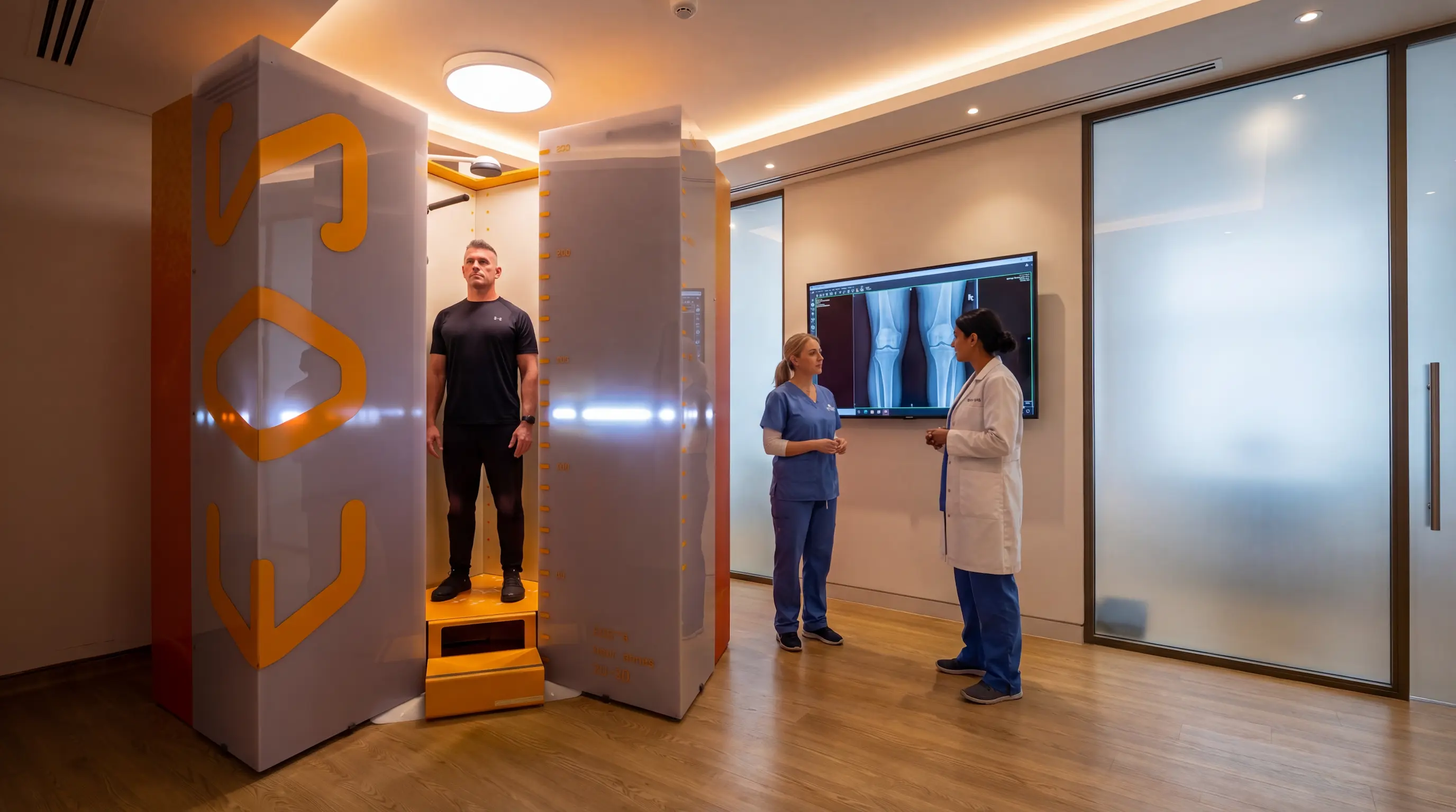
At ScanAlign, based at Harley Street Hospital in London, you stand inside a vertical scanning booth. The EOS system scans from head to toe in a single 15-second exposure. No injection is required, there is no lying down, and most patients find it entirely comfortable.
Scan time and standing position benefits
The full scan takes 15 seconds. Because you stand naturally, the images reflect how your skeleton actually functions during daily life, giving clinicians data they can directly act on. The entire appointment, including consultation and report review, typically takes around 45 minutes.
Hip alignment results and clinical insights
Your scan generates four detailed assessments prepared by UK-trained consultant radiologists, including a 3D skeletal model. Results are shared with you in a follow-up doctor-led consultation. A clinical summary is also provided for your GP or specialist.
Benefits of EOS Imaging for Hip Pain
EOS is designed to make hip assessment both safer and more informative. It gives clinicians detailed alignment data while keeping radiation exposure much lower than CT-based imaging.
Low radiation hip imaging
EOS uses up to 90% less radiation than a CT scan. The dose is comparable to a short-haul flight and is safe for routine use, including in patients who need repeat imaging over time.
Realistic weight-bearing hip assessment
Because EOS captures the body standing, the images reflect real-life loading conditions. This makes findings directly relevant to the pain patients experience during daily activity, not just how they look lying on a table.
Full-body alignment impact on hip diagnosis
EOS shows the whole kinetic chain — feet, knees, pelvis, and spine — in one image. This full-body alignment context means clinicians can identify root causes rather than just local symptoms.
Improved hip pain diagnosis and planning
Accurate imaging leads to more targeted treatment. Whether the plan involves physiotherapy, orthotics, or surgery, EOS gives both patients and clinicians a reliable foundation to make confident, well-informed decisions.
Is EOS Imaging Right for Your Hip Pain?
When EOS is most useful for hip conditions
- Hip pain that worsens with standing or walking
- Normal routine scan results despite ongoing symptoms
- Suspected pelvic tilt, leg length discrepancy, or hip dysplasia
- Pre-surgical planning or second opinion for hip replacement
When other hip imaging methods are needed
EOS is a skeletal imaging system. If soft tissue damage is suspected — such as a labral tear, cartilage injury, or muscle problem — an MRI will still be required. In many cases, EOS and MRI are used together to build a complete picture.
Choosing the right scan for hip diagnosis
The best scan depends on what your clinician needs to assess. For alignment, posture, leg length, and surgical planning, EOS leads. For soft tissue detail, MRI leads. Many patients benefit from both.
Conclusion
Hip pain is rarely just about the hip joint. The pelvis, spine, posture, and leg length all play a role, and understanding the real cause often requires imaging that shows the body under natural load.
EOS imaging for hip pain provides a standing, full-body, low-radiation view of skeletal alignment. It can reveal pelvic tilt, spinopelvic imbalance, leg length discrepancy, and other hidden factors that routine scans may miss, helping support more accurate diagnosis and treatment planning.
Book Your EOS Scan Today →FAQs
- Is EOS better than a standard hip X-ray for hip pain? Yes, for alignment-related hip pain. EOS scans the whole body while you stand, capturing weight-bearing alignment data that a lying-down X-ray cannot provide.
- Can EOS detect leg length difference causing hip pain? Yes. EOS provides precise standing measurements of leg length, distinguishing true structural differences from functional ones caused by pelvic tilt.
- Is EOS useful for adult hip dysplasia imaging? Absolutely. EOS measures hip socket coverage and orientation in the standing position, giving surgeons and specialists the exact data needed for dysplasia assessment and treatment planning.
- What is the difference between EOS and CT for hip imaging? CT provides detailed cross-sectional images but uses much more radiation and is taken lying down. EOS uses up to 90% less radiation, scans the full body while standing, and provides functional alignment data.
- Can EOS help before hip replacement surgery? Yes. EOS provides standing alignment data that helps surgeons plan implant angle and leg length restoration based on functional anatomy rather than resting anatomy.
- Is low-dose EOS imaging safe for hip scans? Yes. EOS is considered one of the safest skeletal imaging systems available. It uses much less radiation than a CT scan and is suitable for repeat imaging when needed.
Continue reading
More articles
Is EOS Scan Safe? Radiation Levels, Risks, and FAQs
Discover if EOS imaging is safe, its radiation levels, risks, and benefits. Learn why it’s preferred for accurate, low-dose skeletal scans.

EOS Scan for Scoliosis: Why Standing Imaging Matters
Discover how EOS for scoliosis provides low-radiation imaging for accurate spinal alignment and better scoliosis diagnosis and monitoring.
Knee Alignment & Osteoarthritis: How EOS Helps Treatment Planning
Struggling with knee pain or osteoarthritis? Discover how EOS imaging reveals knee alignment problems & supports treatment planning.