

Abbas Dhami
Specialist Diagnostic Radiographer
Medical imaging plays an important role in diagnosing bone and joint problems. One modern imaging method becoming more common in orthopaedic care is EOS imaging. This advanced system captures full-body images of the skeleton while the patient stands in a natural position.
Because EOS uses radiation to create internal images, many patients want to know whether it is safe. This guide explains how EOS imaging works, how much radiation it uses, and why it is often considered a safer option than traditional X-rays and CT scans for certain orthopaedic assessments.
What Is EOS Imaging Technology?
EOS imaging is a specialised medical imaging system designed mainly for orthopaedic diagnostics. It produces high-quality images of the musculoskeletal system, including bones, joints, posture, and body alignment.
Doctors use EOS technology to evaluate spinal deformities, scoliosis, posture issues, hip and knee alignment, and bone positioning before or after surgery. One major advantage of EOS is that it captures the body while standing, which helps doctors understand how bones and joints behave under natural body weight.
How EOS Imaging Works
EOS imaging uses advanced slot-scanning technology to create detailed images of the skeleton. Instead of exposing the whole body to radiation at once, the system scans vertically from head to toe using a thin beam. This helps reduce radiation exposure while maintaining image quality.
The system also captures two images at the same time — one from the front and one from the side. These images can then be used to create a 3D reconstruction of the skeleton, which helps doctors analyse spinal curves, joint positioning, and alignment more accurately.
EOS Imaging Radiation: How Much Exposure Is Involved?
Radiation exposure is one of the biggest concerns for patients who need imaging tests. The good news is that EOS imaging is designed to use very low radiation while still producing detailed full-body images.
The EOS scan radiation dose is significantly lower than many traditional imaging methods. This is one reason why it is often used for patients who need repeated monitoring, especially children and teenagers with scoliosis.
- Up to 90% lower radiation than traditional X-rays
- Much lower exposure than CT scans
- Safer for repeated monitoring in selected cases
- Useful for long-term orthopaedic follow-up
Comparison With Traditional Imaging Radiation
It is helpful to compare EOS imaging with other common imaging methods to understand where it fits best.
| Imaging Method | Typical Radiation Dose | Exposure Level | Key Notes |
|---|---|---|---|
| EOS Scan | ~0.05–0.1 mSv | Very Low | Low-dose full-body imaging, ideal for repeated monitoring |
| Traditional X-Ray | ~0.1–0.7 mSv | Low to Moderate | Common and widely available, but often higher radiation than EOS |
| CT Scan | ~5–10 mSv | High | Detailed cross-sectional imaging with significantly higher radiation |
Why Lower Radiation Matters
Lower radiation exposure is important for many patients, especially those who need imaging more than once. This includes children, teenagers, and people with long-term orthopaedic conditions.
- Reduces long-term radiation exposure
- Safer for children and younger patients
- Supports repeated monitoring when needed
- Provides reassurance for patients and families
EOS Scan vs X-Ray and CT Scan
Many patients want to know how EOS compares with traditional X-rays and CT scans. While all three methods are useful, they serve different purposes.
| Feature | EOS Scan | Traditional X-Ray | CT Scan |
|---|---|---|---|
| Purpose | Posture and skeletal alignment | Basic bone imaging | Detailed internal imaging |
| Radiation | Very low | Low to moderate | High |
| Full-Body Imaging | Yes | Usually requires multiple images | No |
| Weight-Bearing Analysis | Yes | Limited | No |
| Best Use | Orthopaedic planning and scoliosis monitoring | General bone checks | Complex injuries and internal detail |
Because EOS combines low radiation with standing full-body imaging, many orthopaedic specialists prefer it for posture analysis, scoliosis monitoring, and treatment planning.
EOS Scan Safety: What Medical Research Suggests
Medical research and clinical practice have shown that one of the biggest advantages of EOS imaging is its low radiation exposure. It has been widely adopted in orthopaedic centres for spinal deformity monitoring, paediatric imaging, and surgical planning.
Experts often consider EOS safe for repeated monitoring because it delivers much lower radiation than many older imaging methods while still producing high-quality orthopaedic images.
Possible EOS Scan Risks and Side Effects
Overall, EOS scan risks are minimal. Like any imaging test that uses radiation, there is still some exposure, but the amount is considered very low.
- Minimal radiation exposure
- Usually avoided during pregnancy unless medically necessary
- Patients need to remain still during the scan
- No known major side effects from the scan itself
The procedure is painless, quick, and non-invasive. For children and younger patients, doctors carefully assess whether imaging is needed and aim to use the lowest reasonable dose.
Who Should Consider an EOS Scan?
EOS imaging may be recommended for patients who need a detailed assessment of skeletal alignment, posture, or joint positioning in a natural standing position.
- Patients with spinal deformities
- Individuals with scoliosis
- People with posture or skeletal alignment issues
- Patients preparing for orthopaedic surgery
- Patients needing post-surgical monitoring
Because the scan captures the body while upright, it provides valuable information about how bones and joints work together under natural body weight.
Benefits of EOS Imaging Over Traditional Scans
EOS imaging offers several important advantages compared with older imaging methods. It is designed not only to reduce radiation but also to improve how doctors assess posture and alignment.
- Lower Radiation Exposure: EOS uses one of the lowest radiation doses available in skeletal imaging.
- Full-Body Imaging: The whole skeleton can be captured in a single scan.
- Accurate Measurements: Doctors can assess spinal curves and joint positioning more precisely.
- Natural Standing Position: The scan reflects real posture and weight-bearing alignment.
- Useful for Monitoring: It is especially helpful for follow-up imaging in selected orthopaedic cases.
Preparing for an EOS Scan
Preparing for an EOS scan is simple and usually does not require any special preparation. Most patients can return to normal activities immediately after the scan.
- Before the Scan: Remove metal objects such as belts or jewellery and wear comfortable clothing.
- During the Scan: You stand inside the scanning cabin while the scanner moves slowly from head to toe.
- Scan Time: The scan itself usually takes around 30 seconds.
- After the Scan: The images are reviewed by a specialist, and the full appointment usually takes only a few minutes.
Why This Imaging Option Matters
Conclusion
EOS imaging is considered a safe and effective option for many orthopaedic imaging needs. It uses significantly lower radiation than traditional X-rays and far less than CT scans, while still providing high-quality full-body images.
Its ability to scan patients in a natural standing position makes it especially useful for posture analysis, scoliosis monitoring, and treatment planning. For patients who need accurate skeletal imaging with lower radiation exposure, EOS can be an excellent option.
Book Your EOS Scan Today →FAQs
- Is an EOS scan safe? Yes, EOS imaging is generally considered safe because it uses very low radiation compared with many traditional imaging methods.
- How much radiation does an EOS scan produce? The EOS scan radiation dose is typically up to 90% lower than traditional X-rays and significantly lower than CT scans.
- Is EOS imaging safer than a CT scan? In terms of radiation exposure, yes. EOS uses far less radiation than a CT scan, making it more suitable for repeated monitoring in selected orthopaedic cases.
- Does an EOS scan hurt? No. The scan is painless, and patients simply need to stand still while the images are captured.
- How long does an EOS scan take? The scan itself usually takes around 30 seconds, while the full appointment may take a few minutes.
Medical Disclaimer: This content is for general informational purposes only and does not constitute medical advice, diagnosis, or treatment. Always consult a qualified doctor or healthcare professional for personalised medical guidance based on your symptoms and condition.
Continue reading
More articles

EOS Scan for Scoliosis: Why Standing Imaging Matters
Discover how EOS for scoliosis provides low-radiation imaging for accurate spinal alignment and better scoliosis diagnosis and monitoring.
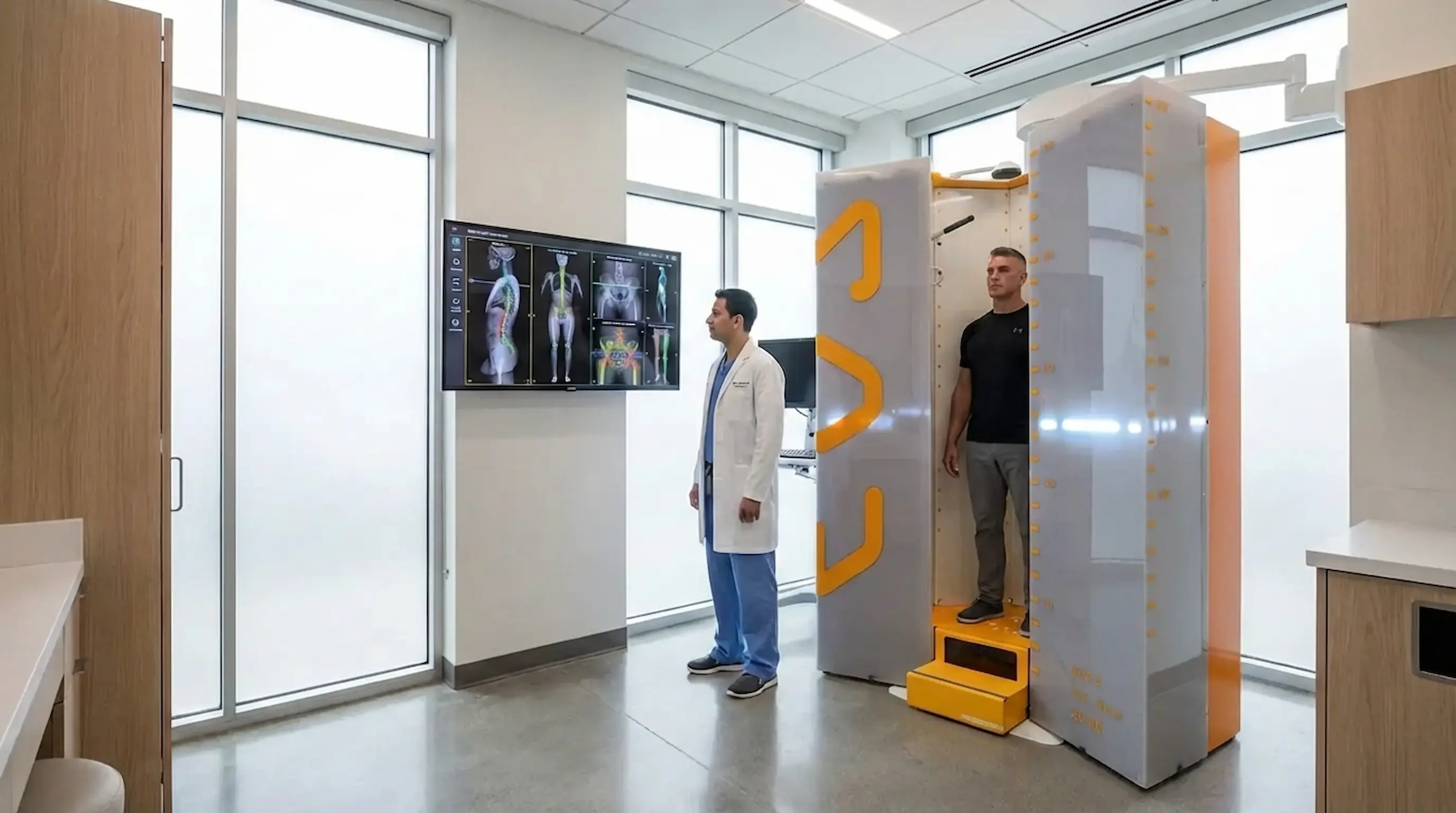
Hip Pain & Alignment: EOS Imaging for Better Diagnosis
EOS imaging for hip pain reveals pelvic tilt, leg length discrepancy & spinopelvic alignment that routine scans miss. Book Appointment now.
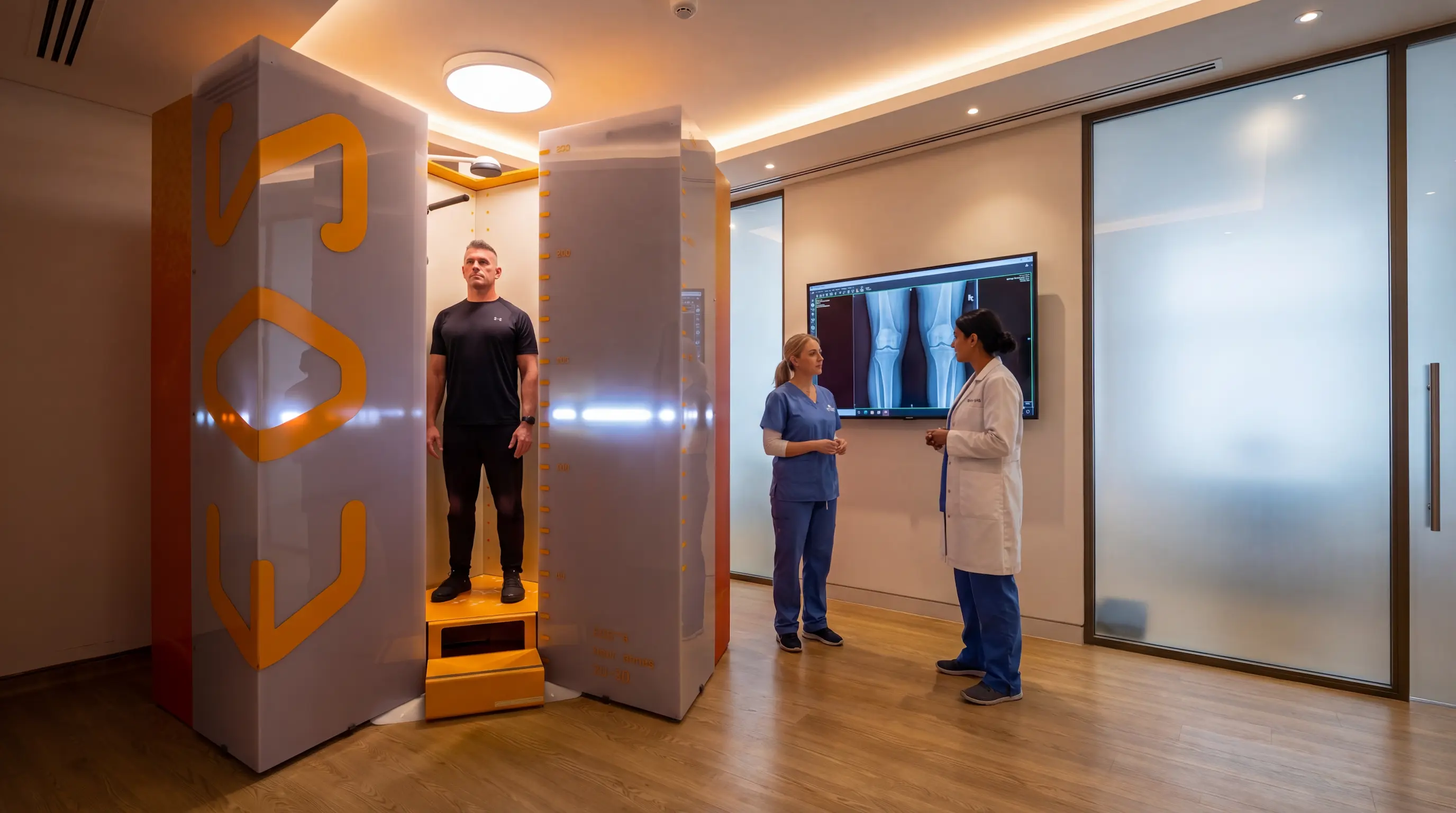
Knee Alignment & Osteoarthritis: How EOS Helps Treatment Planning
Struggling with knee pain or osteoarthritis? Discover how EOS imaging reveals knee alignment problems & supports treatment planning.