

How Is Adult Scoliosis Different From the Teenage Kind?
Adult scoliosis is a sideways curve of the spine that develops or worsens after skeletal maturity. Unlike the teenage form, which usually appears during a growth spurt with no clear cause, adult scoliosis is most often driven by wear and tear in the discs and joints. It tends to cause more pain, stiffness and balance problems than adolescent scoliosis.
Key takeaways
- Adult scoliosis is commonly caused by age-related disc and joint degeneration, not growth.
- Pain, stiffness and balance changes are far more common in adults than teenagers.
- Standing, weight-bearing imaging reveals curve severity and overall balance that lying-down scans can miss.
- Treatment decisions depend on accurate whole-body alignment data, not curve size alone.
- An EOS scan uses about 90% less radiation than a standard X-ray, around ten times less.
What exactly is adult scoliosis?
Adult scoliosis is any lateral (sideways) spinal curve present after the skeleton has finished growing, usually after age 25. Some adults carry a curve that first appeared in their teens. Others develop a completely new curve later in life, often called de novo or degenerative scoliosis. Either way, the impact on daily life tends to be quite different from what a teenager experiences.
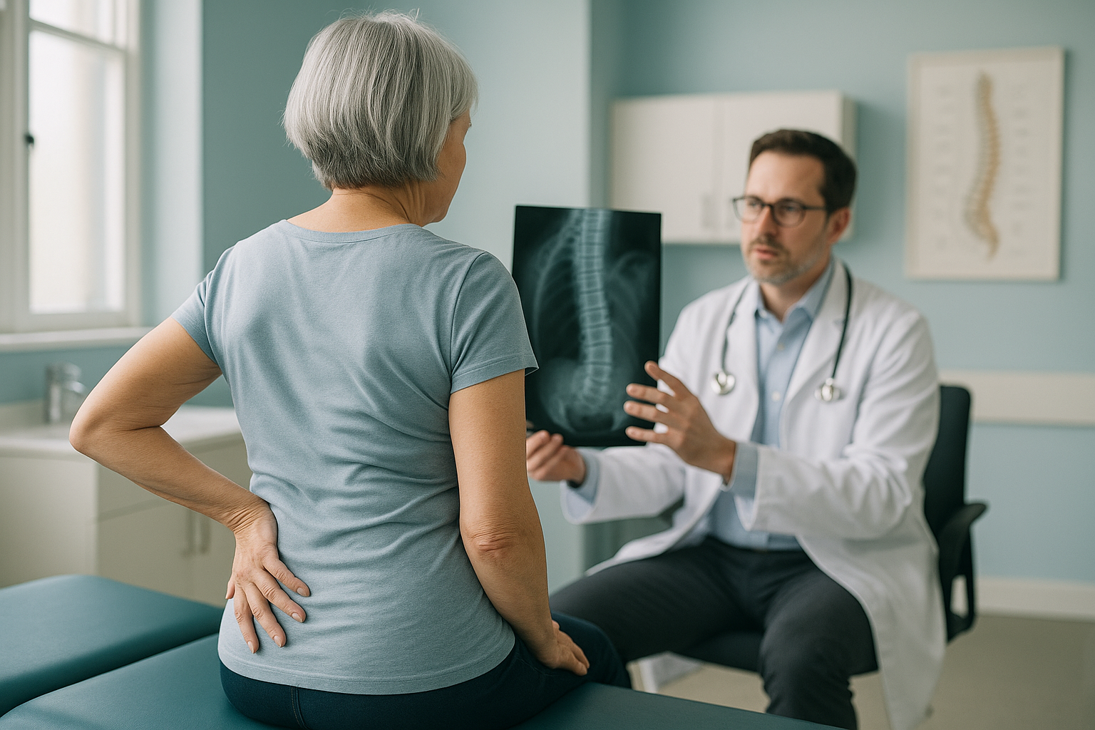
In adolescence, scoliosis is usually painless and discovered by chance. The main concern is whether the curve will worsen during growth. In adults, the picture shifts. Pain, fatigue and changes in balance become the leading issues, and the underlying cause is usually degeneration rather than growth.
What are the different types of adult scoliosis?
There are two main categories, and they behave quite differently.
Progressed adolescent scoliosis means the curve began during the teenage years and has gradually worsened over decades. These curves tend to be smoother and larger. Many people live with them for years without significant symptoms, then notice increasing stiffness, fatigue or a visible trunk shift as the spine ages.
De novo (degenerative) scoliosis develops entirely in adulthood, usually after age 40 or 50. It is driven by asymmetric disc and facet joint wear in the lumbar spine. These curves are typically shorter and smaller, but they can cause significant pain, nerve compression and balance problems because they develop in a spine that is already stiff and arthritic.
In both types, understanding how the whole body balances around the curve is at least as important as measuring the curve itself. That is where Cobb angle measurement fits in. It quantifies the curve, but balance and alignment tell the real story.
What symptoms does adult scoliosis cause?
Pain is the number-one reason adults seek help for scoliosis. Teenagers with scoliosis rarely have pain. Adults almost always do. The symptoms tend to build gradually and may include the following.
Urgent symptoms. If you develop sudden numbness or weakness in your legs, or lose bladder or bowel control, seek immediate medical attention. Contact your GP, call 111, or go to A&E. These symptoms need urgent assessment, not a routine scan.
What causes scoliosis to develop or worsen in adults?
Degenerative scoliosis is caused by uneven wear in the spine's discs and facet joints. Over time, one side of a disc loses height faster than the other, and the vertebra above tilts. Multiply that across several levels and a new curve forms.
If you already had a teenage curve, it can progress slowly through adult life as these same degenerative changes layer on top of the existing curve. Progression tends to be gradual, sometimes less than a degree per year, but over decades that adds up.
Why does a standing scan matter for adult scoliosis?
A standing, weight-bearing image shows how your spine actually behaves under gravity. This is essential because adult scoliosis is as much about balance as it is about the curve itself.
When you lie down for a standard X-ray, CT or MRI, gravity is removed, muscles relax, and the curve can appear smaller or straighter than it really is. Your body may look aligned on a lying-down scan while you are actually leaning significantly to one side when upright. Specialists call this your coronal and sagittal balance, and it is one of the strongest predictors of how much a scoliosis affects your quality of life.
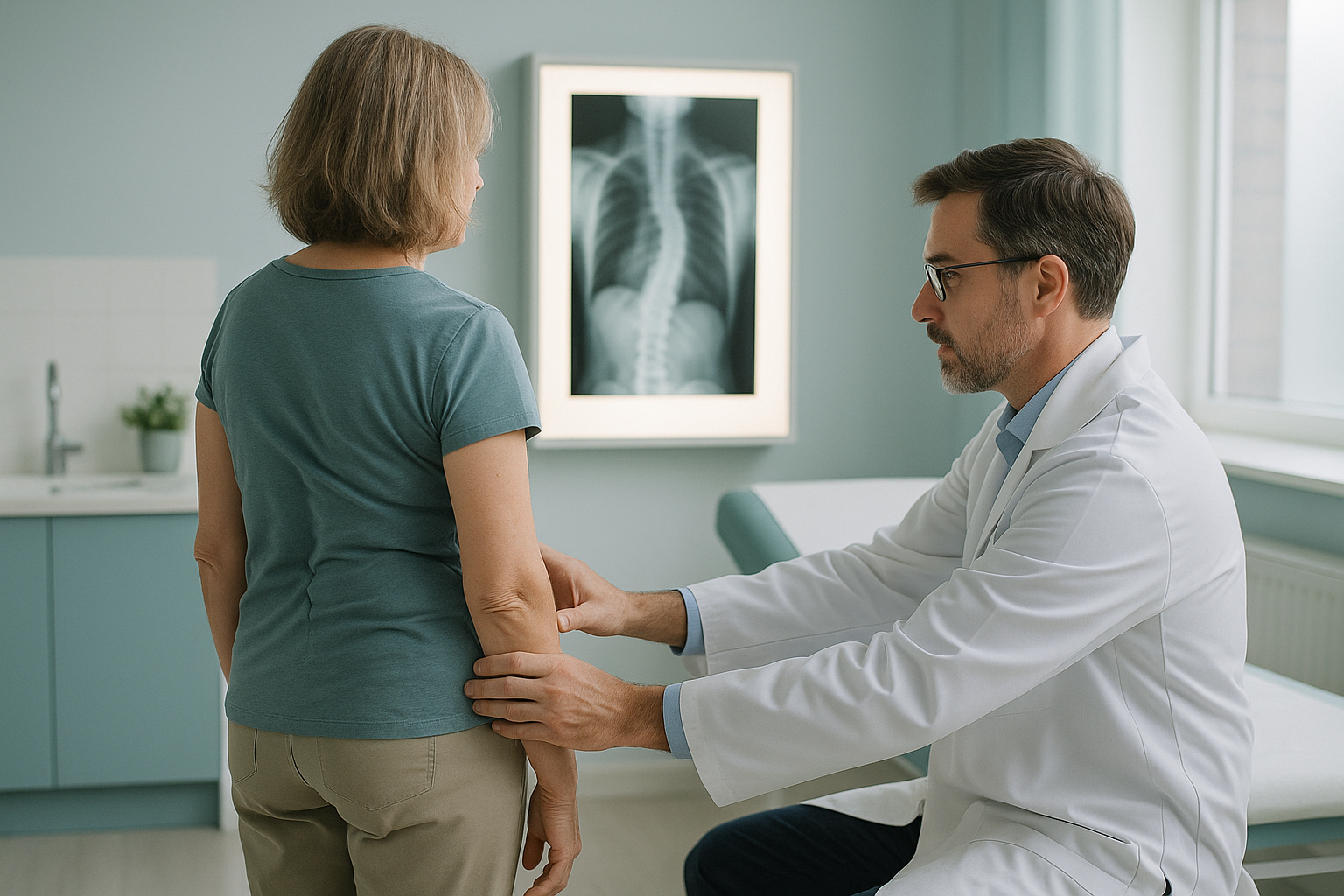
An EOS scan captures the entire skeleton from head to feet in a single standing image, using about 90% less radiation than a standard X-ray, around ten times less. That low dose matters because scoliosis monitoring often requires repeat imaging over the years. Read more about why standing imaging matters in scoliosis.
Your body is a connected chain from feet to head. If your spine curves to one side, your pelvis, hips and even knees compensate to keep you upright. A single-area scan of the lumbar spine alone cannot show those compensations. ScanAlign standing EOS imaging captures the full picture in one visit, giving your specialist the alignment data they need to plan the right next step.
When MRI is the better choice. EOS shows bones, joints and whole-body alignment. For soft-tissue detail, such as compressed nerves, disc herniations or spinal cord problems, MRI is usually the better tool. Many adults with scoliosis benefit from both types of imaging, each answering a different question.
What helps manage adult scoliosis?
The right approach depends on the severity of the curve, the degree of imbalance, the level of pain and how much it affects daily activities. ScanAlign is a diagnostic imaging clinic, not a treatment centre, so we focus on providing the precise alignment data that helps your specialist make informed decisions.
Broadly, management options discussed with your treating clinician may include physiotherapy and targeted exercise, pain management, or in some cases, surgical correction. What all of these have in common is that they work best when based on accurate, standing alignment data rather than a lying-down image that may understate the problem.
Wondering whether a standing alignment scan could help clarify your scoliosis? You can book a standing scoliosis scan or start with a free video consultation to discuss your situation first.
Book a free video consultationFrequently asked questions about adult scoliosis
Yes. Degenerative or de novo scoliosis can develop entirely in adulthood, typically after age 40 or 50. It is caused by asymmetric wear in the spinal discs and joints, not by growth.
Not always, but pain is far more common in adults than in teenagers with scoliosis. Back pain, leg pain and fatigue are the usual reasons adults seek assessment.
Many adult curves progress slowly, sometimes less than a degree per year. Monitoring with periodic standing imaging helps track any change before it becomes a bigger problem.
When you lie down, gravity is removed and muscles relax. The curve can appear smaller than it really is. A standing scan shows the true curve and your overall balance under real-world conditions.
An EOS scan uses about 90% less radiation than a standard X-ray, around ten times less. This is especially relevant for scoliosis patients who may need repeat imaging over time.
No. ScanAlign accepts self-referrals. You can book directly without a GP or consultant letter. You are also welcome to bring a referral if you have one.
We do not list prices on individual pages to keep them up to date. You can find current pricing and details on our FAQs and cost page.
EOS excels at showing bone alignment, joint position and overall balance. For soft-tissue detail like nerve compression or disc herniations, MRI is usually the better tool. Many scoliosis patients benefit from both.
You stand naturally inside the EOS scanner while it captures a full-length image of your skeleton from head to feet. It takes less than a minute and there is no enclosed tunnel.
Surgery is considered when pain and disability are severe and non-surgical options have not helped. Many adults manage well without surgery. Accurate standing imaging data helps both you and your specialist decide whether surgery is worth considering.
Understand your scoliosis, standing up
A free video consultation lets you discuss your symptoms and find out whether a standing EOS scan is the right next step for you.
Book a free video consultationSources
- Aebi, M. "The adult scoliosis." European Spine Journal, 14(10), 2005, pp. 925–948.
- Schwab, F. et al. "Adult scoliosis: prevalence, SF-36, and nutritional parameters in an elderly volunteer population." Spine, 30(9), 2005, pp. 1082–1085.
- Scoliosis Research Society (SRS). "Adult Scoliosis." srs.org.
Written by Abbas Dhami (Specialist Diagnostic Radiographer)
This page is general information and not a diagnosis.
ScanAlign is the trading name of CoreMed Solutions Ltd and operates under The Harley Street Hospital's CQC licence.