
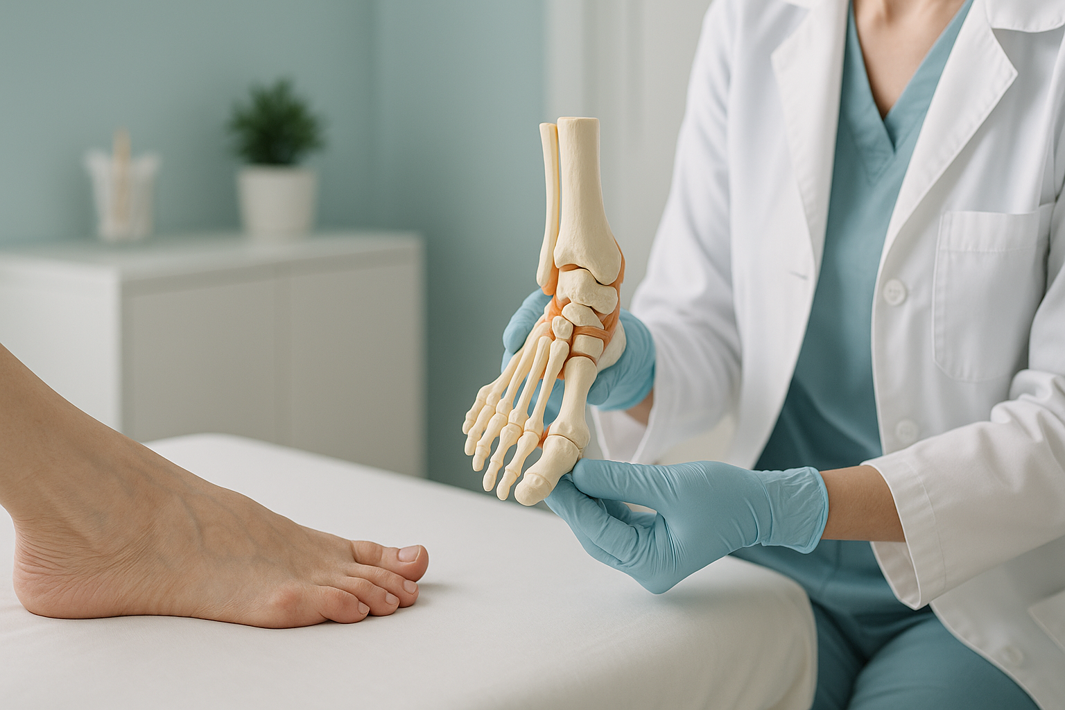
What Causes Bunions (Hallux Valgus), and Can You Stop Them Getting Worse?
A bunion forms when the big toe joint drifts out of line, pushing a bony bump to the side of the foot. Genetics, foot mechanics and footwear all play a part. You cannot reverse a bunion without surgery, but understanding your alignment can help you slow its progress and manage pain.
Key takeaways
- Bunions (hallux valgus) are a progressive shift in the big toe joint, not just a bump of extra bone.
- Flat feet, overpronation and tight shoes are common contributing factors.
- Lying-down imaging can miss how much the joint shifts under body weight.
- A standing weight-bearing x-ray shows the true position of the first metatarsal and big toe when loaded.
- Conservative measures can slow progression, but imaging gives you a clear starting point.
If you have a bony bump at the base of your big toe, chances are you already know what a bunion looks like. What most people do not realise is that the visible bump is only part of the story. Hallux valgus is a structural misalignment of the entire first metatarsal and big toe joint. It tends to get worse over time, and the rate of change depends on how your whole foot loads when you stand and walk.
How do you know if you have a bunion?
You can usually spot a bunion by looking at your foot, but the symptoms go beyond appearance. The big toe gradually angles toward the second toe, and the joint at the base pushes outward. Over time, the joint can become stiff, swollen and painful, especially in shoes.
Some people have a visible bump but no pain. Others find that even mild-looking bunions cause significant discomfort. The outward appearance does not always match the amount of joint change happening underneath.
What causes bunions to develop?
Bunions develop when the first metatarsal bone gradually shifts away from the second, and the big toe tilts inward to compensate. There is rarely a single cause. It is usually a combination of inherited foot shape, the way you walk and the shoes you wear.

Your body is a connected chain from feet to head. When the forefoot loads unevenly, it changes how force travels up through the ankle, knee and hip. A bunion is not an isolated foot problem. It can alter your lower limb alignment and contribute to pain elsewhere.
Why does a bunion need a standing x-ray?
A standard x-ray taken while you are lying down relaxes the muscles and ligaments around the foot. This can make the joint look better aligned than it really is. When you stand, your full body weight presses through the forefoot and the true separation between the first and second metatarsals becomes visible.
With no weight on the foot, the forefoot narrows. The angle between the first metatarsal and the big toe can appear smaller than it actually is during walking. Surgeons and podiatrists may not get the full picture of how the bunion behaves under load.
Under weight, the arch flattens, the forefoot spreads and the first metatarsal fans outward. A standing image captures the position that matters, the one your foot is in every time you take a step. This gives your clinician a realistic baseline and helps them plan the right approach.
At ScanAlign, imaging is taken while you stand in a natural position. The EOS system uses about 90% less radiation than a standard x-ray, around ten times less. It captures the full lower limb in a single standing image, so your clinician can see how the bunion relates to your ankle, knee and hip alignment above it.
Want to see how your bunion looks under real load? A standing scan shows what lying-down imaging misses.
Book a free video consultationHow can you stop bunions getting worse?
You cannot reverse a bunion without surgery. However, there is good evidence that managing the factors that drive progression can slow it down and reduce pain. The first step is understanding where your alignment stands right now.
Conservative measures that may help
- Wider footwear. Shoes with a broad toe box reduce pressure on the bump and give the toes room to sit in a more natural position.
- Toe spacers. Silicone separators worn between the big and second toe can ease discomfort and may limit further drift during daily activities.
- Supportive insoles. If flat feet or overpronation contribute to forefoot loading, custom orthotics may help distribute pressure more evenly.
- Strengthening exercises. Targeted exercises for the muscles around the big toe and arch can improve stability. A podiatrist or physiotherapist can guide you.
- Bunion splints. Night splints hold the toe in a straighter position. Evidence on whether they slow progression is mixed, but some people find them comfortable.
None of these measures replace surgery for a bunion that is already severe. If the joint has stiffened significantly or the toe is crossing over, a surgical opinion is usually the next step. ScanAlign does not provide treatment, but the imaging we produce is exactly what surgeons and podiatrists need to plan their approach.
When is MRI more appropriate? EOS shows bones, joints and alignment. If your clinician suspects soft-tissue damage, nerve irritation or inflammation inside the joint, MRI is usually the better tool. Both types of imaging can work together to give the full picture.
Who should get a standing scan for a bunion?
A standing foot assessment is most useful when you need a clear, objective picture of your alignment before deciding on next steps.
- Your bunion is getting worse and you want a baseline measurement
- A surgeon has asked for weight-bearing imaging before planning an operation
- You have pain in your knee, hip or back and suspect your foot alignment is involved
- You want to track change over time with comparable images
- Your main concern is soft-tissue swelling or nerve pain (MRI is likely more useful)
- You have sudden redness, heat and severe pain in the joint (see your GP to rule out gout or infection first)
- You simply need wider shoes and have not tried conservative measures yet
If you are unsure, book a weight-bearing foot assessment starting with a free video consultation. We will let you know whether imaging is likely to add value for your situation.
Frequently asked questions about bunions
No. Once the bone has shifted, conservative measures can slow progression and manage pain, but they cannot reverse the structural change. Surgery is the only way to realign the joint.
Foot shape and joint flexibility are inherited, and these traits raise the likelihood of developing bunions. Having a parent with hallux valgus does not guarantee you will get one, but it does increase the risk.
Splints may help with comfort and may limit further drift during rest, but current evidence does not show they can permanently straighten the toe. They are best used as part of a broader management plan.
When you lie down, the muscles relax and the forefoot narrows. A standing image captures the true alignment under body weight, which is the position your foot is in every time you walk. This gives surgeons and podiatrists a more accurate view for planning.
Yes. The EOS system uses about 90% less radiation than a standard x-ray, around ten times less. It is a very low-dose system designed for standing whole-body imaging.
No. ScanAlign is a private, self-pay clinic. You can refer yourself directly. We start with a free video consultation to make sure imaging is appropriate for you.
Pricing details are on our FAQs page. We are transparent about costs before you commit to anything.
It can contribute. When the big toe joint is out of line, it changes how you load the foot, which can alter the way force travels up through the ankle, knee and hip. Pain can show up well away from its original cause.
A bunion (hallux valgus) is a sideways shift of the big toe. Hallux rigidus is stiffness and arthritis of the same joint, often with bone spurs on top. They can occur together, but they are distinct conditions.
A specialist radiographer reviews your concern, discusses your symptoms and checks whether standing imaging would add useful information for your case. There is no obligation to book a scan.
See your bunion under real weight
A standing scan captures what lying-down imaging misses. Start with a free video consultation to find out if it is right for you.
Book a free video consultationSources
- Nix S, Smith M, Vicenzino B. Prevalence of hallux valgus in the general population: a systematic review and meta-analysis. Journal of Foot and Ankle Research. 2010;3:21.
- NHS. Bunions. nhs.uk. Available at: https://www.nhs.uk/conditions/bunions/
- Illés T, Somoskeöy S. The EOS imaging system and its uses in daily orthopaedic practice. International Orthopaedics. 2012;36(7):1325-1331.
Written by Abbas Dhami (Specialist Diagnostic Radiographer)
This page is general information and not a diagnosis.
ScanAlign is the trading name of CoreMed Solutions Ltd and operates under The Harley Street Hospital's CQC licence.