
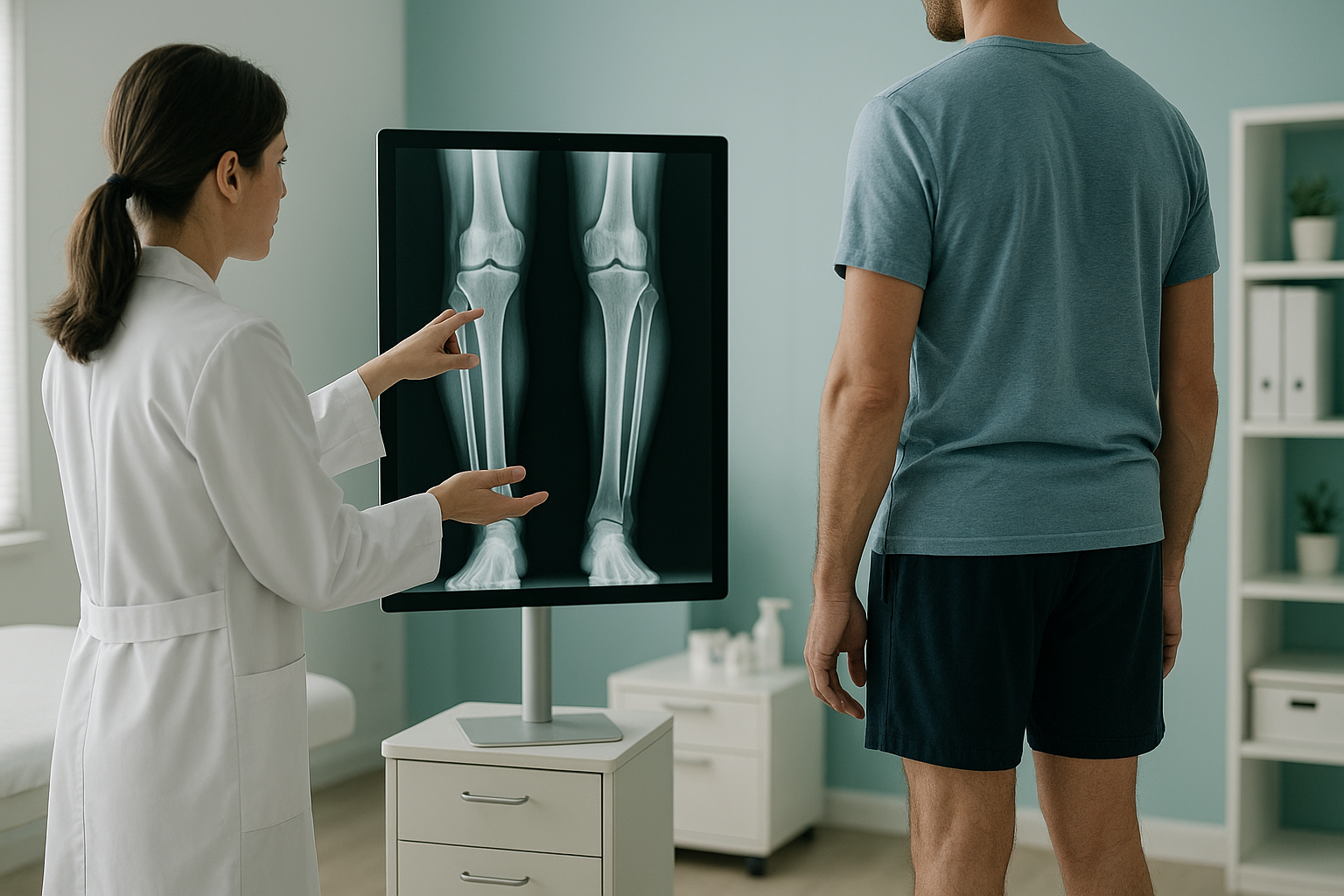
Weight-Bearing X-ray: Why Does Standing Up Change What Your Scan Shows?
A weight-bearing x-ray is taken while you stand, so your bones and joints carry your full body weight. This reveals how your skeleton truly aligns under load. Lying-down scans relax muscles and decompress joints, which can hide the narrowing, tilting or shifting that causes your pain in everyday life.
Key takeaways
- Standing x-rays show your joints under real load, not relaxed on a table.
- Problems like joint narrowing, pelvic tilt and spinal curves can look normal when lying down.
- Your body is a connected chain. A problem at one level often causes pain somewhere else.
- EOS whole-body standing imaging captures your full alignment in a single, low-dose scan.
- No referral is needed. You can self-refer for a standing scan at ScanAlign.
What is a weight-bearing x-ray?
A weight-bearing x-ray is an image taken while you stand upright, with gravity pulling on your body exactly as it does when you walk, stand or sit. Standard x-rays, CT scans and MRI are usually performed lying down. When you lie flat, your muscles relax and your joints decompress slightly. That can make a problem look less severe than it really is, or even hide it altogether.
Standing imaging captures the truth of how your skeleton behaves under load. Joint spaces narrow, curves deepen, and any tilt or shift becomes visible. For anyone with ongoing pain that does not match their lying-down scan results, a weight-bearing x-ray often fills in the missing piece.
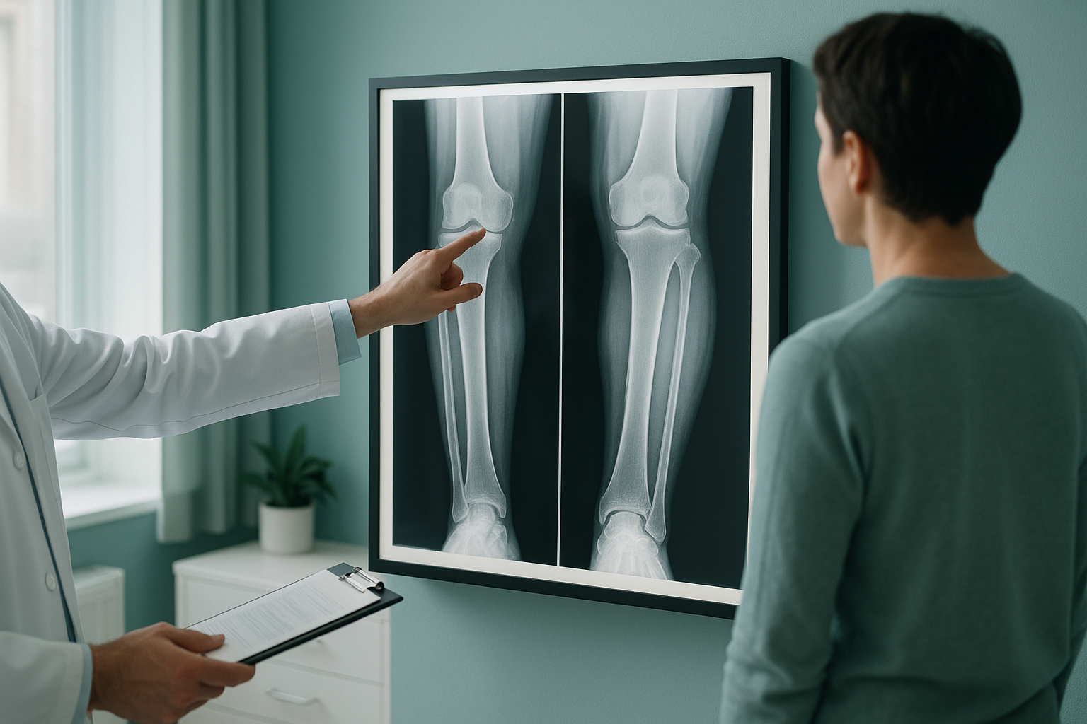
Why are weight-bearing x-rays used instead of lying-down scans?
Weight-bearing x-rays are used because they show your body the way it actually functions. When you lie on a scan table, gravity no longer compresses your joints. Disc spaces open up, a tilted pelvis can level out, and a knee that grinds bone-on-bone when you walk may look fine on the image.
Think of your body as a chain that runs from your feet through your knees, hips and spine up to your head. Every link affects the ones above and below it. If one link is off, other areas compensate. You might feel pain in your lower back, but the real cause could be a tilted pelvis or uneven leg length. A lying-down scan of just your lower back would never show that.
A standing, weight-bearing image captures the whole chain under real conditions. It lets clinicians see the true joint spacing, the actual spinal curves and any compensatory shifts your body has developed over time.
How do you know if you need a weight-bearing x-ray?
You may benefit from a standing, weight-bearing x-ray if you recognise any of the situations below. These are common reasons people come to ScanAlign after standard scans have not given clear answers.
Your MRI or x-ray was reported as normal, but your symptoms persist. The lying-down position may have masked the problem.
If your symptoms ease when you sit or lie down, weight-bearing imaging is designed to capture what triggers them.
One shoulder sitting higher, a hip that drops, or a feeling that you lean to one side. A standing scan shows how your alignment shifts under load.
Surgeons planning joint replacement or spinal surgery need to see your true alignment under load. Weight-bearing images provide the detail they require.
Which body areas benefit most from a standing x-ray?
Any joint that carries your weight can look different standing compared with lying down. The areas below are the most common reasons people seek weight-bearing imaging.
Curves, disc narrowing and vertebral shifts are often more obvious standing up.
Pelvic tilt and leg-length differences only become clear under full body weight.
Weight-bearing knee x-rays reveal true joint space narrowing that lying-down scans can underestimate.
Whole-body standing imaging shows how all of these areas connect and influence each other.
When MRI is the better choice: EOS and standing x-rays show bones, joints and overall alignment. If your clinician suspects a disc bulge, nerve compression, ligament tear or other soft-tissue problem, MRI is usually the more appropriate tool. The two types of imaging often work best together.
How does EOS standing imaging differ from a standard weight-bearing x-ray?
A standard weight-bearing x-ray typically focuses on one area, such as a single knee or part of the spine. EOS is an advanced form of standing, weight-bearing imaging that captures your entire skeleton from head to toe in a single scan. This means your clinician can see how your feet, knees, hips, pelvis and spine all relate to each other, rather than looking at one joint in isolation.
EOS also uses about 90% less radiation than a standard x-ray, around ten times less. The images are detailed enough for surgical planning and alignment measurements, but at a fraction of the dose.
At ScanAlign, the EOS scan is performed at 19 Harley Street, London. You stand naturally in the scanner for about 20 seconds while it captures a full-length image. No referral is needed, and you can self-refer directly.
Want to find out whether a standing whole-body scan could answer your questions? Speak with our team at no cost.
Book a free video consultationWhat should you expect during a weight-bearing scan at ScanAlign?
The process is straightforward. Here is a step-by-step overview.
A specialist radiographer reviews your history and symptoms to confirm a standing scan is appropriate for you.
At the clinic on Harley Street, you stand naturally in the EOS unit. The scan takes about 20 seconds. No injections, no enclosed tunnel.
Your images are reviewed by a specialist. You receive a detailed report covering your alignment from head to toe.
Share your report with your GP, surgeon, osteopath or physiotherapist so they can plan the right next step.
Frequently asked questions about weight-bearing x-rays
It is an x-ray taken while you stand upright, so your bones and joints carry your full body weight. This shows how your skeleton truly aligns under the same forces you experience in daily life.
Lying down relaxes muscles and decompresses joints, which can hide problems like joint narrowing, pelvic tilting or spinal shifting. Standing imaging captures these issues as they actually occur when you are upright and weight-bearing.
EOS is a type of standing, weight-bearing imaging. The key difference is that EOS captures your entire body from head to toe in one scan, with about 90% less radiation than a standard x-ray, around ten times less. A standard weight-bearing x-ray usually focuses on just one area.
No. At ScanAlign you can self-refer. You book a free video consultation first, and our specialist radiographer confirms the scan is right for you.
EOS uses about 90% less radiation than a standard x-ray, around ten times less. This makes it one of the lowest-dose options available for full-body skeletal imaging.
Yes. Standing knee x-rays reveal true joint-space narrowing that lying-down scans can underestimate. This helps clinicians assess the severity of cartilage loss more accurately.
EOS and standing x-rays show bones, joints and alignment. For soft-tissue detail such as disc bulges, ligament tears or nerve compression, MRI is usually the better tool. The two types of scan often complement each other well.
The EOS scan itself takes about 20 seconds. You simply stand still in the scanner. The whole appointment, including positioning, is quick and straightforward.
Pricing details are available on our FAQs and cost page. You can also ask during your free video consultation.
You receive a detailed alignment report that you can share with your GP, surgeon, physiotherapist or other clinician. They can use it to guide treatment decisions. ScanAlign is a diagnostic imaging clinic and does not provide treatment itself.
Ready to see how your body really aligns?
A free video consultation with our specialist radiographer can help you decide whether a standing, whole-body scan is right for you.
Book a free video consultation Learn more about EOS imagingWritten by Abbas Dhami (Specialist Diagnostic Radiographer)
Sources
- Illés, T. and Somoskeöy, S. (2012). The EOS imaging system and its uses in daily orthopaedic practice. International Orthopaedics, 36(7), pp.1405–1411.
- Rossi, A. et al. (2007). Weight-bearing versus supine MRI of the knee: systematic review. European Radiology, 17(6), pp.1563–1574.
- NHS (2023). X-ray overview. Available at: nhs.uk/conditions/x-ray.
This page is general information and not a diagnosis.
ScanAlign is the trading name of CoreMed Solutions Ltd and operates under The Harley Street Hospital's CQC licence.