
What Causes Flat Feet in Adults, and Can They Be Corrected?
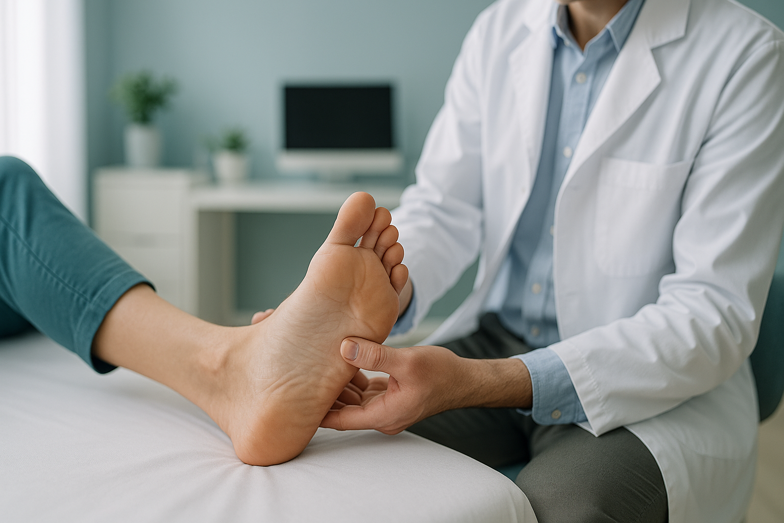
Flat feet in adults happen when the arch of the foot collapses, often because the posterior tibial tendon weakens over time. Some people are born with low arches that only become painful later. Full correction is not always possible, but the right support and strengthening can reduce symptoms significantly. A standing scan reveals how flat feet affect your whole alignment.
Key takeaways
- Fallen arches in adults are common and often develop gradually over years.
- The posterior tibial tendon is the most frequent cause of adult acquired flatfoot.
- Flat feet can cause pain in your knees, hips and lower back, not just your feet.
- Lying-down scans can miss alignment changes that only appear when you stand and bear weight.
- Diagnosis guides the right next step, whether that is orthotics, physiotherapy or surgery.
What are flat feet, and what does pes planus mean?
Flat feet, also called fallen arches or pes planus, means the arch on the inner side of your foot sits very low or touches the ground when you stand. Everyone is born with flat feet. Most children develop an arch by about age six. In adults, the arch can collapse again later in life.
There are two broad types. Flexible flat feet still have some arch when you sit or stand on tiptoe, but the arch flattens under your full body weight. Rigid flat feet stay flat regardless of position. Flexible flat feet are far more common and are the type most adults develop.
How do you know if you have flat feet?
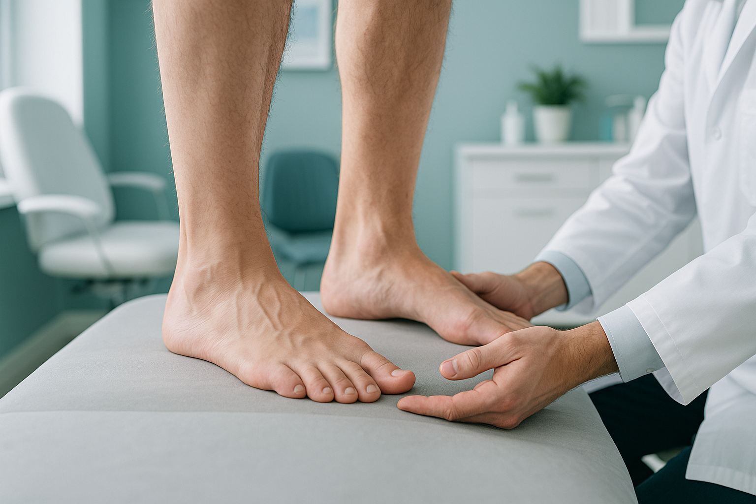
You might notice it when you look at your footprint after a shower, or when your shoes wear down unevenly on the inside edge. But symptoms often go beyond the foot itself.
Pain or tiredness along the inner arch or heel, especially after standing or walking for a while.
When you look from behind, the heel leans outward. This is called hindfoot valgus.
Shoes wearing down heavily on the inner edge, or feeling that your ankles roll inward when you walk.
Flat feet change how load travels up through your legs, sometimes causing knee pain, hip discomfort or lower back stiffness.
What causes flat feet to develop in adults?
Adult acquired flatfoot usually develops slowly. The most common cause is weakening of the posterior tibial tendon, the tendon that supports your arch from the inside of your ankle. But several other factors play a role.
The posterior tibial tendon stretches or tears gradually, losing its ability to hold the arch up.
Carrying extra body weight and the natural ageing of ligaments both increase load on the arch.
A fracture, sprain or inflammatory arthritis can damage the joints and ligaments that maintain the arch.
Naturally loose ligaments let the bones shift more easily under load, flattening the arch over time.
Can flat feet cause knee pain and other problems?
Yes. Your body is a connected chain from your feet to your head. When the arch drops, your foot rolls inward. This is called overpronation. The shin bone follows, rotating inward, which pushes the knee into a knock-kneed position known as knee valgus. Your hip, pelvis and lower back then have to compensate to keep you balanced.
This is why flat feet causing knee pain is so common, and why some people with collapsed arches end up with hip or back symptoms that seem unrelated to their feet. Understanding your full lower limb alignment explained is the key to finding where the real problem starts.
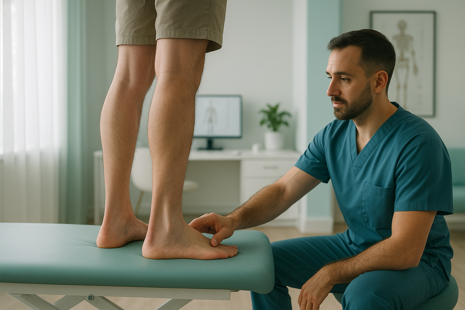
A clinical gait analysis can show how your walking pattern changes when the arch collapses, revealing compensations you may not feel until they become painful.
Why does a standing scan matter for flat feet?
Standard X-rays and MRI scans are taken lying down. When you lie flat, gravity is removed and your muscles relax. A flexible flat foot can look almost normal in that position because the arch is not under load. The alignment problems only appear when you stand and your full body weight presses through the arch.
A standing EOS scan captures your feet, ankles, knees, hips and spine in one single image while you bear weight. It shows exactly how much the arch drops, how the heel tilts, and how those changes affect your knees, hips and back. This full picture helps clinicians see the cause, not just the symptom.
Lying-down imaging relaxes the muscles and removes gravity. The arch may appear partially normal, and alignment shifts further up the chain are hidden. This can lead to incomplete assessments that miss the connection between your flat feet and symptoms elsewhere.
ScanAlign standing EOS imaging uses about 90% less radiation than a standard X-ray, around ten times less. That makes it practical for the full-length weight-bearing images needed to assess flat feet properly.
When MRI is the better choice: EOS shows bones, joints and alignment under load. If your clinician suspects a posterior tibial tendon tear, inflammation or other soft-tissue damage, MRI is usually the better tool for that. The two scans complement each other.
Can flat feet be corrected in adults?
It depends on the type and severity. Full structural correction of a rigid flat foot usually requires surgery. But most adults with flexible flat feet respond well to non-surgical approaches that reduce symptoms and improve function. ScanAlign does not provide treatment. It provides the diagnostic imaging that helps your clinician decide on the right path.
Common approaches your clinician may consider
- Supportive orthotics that prop up the arch and reduce overpronation. Learn more about how orthotics work for overpronation.
- Strengthening exercises targeting the posterior tibial tendon and the small muscles of the foot.
- Footwear changes such as structured shoes with good arch support.
- Physiotherapy to improve gait pattern and reduce compensatory strain on the knees and hips.
- Surgery in more advanced cases, especially if the tendon is badly damaged or the deformity is rigid.
The right option depends on accurate imaging. A weight-bearing scan shows how your foot behaves under real conditions, giving your specialist the information they need.
Flat feet that have not responded to generic insoles or exercises may need a closer look at your whole alignment. A standing scan can reveal what standard imaging misses.
Book a free video consultationFrequently asked questions about flat feet
No. Many adults have flat feet with no pain at all. Flat feet only need attention when they cause symptoms in the feet, knees, hips or back, or when they are getting noticeably worse over time.
Adult acquired flatfoot is when someone who previously had a normal arch develops flat feet later in life. It is usually caused by progressive weakening of the posterior tibial tendon and is more common after age 40.
Yes. When the arch collapses, the chain reaction of misalignment travels upward through the knees and hips to the pelvis and lower back. This is one of the reasons a full standing scan is valuable, because it shows the complete chain in one image.
No. ScanAlign is a private, self-pay clinic. You can book a standing alignment scan directly without a referral. You start with a free video consultation to check that a scan is appropriate for you.
Yes. An EOS scan uses about 90% less radiation than a standard X-ray, around ten times less. It is considered very low-dose imaging.
Pricing details are available on the ScanAlign FAQs and cost page.
Custom orthotics can support the arch, reduce overpronation and ease symptoms. They do not permanently change the bone structure, but for many people they provide significant relief and improved function.
Flexible flat feet have an arch that appears when you sit or go up on tiptoe but flattens under weight. Rigid flat feet remain flat in all positions. Flexible flat feet are far more common and generally respond better to non-surgical treatment.
Lying down removes gravity. The arch is not under load, so it can appear more normal than it actually is. A standing scan captures your foot and entire lower limb alignment under real weight-bearing conditions.
See your GP if you have persistent foot, ankle or knee pain, if one foot is becoming noticeably flatter than the other, or if your symptoms are worsening. If you develop sudden severe pain, swelling or difficulty walking, seek medical attention promptly.
See the full picture, standing up
A free video consultation is the first step. We will discuss your symptoms and explain whether a standing alignment scan is right for you. No referral needed.
Book a free video consultationSources
- Myerson, M.S. (1997). Adult acquired flatfoot deformity. The Journal of Bone and Joint Surgery, 78-A(5), 780-792.
- NHS. Flat feet. nhs.uk. Available at: https://www.nhs.uk/conditions/flat-feet/
- Deschamps, K. et al. (2011). Body of evidence supporting the clinical use of 3D multisegment foot models. Gait & Posture, 33(3), 338-349.
Written by Abbas Dhami (Specialist Diagnostic Radiographer)
This page is general information and not a diagnosis.
ScanAlign is the trading name of CoreMed Solutions Ltd and operates under The Harley Street Hospital's CQC licence.