
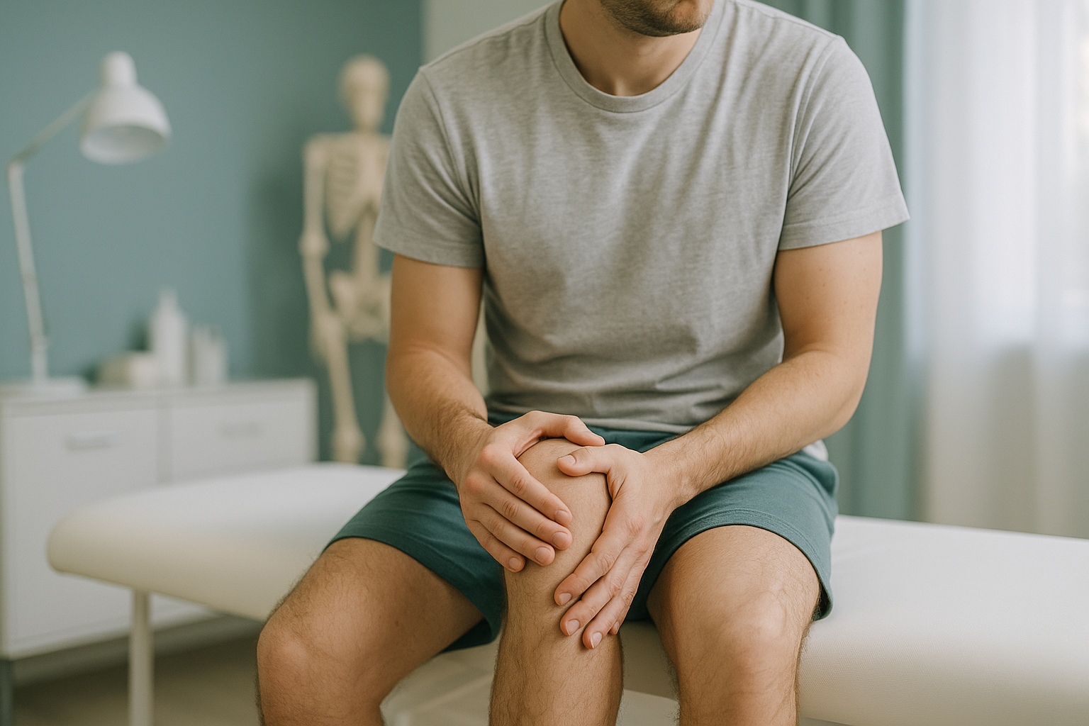
Why Do I Always Get Injured on the Same Side?
Recurring injuries on one side usually mean that side is carrying more load than the other. A subtle difference in leg length, a pelvic tilt, or a shift in your spine can force one hip, knee or ankle to work harder. Finding and measuring that asymmetry is the first step toward breaking the cycle.
Key takeaways
- One-sided injuries are rarely bad luck. They usually point to a structural or alignment cause.
- Your body is a connected chain. A tilt at the pelvis can overload a knee or ankle below it.
- Lying-down scans can miss alignment problems because gravity is removed.
- A standing, full-body image captures both sides on one picture so asymmetry can be measured precisely.
- Diagnosis is the first step. A clear picture helps your clinician target the real source, not just the symptoms.
What does it mean when you keep getting injured on one side?
It means one side of your body is absorbing more force than it should. When your skeleton is perfectly symmetrical, each leg, hip and shoulder shares the load roughly equally. But very few people are perfectly symmetrical. Even a small difference, one that you cannot see in a mirror, can tip the balance.
Over time, the overloaded side wears down faster. You might pull a calf muscle, develop a sore knee, or strain a hip flexor, and it is always the same leg. You recover, return to activity, and the same thing happens again. The injury itself heals, but the underlying reason it happened has not been addressed.
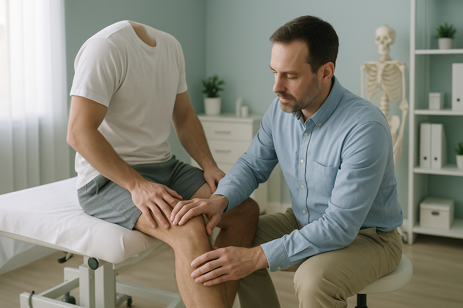
What are the signs that one side of your body is taking more strain?
The signs are often things you have noticed for years but never connected together. Here are the most common patterns.
You always pull the same calf, hamstring or Achilles, even after full rehab.
One shoe sole wears down faster, especially on the outer or inner edge.
After running, walking or sitting for a while, it is always the same joint that stiffens.
A friend or trainer has pointed out that your shoulders are uneven, or your clothes hang unevenly.
What causes one-sided injuries to keep coming back?
Recurring injuries on the same side are rarely caused by a single event. They are almost always driven by an underlying structural or postural factor that has never been identified. The most common causes include the following.
Even a small difference in leg length discrepancy tilts the pelvis, shifting load to the longer side.
A tilted pelvis changes how force travels through the hip joints and down each leg.
A sideways curve in the spine shifts the trunk off-centre, adding load to one side of the body.
When muscle imbalance develops, the weaker side gives way first under load.
How does a problem in one area cause injury somewhere else?
Your body is a connected chain from your feet to your head. When one link is slightly off, the links above and below it compensate. A tilted pelvis, for example, may not cause hip pain at all. Instead, it forces the knee below it to twist inwards slightly with every step, and that knee is the one that keeps getting injured.
This is why treating only the painful area often fails. The injury heals, but the tilt remains, so the same pattern repeats. The real cause and the pain are in different places.
A proper gait analysis or full-body alignment assessment can trace these connections and show exactly where the chain breaks down.
Wondering if alignment could be behind your pattern? A free video consultation lets you talk through your history before committing to a scan.
Book a free video consultationWhy does lying-down imaging miss alignment problems?
Standard X-rays, CT and MRI are usually taken while you lie flat. In that position, gravity is removed, your muscles relax, and your joints settle into a neutral shape. Any tilt, twist or shift that appears when you stand and bear weight simply disappears on the image.
A standing, weight-bearing scan captures your skeleton the way it actually works, under load, with gravity pulling on every joint. Both legs appear on the same image, so a difference in leg length, a pelvic tilt, or an uneven hip height can be measured directly rather than estimated.
Captures your body under load. You stand naturally while a low-dose image is taken from head to foot. Both sides are visible on one picture, so left-to-right differences are measured precisely. The EOS system uses about 90% less radiation than a standard X-ray, around ten times less.
Removes gravity from the picture. Lying flat relaxes your muscles and lets your pelvis settle into a neutral position. A tilt or rotation that causes problems when standing may not appear at all. Each body part is usually imaged separately, making side-to-side comparison harder.
At ScanAlign, full-body standing imaging captures your entire skeleton in one upright scan, giving your clinician a complete map of your alignment from feet to skull.
When is MRI more useful? EOS shows bones, joints and alignment. For soft-tissue problems like disc injuries, ligament tears or nerve compression, MRI is usually the better tool. If your clinician suspects a soft-tissue cause, they may recommend MRI instead of, or alongside, a standing scan.
What can be done once the asymmetry is found?
The answer depends on what the imaging reveals. ScanAlign is a diagnostic imaging clinic, not a treatment provider, so the scan and report give you and your clinician the information needed to make the right decision. Common next steps might include targeted physiotherapy, a heel lift or orthotic, a change in training load, or a referral to an orthopaedic specialist.
The important point is this: without accurate, weight-bearing measurements, any treatment is based on guesswork. A clear picture of the asymmetry turns guesswork into a plan.
- Keep injuring the same leg, hip or knee
- Have been told one leg is shorter
- Notice uneven shoe wear or a lopsided posture
- Have had multiple courses of physio with no lasting result
- Sudden numbness or weakness in a limb
- Loss of bladder or bowel control
- Severe pain after an acute injury or fall
- Contact your GP, call 111, or attend A&E immediately
Frequently asked questions
Recurring injuries in one leg usually mean that leg is carrying more load. A difference in leg length, a pelvic tilt, or an alignment shift can force one side to work harder, making it more vulnerable to strain and overuse.
Yes. Even a subtle difference can tilt the pelvis and change how force travels through the hip, knee and ankle on that side. Over hundreds of thousands of steps, the effect adds up.
Some degree of asymmetry is very common. Problems arise when the asymmetry is large enough to shift load unevenly and the body can no longer compensate without strain.
Standard X-rays are usually taken lying down and focus on one body part at a time. This removes gravity and makes it difficult to compare both sides or see how the whole skeleton lines up under load.
It is an X-ray taken while you stand naturally. The EOS system captures your entire skeleton, from head to feet, in one image with about 90% less radiation than a standard X-ray, around ten times less.
No. ScanAlign accepts self-referrals. You can book a full-body standing scan directly, starting with a free video consultation to discuss your situation.
Pricing details are available on our frequently asked questions page. Visit the FAQs page for the latest information.
The EOS system uses about 90% less radiation than a standard X-ray, around ten times less. It is one of the lowest-dose skeletal imaging systems available.
No. A standing scan shows bones, joints and alignment. For soft-tissue problems such as ligament tears, disc injuries or nerve issues, MRI is usually the better tool.
You receive a detailed report showing your alignment measurements. You can share this with your physiotherapist, osteopath, or orthopaedic surgeon so they can plan treatment based on accurate, weight-bearing data.
Stop guessing. See the full picture.
A free video consultation lets you discuss your injury pattern with a specialist radiographer before committing to anything.
Book a free video consultationWritten by Abbas Dhami (Specialist Diagnostic Radiographer)
Sources
- Khamis, S. & Yizhar, Z. (2007). Effect of feet hyperpronation on pelvic alignment in a standing position. Gait & Posture, 25(1), 127-134.
- Gurney, B. (2002). Leg length discrepancy. Gait & Posture, 15(2), 195-206.
- NHS. (2023). Back pain: causes, treatment and prevention. NHS website. Available at: nhs.uk/conditions/back-pain.
This page is general information and not a diagnosis.
ScanAlign is the trading name of CoreMed Solutions Ltd and operates under The Harley Street Hospital's CQC licence.