
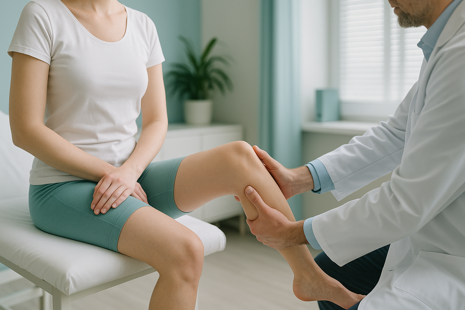
What Are Hyperextended Knees (Genu Recurvatum), and Are They a Problem?
Knee hyperextension, also called genu recurvatum, means the knee bends further back than its normal straight position when you stand. In mild cases it may cause no symptoms at all. But when the knee regularly pushes backwards under load, it can stress the joint, alter your whole leg alignment, and cause pain in places you would not expect.
Key takeaways
- Hyperextended knees curve backwards beyond straight when you bear weight.
- Common causes include joint hypermobility, quadriceps weakness, and habitual locking when standing.
- Pain often appears in the hip, lower back or ankle rather than the knee itself.
- The problem only shows up properly on imaging taken while you are standing.
- An EOS standing scan captures full leg alignment in a single low-radiation image.
How do you know if your knees hyperextend?
The simplest test is to stand naturally and look at your leg from the side. If the knee appears to bow backwards, it is hyperextending. You may also notice the following.
You push your knees fully back to feel "stable", rather than holding them slightly soft.
The posterior capsule and ligaments stretch under repeated backward pressure, causing a dull ache.
The quadriceps switch off when the knee locks back, so the muscles lose conditioning over time.
The hip, lower back or ankle may hurt because they are compensating for the unstable knee position.
What causes knees to bend backwards?
Genu recurvatum happens when the structures that normally stop the knee at straight are too loose, too weak, or both. Several factors can contribute.
Naturally loose connective tissue lets all joints move further than average. This is common in people with connective tissue disorders or hypermobile Ehlers-Danlos syndrome.
When the front thigh muscles cannot hold the knee steady, the joint drifts back into a locked position for passive support.
Years of locking the knees back while standing, especially in jobs that involve long periods on your feet, gradually stretches the posterior capsule.
A ligament injury (particularly the ACL or PCL) or a fracture that changed the bone shape can leave the knee unable to stop at straight.
Dancers, gymnasts and people who train heavily in end-range flexibility are also at higher risk, because their loading strategy regularly pushes the knee beyond its comfortable range.
Why does knee hyperextension cause pain somewhere else?
Your body works as a connected chain from your feet to your head. When the knee pushes backwards, the pelvis tilts, the lower back curve changes, and the ankle rolls to compensate. Pain often ends up in the hip or spine rather than the knee, which is why scans of a single area can miss the real cause.
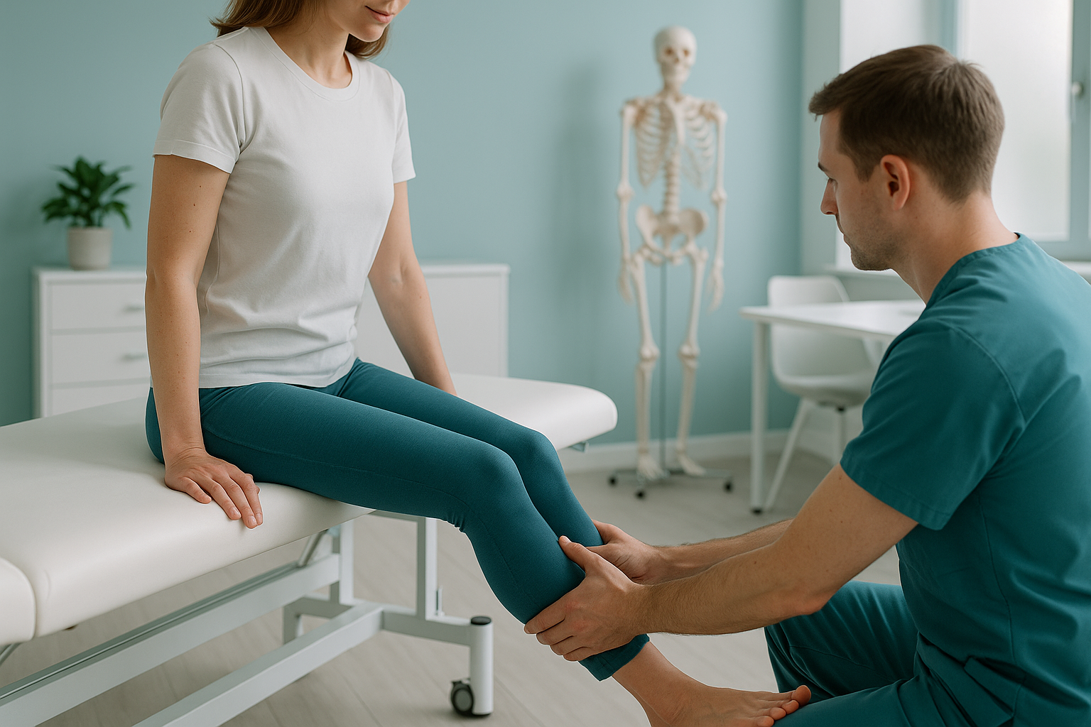
This compensation effect is why many people with persistent hip or lower back pain have never had their knee alignment checked. The body adapted, but the adaptation itself became the source of symptoms.
How is knee hyperextension assessed with a standing scan?
Knee hyperextension is a weight-bearing problem. It only appears when gravity is acting on the joint. Lying-down scans (standard X-ray, CT, MRI) relax the muscles and let the knee settle into a neutral position, so the backward bend simply disappears from the image.
An EOS scan at ScanAlign standing EOS imaging captures a full-length image of both legs and the spine while you stand naturally. The sagittal knee angle, the pelvic tilt, and the spinal curves are all visible in a single, weight-bearing picture. This lets a clinician see the entire compensation chain rather than a cropped snapshot of one joint.
The EOS system uses about 90% less radiation than a standard X-ray, around ten times less. That makes it practical for follow-up scans to track whether rehabilitation or bracing is making a measurable difference.
When is MRI the better choice? EOS shows bones, joints and alignment. If your clinician suspects soft-tissue damage inside the knee, such as a torn ligament, cartilage injury or a Baker's cyst, MRI is usually the better tool. The two types of scan answer different questions and are often used together.
What helps manage hyperextended knees?
Management depends on the cause and severity. ScanAlign is a diagnostic imaging clinic, not a treatment provider, so the suggestions below are general guidance that a physiotherapist or orthopaedic specialist would typically consider.
Rebuilding the front thigh muscles gives the knee active control so it does not need to lock backwards for stability.
Balance exercises help retrain the brain's sense of where the knee is in space, reducing the habit of pushing it back.
Learning to stand with a slight "micro-bend" at the knee, rather than locking out, takes pressure off the posterior capsule over time.
A standing alignment scan provides the measurements a specialist needs to plan bracing, orthotics or surgical strategy if conservative care is not enough.
Want to find out whether your knee alignment is contributing to your symptoms? A standing EOS scan can show the full picture.
Book a free video consultationWhen should you see a doctor urgently?
Most knee hyperextension is a long-standing postural or flexibility issue, not an emergency. However, if you experience any of the following after an acute injury, seek urgent medical attention through your GP, 111, or A&E.
- Sudden severe swelling or an inability to bear weight after a fall or twist.
- Numbness, tingling or weakness in the lower leg or foot.
- The knee giving way completely without warning.
These may indicate a ligament rupture or nerve injury that needs prompt assessment.
Frequently asked questions about knee hyperextension
Not exactly. "Double-jointed" is an informal term for generalised joint hypermobility. Knee hyperextension is one specific sign of hypermobility, but you can hyperextend your knees without being generally hypermobile, for example after an injury or from a standing habit.
Over time, yes. Repeatedly forcing the knee into full extension stretches the posterior capsule and ligaments. It also lets the quadriceps switch off, which weakens them and makes the locking habit harder to break.
A standard X-ray is usually taken lying down, which relaxes the muscles and hides the backward bend. A weight-bearing image taken while you stand is far more reliable for showing how far the knee actually extends under load.
Yes. Knee hyperextension is one of the criteria assessed in the Beighton score, which is used to screen for generalised hypermobility. People with conditions such as hypermobile Ehlers-Danlos syndrome or other connective tissue disorders are more likely to have it.
Targeted strengthening (especially the quadriceps and hamstrings) and proprioception exercises can significantly reduce the degree of hyperextension and improve symptoms. A physiotherapist can design a programme based on your specific alignment findings.
When the knee pushes backwards, the pelvis tilts and the spinal curves shift to compensate. This compensation can overload the lower back or hip. A full standing scan reveals the connection by capturing the knees, pelvis and spine together.
The EOS system uses about 90% less radiation than a standard X-ray, around ten times less. This makes it particularly suitable when repeated scans are needed to monitor progress.
No. ScanAlign accepts self-referrals. You can book a free video consultation directly to discuss whether a standing scan is appropriate for your symptoms.
Yes. The EOS scan is taken while you stand in your natural posture, which is exactly what your clinician needs to see. You do not need to hold any unusual position.
Pricing details are available on the ScanAlign FAQs and pricing page. You can also ask about costs during your free video consultation.
See your full alignment, standing up
A single EOS image captures your knees, hips and spine under real weight-bearing conditions, with about 90% less radiation than a standard X-ray.
Book a free video consultationWritten by Abbas Dhami (Specialist Diagnostic Radiographer)
Sources
- Loudon JK, Goist HL, Loudon KL. Genu recurvatum syndrome. Journal of Orthopaedic & Sports Physical Therapy. 1998;28(5):361-367.
- Beighton P, Solomon L, Soskolne CL. Articular mobility in an African population. Annals of the Rheumatic Diseases. 1973;32(5):413-418.
- NHS guidance on joint hypermobility. nhs.uk. Available at: https://www.nhs.uk/conditions/joint-hypermobility/
This page is general information and not a diagnosis.
ScanAlign is the trading name of CoreMed Solutions Ltd and operates under The Harley Street Hospital's CQC licence.