

Why Do My Knees Cave In When I Squat?
Your knees cave inward during a squat, a pattern called dynamic knee valgus, because of weakness in the hip muscles, limited ankle mobility, or the way your bones are shaped. It puts extra stress on your knees and can cause pain. A standing whole-body scan helps identify whether bone alignment, muscle imbalance, or both are driving the problem.
Key takeaways
- Knees caving inward under load is called dynamic knee valgus, and it is very common.
- Weak glute medius muscles, tight ankles and foot pronation are the most frequent causes.
- Bone shape, including hip anteversion and retroversion, can make valgus worse.
- Lying-down scans miss alignment problems that only appear when you are standing and weight-bearing.
- A standing EOS scan shows your full leg alignment from hip to ankle in one image.
What is dynamic knee valgus?
Dynamic knee valgus is when your knees collapse inward during movement, most obviously during a squat, a lunge, or when landing from a jump. It is different from structural knock knees, which is a fixed bone position. Dynamic valgus happens only when you load the joint. It may look like a small wobble or a dramatic inward cave, depending on the cause.
This movement pattern increases stress on the inner side of the knee, the kneecap, and the ligaments that hold the joint stable. Over time, repeated valgus loading can contribute to knee pain, patellofemoral problems and even injury.
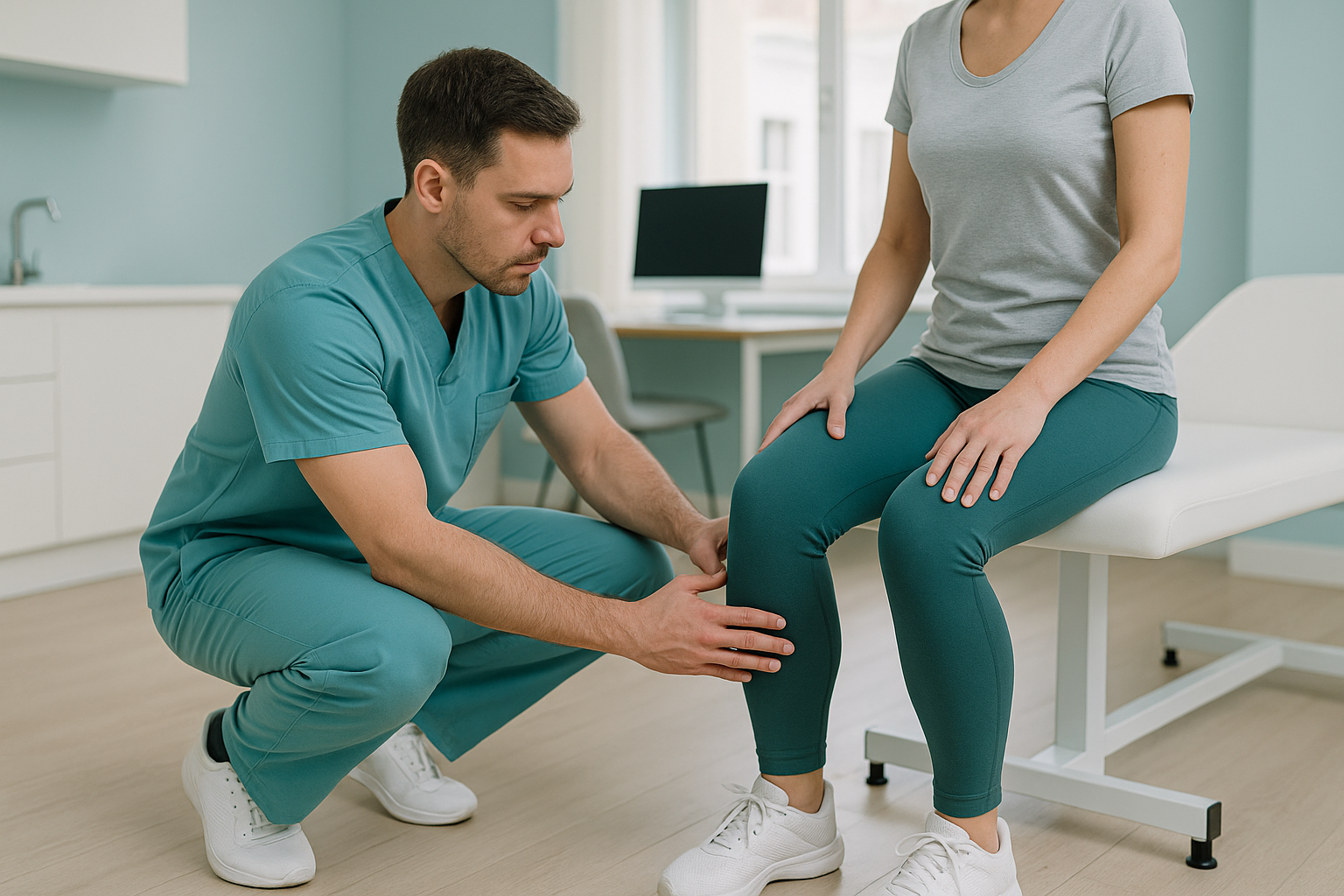
How do I know if my knees are caving in?
You may spot it yourself in a mirror, or someone else may point it out. These are the most common signs.
Watching yourself squat, you notice one or both knees drift toward the midline instead of tracking over your toes.
Aching around or behind the kneecap during or after loaded squats, lunges or stair climbing.
The inner edges of your shoes wear down faster than the outer edges, a sign of foot pronation linked to valgus.
Your knees track well for the first few reps but cave inward as the set goes on and muscles tire.
What causes knees to cave in during a squat?
Several factors can make your knees collapse inward, and most people have more than one at play. The causes fall into two groups: soft tissue (muscles and tendons) and structure (the shape of your bones).
The glute medius controls hip external rotation. When it is weak, the thigh rolls inward and the knee follows.
If your ankle cannot bend far enough, your body compensates by letting the knee drop inward to get lower.
Flat or overpronating feet tilt the shin inward, which pushes the knee into valgus from the ground up.
The natural twist of the thigh bone varies between people. More anteversion can predispose the knee to caving in.
Stance width and cueing also matter. A very narrow squat stance or simply not knowing where your knees should track makes valgus more likely. As load or fatigue increases, even people with good technique can lose control.
Why does the whole leg matter, not just the knee?
Your body is a connected chain from your feet to your head. A problem at one level makes another level compensate. Knee valgus rarely starts at the knee itself. It is usually driven from above (the hip) or below (the ankle and foot).
This is why a scan of just the knee often misses the real issue. It might show the joint looks normal, while the reason for your pain, perhaps a rotational pattern in the hip or poor alignment through the whole leg, stays hidden.
Lying-down imaging relaxes the muscles and removes gravity. A knee that collapses inward only when standing and loaded will look perfectly fine on a scan taken while you are flat on your back. Standing imaging captures what actually happens under your own body weight.
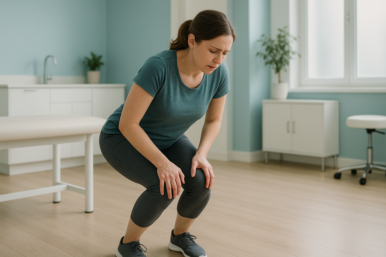
How does a standing EOS scan help with knee valgus?
An EOS scan captures your full leg alignment, from the hip joint down through the knee and ankle, in a single standing image. Because you are upright and weight-bearing, it shows the alignment your body actually uses, not the relaxed position that appears when you lie down.
The scan uses about 90% less radiation than a standard X-ray, around ten times less. It takes seconds to capture and requires no special preparation.
With ScanAlign standing EOS imaging, a specialist radiographer assesses the relationship between your hip rotation, knee angle and ankle position in one view. This can reveal whether the valgus pattern is driven by bone shape, by soft-tissue factors, or by a combination of both.
Want to understand what is driving your knee valgus? A free video consultation can help you decide whether a standing scan is the right next step.
Book a free video consultationWhen MRI is the better tool: EOS shows bones, joints and alignment. If your clinician suspects a soft-tissue injury, such as a meniscus tear, cartilage damage or ligament strain, MRI is usually the better choice. The two scans answer different questions and can work well together.
What helps stop knees collapsing inward?
Addressing knee valgus usually means working on the areas that cause it, not the knee itself. Common approaches recommended by physiotherapists and strength coaches include the following.
- Glute medius strengthening. Exercises such as banded side-walks, clamshells and single-leg work build the hip muscles that control inward rotation of the thigh.
- Ankle mobility drills. Improving dorsiflexion lets you squat deeper without the knee compensating.
- Cueing and technique. Thinking "knees out" during a squat, or using a light resistance band around the knees as a reminder, helps retrain the movement pattern.
- Stance width adjustment. A slightly wider stance with toes turned out can reduce the demand on ankle mobility and make it easier to keep the knees tracking well.
- Foot support. If foot pronation is a factor, appropriate footwear or insoles may help control the chain reaction from the ground up.
These strategies work well when the cause is muscular. But if a structural issue, such as the way your thigh bone or shin bone is shaped, is contributing, exercises alone may not fully resolve it. That is where imaging becomes important, so you and your clinician know exactly what you are dealing with before deciding on a plan. You can book a leg alignment scan to get a clear picture of your structure.
Frequently asked questions
Pain during squats often comes from the kneecap being pulled out of its ideal track. When the knee caves inward (dynamic valgus), it increases pressure on the inner joint and the underside of the kneecap. Weak hip muscles, limited ankle flexibility and bone alignment can all contribute.
Not exactly. Knock knees (genu valgum) is a structural alignment where the knees sit inward even when standing still. Dynamic knee valgus only appears during loaded movement like squatting or jumping. Some people have both. You can learn more about the structural side on our knock knees page.
Yes. The glute medius controls hip external rotation. When it is weak, the thigh rotates inward under load, pulling the knee with it. This is one of the most common and well-documented causes of dynamic valgus and related knee pain.
A band is a useful cueing tool. It reminds you to push your knees outward and activates the glute medius. But it addresses the symptom, not the root cause. If bone shape or a significant muscle imbalance is involved, you may need a fuller assessment to guide your approach.
It can. As you squat deeper, the demand on ankle dorsiflexion and hip control increases. If either is limited, the knee is more likely to cave. Adjusting stance width or heel elevation can help you train through a comfortable range while you work on the underlying causes.
Yes. Flat or overpronating feet tilt the shin inward, which pushes the knee into valgus from the ground up. This is why a whole-leg assessment, from the hip to the foot, matters. Fixing just one level may not be enough if the chain reaction starts at the foot.
No. ScanAlign is a self-referral clinic. You can book directly without needing a GP or consultant letter. The process starts with a free video consultation to make sure the scan is appropriate for you.
EOS uses about 90% less radiation than a standard X-ray, around ten times less. It is one of the lowest-dose skeletal imaging methods available.
Pricing details are on our FAQs and cost page. The free video consultation will also confirm which scan package suits your situation.
It can be. The natural twist of the thigh bone (femoral version) varies between people. Greater anteversion angles the thigh inward, making valgus more likely regardless of muscle strength. A standing EOS scan can assess this. Our page on hip anteversion and retroversion covers this in more detail.
Find out what is behind your knee valgus
A free video consultation with a specialist radiographer can help you understand whether a standing alignment scan is the right next step for you.
Book a free video consultationWritten by Abbas Dhami (Specialist Diagnostic Radiographer)
Sources
- Powers CM. The influence of abnormal hip mechanics on knee injury: a biomechanical perspective. Journal of Orthopaedic & Sports Physical Therapy. 2010;40(2):42-51.
- Hewett TE, Myer GD, Ford KR et al. Biomechanical measures of neuromuscular control and valgus loading of the knee predict anterior cruciate ligament injury risk in female athletes. American Journal of Sports Medicine. 2005;33(4):492-501.
- NHS. Knee pain and what to do about it. nhs.uk. Available at: https://www.nhs.uk/conditions/knee-pain/
This page is general information and not a diagnosis.
ScanAlign is the trading name of CoreMed Solutions Ltd and operates under The Harley Street Hospital's CQC licence.