
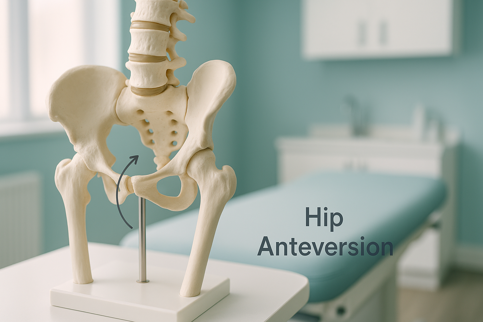
Hip Anteversion and Retroversion: What Are They and Could They Be Causing Your Pain?
Hip anteversion means the top of the thigh bone (femur) or the hip socket angles too far forward. Hip retroversion means it angles too far back. Both change the way your leg sits and moves, which can cause hip, knee or lower back pain, especially when you are standing and weight-bearing.
Key takeaways
- Anteversion and retroversion describe the twist angle of the femur or hip socket.
- They often show up as in-toeing (feet turning in) or out-toeing (feet turning out) when you walk.
- Pain may appear in the hip, knee or back because the rest of the body compensates.
- Lying-down scans can miss these alignment issues because gravity and muscle tension are removed.
- A standing whole-body EOS scan shows how the hip angle affects your entire chain of alignment.
What is hip anteversion and retroversion?
Hip anteversion is a twist in the femur or the acetabulum (hip socket) that makes the joint point more forward than normal. Hip retroversion is the opposite, where the joint points more backward. Everyone has a natural angle of twist. Problems arise when that angle sits outside the usual range and begins to affect how you stand, walk and distribute weight.
The twist can involve the femur itself (femoral anteversion or femoral retroversion) or the socket it sits into (acetabular retroversion or anteversion). Sometimes both are affected at once.
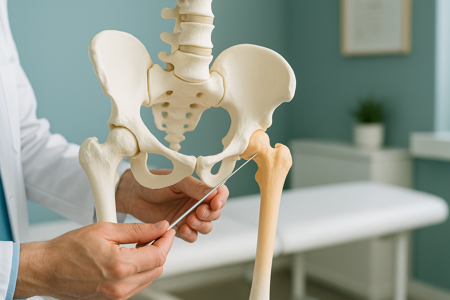
Think of the femur like a key fitting into a lock. If the key is twisted slightly, it still fits, but it does not turn smoothly. Over time the joint may pinch (impingement) on one side or sit too loosely on the other, leading to cartilage wear and pain.
What are the signs of femoral anteversion or retroversion?
The signs depend on which direction the twist goes. Many people have lived with these patterns their whole lives without realising there is a structural reason behind them.
Feet that naturally point inward (anteversion) or outward (retroversion) when you stand or walk.
A deep ache in the front of the hip or groin, often worse after long walks or sitting cross-legged.
The twist higher up changes how your kneecap tracks, causing pain that scans of the knee alone may not explain.
Your pelvis tilts to compensate for the hip angle, which can overload the lower spine.
If you notice any combination of in-toeing and out-toeing with hip, knee or back discomfort, the cause may sit in the angle of your femur or socket rather than in the area where you feel pain.
What causes hip anteversion or retroversion in adults?
In most adults, femoral anteversion or retroversion is a structural trait that developed during growth. The femur gradually twists into its final position during childhood and adolescence, and sometimes that process leaves a greater or lesser twist than average.
The twist angle is set during growth and remains into adulthood.
The socket itself may face too far forward or backward, independent of the femur.
Years of compensating for the twist can lead to cartilage wear and early joint changes.
Because these patterns are structural, they tend to be long-standing. You may have been told your hip "looks normal" on a standard X-ray taken while lying down. That is because the twist is best seen when you are standing and your muscles are working to hold you upright.
How is hip version assessed with a standing EOS scan?
A standing EOS scan captures your entire skeleton, from head to feet, in a single upright image. This is the key difference. When you lie down for a standard X-ray, CT or MRI, gravity is removed and your muscles relax. That can mask the true angle of the hip and the knock-on effects it has further up or down the body.
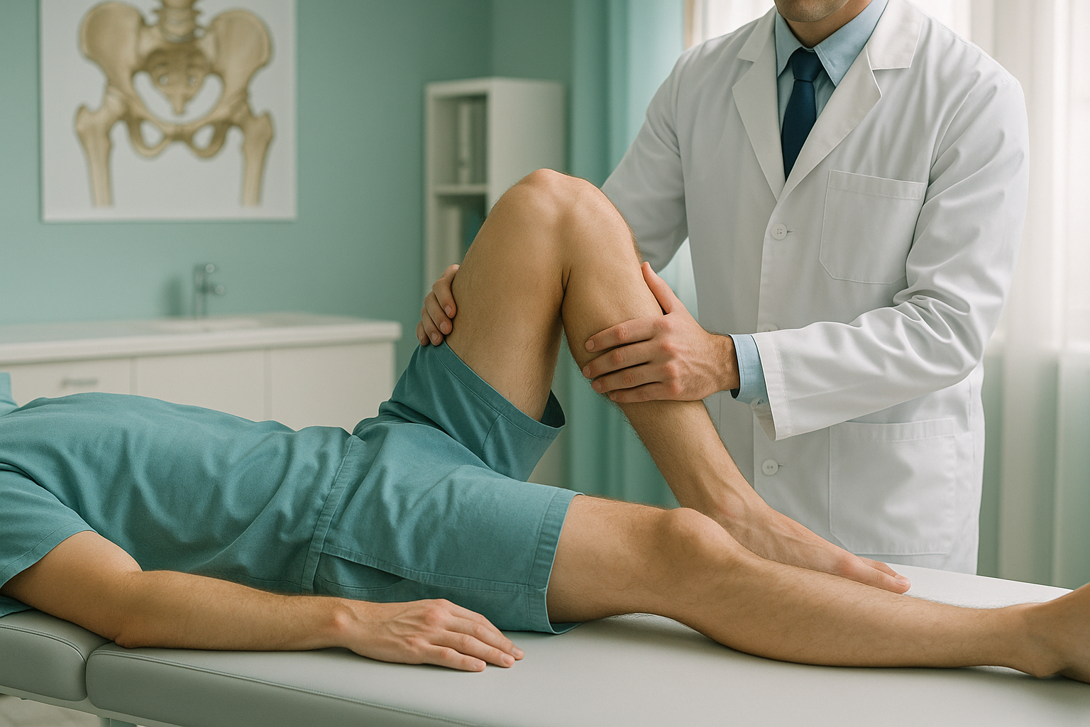
With EOS, your clinician can see how the hip twist affects your knee alignment, pelvic tilt, spine curvature and even how your weight is distributed across both legs. Because it captures the full picture in one scan, it removes the guesswork of piecing together separate images of separate body parts.
EOS also uses about 90% less radiation than a standard X-ray, around ten times less. That makes it a practical choice when a full standing assessment is needed.
Wondering whether your hip angle could be behind your symptoms? A standing EOS scan shows the full picture.
Book a free video consultationWhat can be done about hip anteversion or retroversion?
The first step is getting an accurate picture of how far the twist goes and what effect it has on the rest of your body. Without that information, treatment often targets the wrong area.
Once the alignment picture is clear, your treating clinician (orthopaedic surgeon, physiotherapist or sports medicine specialist) can tailor a plan. Options might include targeted physiotherapy to strengthen the muscles around the hip, gait retraining, or in more significant cases, surgical correction. ScanAlign provides the diagnostic imaging that informs those decisions. It does not provide treatment itself.
When MRI is the better tool: EOS is excellent at showing bone position, joint angles and whole-body alignment. If your clinician suspects a labral tear, cartilage damage or soft-tissue injury inside the hip, MRI is usually the better choice for those specific details. The two scans complement each other.
Your body is a connected chain from your feet to your head. A twist at the hip can tilt the pelvis, curve the spine and change how your knees track. Treating only the painful area without understanding the underlying alignment pattern often means the problem keeps coming back. A whole-body standing image helps make sure the full picture is considered from the start.
Frequently asked questions about hip anteversion and retroversion
They are closely related. Femoral anteversion refers specifically to the twist in the thigh bone. Hip anteversion is a broader term that can also include the angle of the socket (acetabular anteversion). Both affect how the joint sits and moves.
Yes. Acetabular retroversion in particular can cause the front of the socket to pinch against the femoral head during movement, a form of hip impingement. This often shows up as groin or deep front-of-hip pain, especially with bending or sitting for long periods.
In-toeing is one of the most common signs of femoral anteversion, but it can also come from tibial torsion (a twist in the shin bone) or from foot structure. A standing whole-body scan helps identify where in the chain the twist originates.
A single lying-down X-ray gives limited information about the twist angle. The version of the femur is a three-dimensional measurement that is much better captured with imaging that includes standing weight-bearing views or cross-sectional techniques.
EOS captures a full standing image of your skeleton in one go. This lets your clinician see how the hip twist affects your leg rotation, knee alignment, pelvic tilt and spine. It shows the real-world alignment under gravity, which lying-down scans can miss.
Yes. EOS uses about 90% less radiation than a standard X-ray, around ten times less. You can find more details about safety and what to expect on our FAQs page.
No. ScanAlign is a private self-pay clinic and you can self-refer. You start with a free video consultation where a specialist radiographer reviews your history and confirms whether a scan is appropriate.
Pricing details are available on our FAQs and cost page.
It can. When the femur is twisted inward more than usual, the kneecap may track differently, which puts extra stress on the front of the knee. This is why knee pain sometimes persists even after knee-focused investigations come back normal.
The twist angle is mostly set by the time you finish growing. However, the symptoms it causes often become noticeable later in life as joint wear progresses or activity levels change. Previous hip surgery can also alter the version angle.
See how your hip alignment affects your whole body
A standing EOS scan at ScanAlign shows bones, joints and alignment from head to feet, all in one image. Start with a free video consultation.
Book a free video consultationLearn more about hip alignment and EOS imaging, or explore how EOS works on our main EOS scan page.
Author: Abbas Dhami (Specialist Diagnostic Radiographer)
Sources
- Tönnis D, Heinecke A. Acetabular and femoral anteversion: relationship with osteoarthritis of the hip. Journal of Bone and Joint Surgery (American). 1999;81(12):1747-1770.
- Reynolds D, Lucas J, Klaue K. Retroversion of the acetabulum: a cause of hip pain. Journal of Bone and Joint Surgery (British). 1999;81(2):281-288.
- NHS. Hip pain in adults: causes and when to get help. nhs.uk. Available at: https://www.nhs.uk/conditions/hip-pain/
This page is general information and not a diagnosis.
ScanAlign is the trading name of CoreMed Solutions Ltd and operates under The Harley Street Hospital's CQC licence.