

What Is a Microdiscectomy, and When Is It Recommended?
A microdiscectomy is a minimally invasive spinal operation that removes the portion of a herniated disc pressing on a nerve. It is usually recommended when leg pain caused by nerve root compression has not improved after several weeks of conservative treatment such as physiotherapy and pain relief.
Key takeaways
- Microdiscectomy targets disc material that is compressing a spinal nerve, usually in the lower back.
- It is typically a day-case or overnight procedure carried out under general anaesthetic.
- Surgery is considered after conservative measures have been given a fair trial and symptoms persist.
- Most people notice significant leg-pain relief shortly after the operation.
- Pre-operative and follow-up imaging helps your surgeon plan the approach and track your recovery.
What exactly is a microdiscectomy?
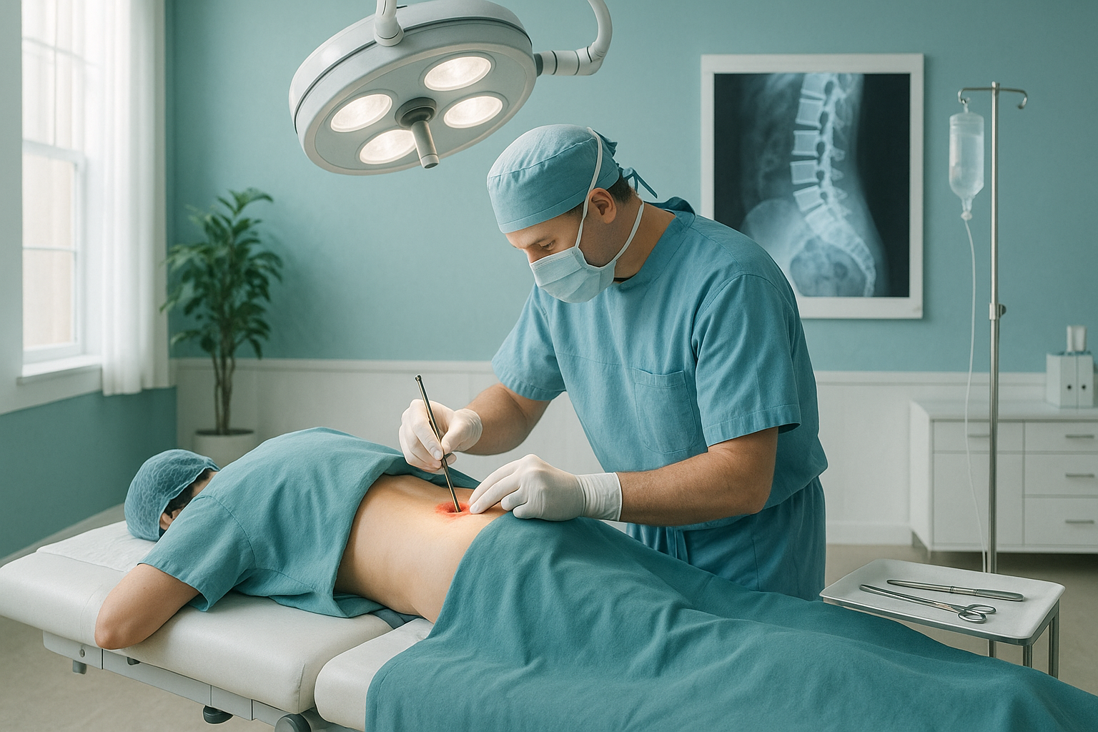
A microdiscectomy is a surgical procedure to remove a small piece of a spinal disc that has bulged or ruptured and is pressing on a nearby nerve root. The surgeon works through a small incision, usually less than a few centimetres, using a microscope or magnifying loupes for precision. Only the fragment of disc causing the problem is removed, not the entire disc.
The operation is sometimes called a lumbar discectomy because it is most commonly performed in the lower (lumbar) spine. It differs from a laminectomy, which removes part of the bony roof of the spinal canal to relieve broader pressure. In practice, a surgeon may trim a tiny amount of bone to gain access, but the main goal is nerve root decompression rather than widening the canal.
When is a microdiscectomy recommended?
A microdiscectomy is usually recommended when a herniated disc is causing persistent leg pain, numbness or weakness that has not responded to conservative treatment over a reasonable period, typically six to twelve weeks. The key symptom is lumbar radiculopathy, where the compressed nerve sends pain, tingling or weakness down one leg. The procedure tends to be more effective for leg pain than for back pain alone.
Conservative measures tried first often include physiotherapy, anti-inflammatory medication, activity modification and sometimes steroid injections. Surgery is generally not the first option. Your specialist will weigh the severity of your symptoms, how much they affect your daily life, and the imaging findings before recommending surgery.
Urgent note. If you develop sudden loss of bladder or bowel control, numbness around your groin or saddle area, or rapidly worsening weakness in both legs, seek urgent medical attention immediately. Contact your GP, call 111 or go to A&E. These symptoms may indicate a condition called cauda equina syndrome, which requires emergency assessment.
What symptoms suggest you might need a microdiscectomy?
The hallmark symptom is leg pain that follows the path of the compressed nerve, often described as sharp, burning or electric. Below are the common signs that may point towards surgery being considered.
Pain radiating from the lower back into the buttock and down one leg, often worse than the back pain itself.
Altered sensation in the leg, foot or toes supplied by the affected nerve.
Difficulty lifting the foot, pushing off when walking, or a feeling of the leg giving way.
Symptoms that have not improved meaningfully after weeks of physiotherapy, medication and rest.
How is lumbar discectomy surgery performed?
Lumbar discectomy surgery is performed under general anaesthetic, typically as a day case or with an overnight stay. Here is a simplified overview of what happens.
You are put to sleep under general anaesthetic and positioned face down on the operating table.
The surgeon makes a short cut over the affected level of the spine, guided by imaging taken beforehand.
Using a microscope, the surgeon moves the nerve root gently aside and removes the disc fragment pressing on it.
The incision is closed. You are monitored as the anaesthetic wears off and most people can walk the same day.
How long does discectomy recovery take?
Most people feel a noticeable improvement in leg pain within the first few days after surgery. Full recovery varies from person to person, depending on factors like fitness, job demands and how long symptoms were present before the operation.
As a rough guide, many people return to light desk work within two to four weeks and to more physical roles within six to twelve weeks. Your surgeon and physiotherapist will give you a personalised timeline. Gradual return to exercise and lifting is usually encouraged rather than jumping straight back in.
It is common to have some soreness around the incision site for the first few weeks. This is different from the nerve pain you had before and usually settles on its own.
What are the risks of microdiscectomy?
Like any surgery, microdiscectomy carries some risks. Most are uncommon, but it is important to be aware of them before making a decision.
- Recurrence of herniation. A small proportion of people experience another disc herniation at the same level in the months or years after surgery.
- Infection. Wound or deeper infection is possible but uncommon with modern surgical techniques.
- Nerve damage. There is a small risk of damage to the nerve root or its covering (dura) during surgery.
- Ongoing symptoms. Some people find their back pain or leg symptoms do not fully resolve, particularly if the nerve was compressed for a long time before surgery.
- General anaesthetic risks. Blood clots, breathing issues and reactions to medication, though these are rare.
Your surgeon will discuss these risks with you in detail before you consent to the procedure.
How does standing imaging help before or after a microdiscectomy?
Standard pre-operative imaging, such as MRI, is done while you are lying down. This is excellent for seeing soft tissue detail like the disc and nerve. However, lying-down scans relax the muscles and remove the effect of gravity, which can mask alignment issues that only appear when you are upright and weight-bearing.
A standing whole-body EOS scan shows your full spinal alignment under load, from your head down to your feet, in a single image. This can reveal how a disc problem fits into a bigger picture of compensatory posture changes. For example, a herniated disc on one side can cause you to lean away from the pain, shifting your pelvis and changing the curves in your spine. These shifts are invisible on a lying-down scan.
After surgery, a standing image can help your surgeon and physiotherapist see whether your alignment has improved and guide your rehabilitation. ScanAlign standing EOS imaging uses about 90% less radiation than a standard X-ray, around ten times less, making it a practical option for follow-up checks.
A note about MRI. EOS shows bones, joints and alignment clearly. For soft tissue detail, such as discs, nerves and ligaments, MRI is usually the better tool. The two types of imaging answer different questions and often complement each other. Your surgeon may use both when planning or reviewing your surgery.
Considering pre-operative or follow-up imaging for spinal surgery? You can book pre-operative or follow-up imaging at our Harley Street clinic.
Book a free video consultationFrequently asked questions about microdiscectomy
The terms are often used interchangeably. A microdiscectomy uses a microscope or magnifying loupes through a smaller incision, while a traditional discectomy may involve a larger opening. The goal of both is the same: removing disc material that is compressing a nerve.
Many microdiscectomies are performed as day-case surgery, meaning you go home the same day. Some people stay overnight. Your surgical team will advise based on your individual circumstances.
This depends on the physical demands of your job. Desk-based workers often return within two to four weeks. More physical roles may need six to twelve weeks. Your surgeon will give you specific guidance.
Published research suggests that microdiscectomy relieves leg pain in most patients when they are carefully selected for surgery. Individual outcomes depend on many factors, including how long the nerve was compressed and overall health. Your surgeon can discuss what to expect in your case.
Yes. A proportion of patients experience a recurrence of herniation at the same level. Maintaining a healthy weight, staying active and following your physiotherapy programme can help reduce this risk, though it cannot be eliminated entirely.
Microdiscectomy is primarily aimed at relieving leg pain caused by nerve compression. It is less predictable for back pain on its own. If your main complaint is back pain without significant leg symptoms, your specialist may recommend other approaches first.
No. ScanAlign accepts self-referrals. You can book a free video consultation to discuss whether an EOS scan would be helpful for your situation, whether that is before or after spinal surgery.
An EOS scan uses about 90% less radiation than a standard X-ray, around ten times less. This makes it especially practical when follow-up or repeated imaging is needed.
MRI is done lying down and excels at showing soft tissues like discs and nerves. A standing EOS scan shows how your entire skeleton aligns under the force of gravity. Together, they give your surgeon a more complete picture, especially when compensatory posture changes are suspected.
Most guidelines recommend trying conservative treatment first. This usually includes physiotherapy, pain-relief medication, staying as active as you comfortably can, and sometimes guided injections. Surgery is typically considered when these measures have been given a fair trial and symptoms remain severe.
Understand your alignment before or after surgery
A standing whole-body EOS scan shows how your spine, pelvis and legs align under gravity. It can help your surgical team plan the approach or track your recovery.
Book a free video consultationSources
- NHS, "Lumbar decompression surgery," nhs.uk, accessed 2024.
- Weinstein JN et al., "Surgical vs Nonoperative Treatment for Lumbar Disk Herniation: The Spine Patient Outcomes Research Trial (SPORT)," JAMA, 2006;296(20):2441-2450.
- British Association of Spine Surgeons (BASS), patient information on discectomy, spinesurgeons.ac.uk.
Written by Abbas Dhami (Specialist Diagnostic Radiographer)
This page is general information and not a diagnosis.
ScanAlign is the trading name of CoreMed Solutions Ltd and operates under The Harley Street Hospital's CQC licence.