

Nerve Root Compression: What Causes It and How Do You Find Relief?
Nerve root compression happens when a spinal nerve is squeezed where it leaves the spine. This can cause pain, tingling or weakness that travels into an arm or leg. Common causes include disc bulges, bony spurs and spinal narrowing. Identifying exactly where the compression occurs is the key first step toward relief.
Key takeaways
- A compressed nerve root can cause pain, numbness or weakness far from the spine itself.
- Lying-down scans can miss alignment problems that put extra pressure on nerves when you stand.
- EOS whole-body standing imaging shows your skeleton under real weight-bearing load, helping pinpoint the source.
- ScanAlign is a diagnostic imaging clinic. It does not treat nerve compression, but provides the detailed picture that guides treatment elsewhere.
Seek urgent help if you notice: sudden loss of bladder or bowel control, numbness in the groin or saddle area, or rapidly worsening weakness in both legs. Contact your GP, call 111 or go to A&E straight away. These could be signs of a serious condition that needs immediate attention.
Your spine is made up of bones stacked on top of each other, with nerves branching out at every level. Each of these nerve roots carries signals between your brain and a specific part of your body. When something presses on one of these roots, the signals get disrupted. That is nerve root compression.
The tricky part is that the pain often shows up far from the actual problem. A compressed nerve in your lower back might cause aching in your calf. One in your neck might send tingling into your fingertips. This is why finding the exact location matters so much.
What does a pinched nerve in the spine feel like?
A pinched nerve typically causes sharp or burning pain that follows a specific path down an arm or leg, depending on which nerve is affected. You might also notice numbness, tingling or muscle weakness in the area that nerve supplies.
Sharp or burning pain that shoots down your arm or leg, often on one side only.
A pins-and-needles feeling or patches of reduced sensation in your hand, foot or along a limb.
Difficulty gripping objects, lifting your foot or performing movements that used to be easy.
Pain that flares when you sit, stand, cough or turn your head in a particular direction.
When the compression is in the neck (cervical spine), symptoms tend to travel into the shoulder, arm and hand. When it is in the lower back (lumbar spine), symptoms follow the buttock, thigh and down into the foot. This travelling pattern is sometimes called radiculopathy, or more commonly, sciatica when it affects the leg.
What causes nerve root compression?
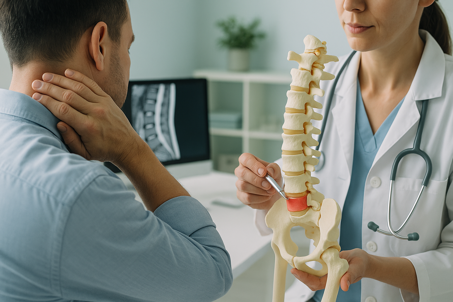
Nerve root compression happens when something narrows the space where a nerve exits the spine. This can be a disc that has bulged out of position, a bony spur that has grown over time, or a general narrowing of the spinal canal.
The soft centre of a spinal disc pushes outward and presses on a nearby nerve root.
Extra bone growth, often from wear and tear, encroaches on the nerve exit channel.
The canal or exit tunnels gradually narrow, leaving less room for nerves to pass through.
An abnormal spinal curve or pelvic tilt shifts load unevenly, increasing pressure at certain nerve exits.
Your body works as a connected chain from your feet to your head. If your pelvis is tilted or your spine curves unevenly, the load shifts. That extra pressure can narrow the gaps where nerves exit, even when each individual joint looks normal on its own. This is one reason why whole-body imaging matters.
Why does standing imaging help find nerve root compression?
Standing imaging captures your skeleton under the same load it carries all day, revealing alignment problems and nerve exit narrowing that lying-down scans can miss. When you lie flat for a standard X-ray, CT or MRI, gravity no longer pulls on your spine. Your muscles relax. Disc bulges may reduce. The gaps between your vertebrae can open slightly. The result is that the very thing causing your nerve compression may look less severe, or disappear altogether, on a scan taken lying down.
An EOS scan images your entire skeleton from head to feet while you stand. This means your spine, pelvis, hips, knees and ankles are all captured in one weight-bearing image. Your clinician can see how the whole chain of alignment relates, and whether a problem further down, such as uneven leg length or a tilted pelvis, is contributing to nerve compression higher up.
The EOS system also uses about 90% less radiation than a standard X-ray, around ten times less. That makes it a practical option if you need follow-up images over time.
Want to understand whether a standing scan could help your symptoms?
Book a free video consultationWhen is MRI the better choice? EOS shows bones, joints and alignment clearly. For soft-tissue detail, such as the condition of a disc, inflammation around a nerve or spinal cord problems, MRI is usually the better tool. In many cases, both types of imaging work together to give the fullest picture. Your clinician can advise which is right for you.
What helps with nerve root compression?
Most people with a compressed nerve improve over time, but the speed and approach depend on finding the underlying cause accurately. ScanAlign does not treat nerve compression. It provides detailed standing imaging so that your treating clinician, whether a physiotherapist, pain specialist or surgeon, can plan the right next step.
General approaches that clinicians commonly consider include:
- Guided exercises and physiotherapy to take pressure off the nerve, strengthen supporting muscles and improve posture.
- Pain management with medication or injections, guided by imaging that shows exactly where the nerve is being squeezed.
- Postural and alignment correction, addressing the wider chain of alignment rather than just the painful spot.
- Surgery, considered when conservative approaches have not provided relief and imaging confirms a specific structural cause.
The common thread is that all of these work better when the clinician knows exactly where and why the compression is happening. That is where accurate, weight-bearing imaging fits in. You can learn more about how EOS imaging works on the ScanAlign homepage.
Frequently asked questions about nerve root compression
Nerve root compression is when a spinal nerve is squeezed where it exits the spine. This can be caused by a bulging disc, bony spur or narrowing of the spinal canal. It often causes pain, tingling or weakness that travels into an arm or leg.
It varies widely depending on the cause and severity. Many people notice improvement within weeks to a few months with appropriate management. Some cases take longer, and a small number may need surgical intervention if the nerve remains compressed.
A standard X-ray can show bony changes such as spurs or narrowed disc spaces that suggest compression. A standing EOS scan adds the advantage of showing your full alignment under weight, which helps identify structural causes that lying-down imaging may miss. For direct views of the nerve and disc, MRI is usually needed.
Sciatica is a symptom, not a diagnosis. It describes pain travelling down the leg along the sciatic nerve path. Nerve root compression in the lower back is one of the most common causes of sciatica, but not the only one.
No. ScanAlign is a private, self-pay clinic. You can book directly without a GP or consultant referral. The process begins with a free video consultation to check the scan is appropriate for you.
Pricing details are available on the ScanAlign FAQs page.
Yes. The EOS system uses about 90% less radiation than a standard X-ray, around ten times less. You can read more about safety on the FAQs page.
Yes. When your spine, pelvis or legs are out of alignment, load is distributed unevenly. Over time, this can narrow the exit channels where nerves leave the spine and increase pressure on specific nerve roots. This is why whole-body standing imaging can be so useful.
Exercise recommendations depend on which nerve is affected and the underlying cause. A physiotherapist can design a programme based on your imaging findings. General advice often includes gentle nerve gliding movements and core strengthening, but exercises should always be guided by a qualified professional.
You stand inside the EOS machine in a natural position for about 20 seconds while a low-dose image is captured from head to feet. There is no tunnel and no need to lie down. The whole visit is quick and straightforward. More details are on the FAQs page.
Find out where the compression is coming from
A free video consultation with ScanAlign helps you understand whether standing whole-body imaging is the right next step for your symptoms.
Book a free video consultationSources
- NHS. "Slipped disc." nhs.uk, 2023. Overview of disc herniation, nerve root compression symptoms and management.
- National Institute for Health and Care Excellence (NICE). "Low back pain and sciatica in over 16s: assessment and management." NICE guideline NG59, 2016 (updated 2020).
- Illés, T. & Somoskeöy, S. "The EOS imaging system and its uses in daily orthopaedic practice." International Orthopaedics, 36(7), 1405-1411, 2012.
Written by Abbas Dhami (Specialist Diagnostic Radiographer)
This page is general information and not a diagnosis.
ScanAlign is the trading name of CoreMed Solutions Ltd and operates under The Harley Street Hospital's CQC licence.