

Laminectomy: What Is It and Do You Really Need One?
A laminectomy is a spinal decompression surgery that removes part of a vertebra called the lamina to create more space for compressed nerves. It is most commonly used for spinal stenosis when other treatments have not helped. Standing, weight-bearing imaging can give your surgeon a clearer picture of how your spine behaves under load before deciding on surgery.
Key takeaways
- A laminectomy removes a small section of bone to relieve pressure on spinal nerves.
- It is usually considered after physiotherapy, injections or medication have not worked.
- Lying-down scans can miss alignment issues that only appear when you stand and bear weight.
- An EOS standing scan shows your full spinal alignment with about 90% less radiation than a standard X-ray.
- ScanAlign provides diagnostic imaging only, not surgery or treatment.
What is a laminectomy?
A laminectomy is a surgical procedure that removes part or all of the lamina, the bony arch at the back of a vertebra, to take pressure off the spinal cord or nerve roots. The goal is to give the nerves more room so they stop being squeezed.
Most laminectomies are carried out on the lower (lumbar) spine, though they can also be done in the neck (cervical) region. Your surgeon may also trim thickened ligaments or remove bone spurs at the same time. In some cases, a spinal fusion is performed alongside the laminectomy to keep the spine stable afterwards.
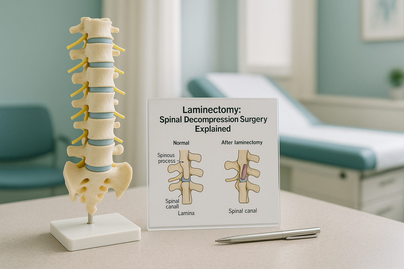
Laminectomy vs laminoplasty
A laminoplasty is a slightly different approach. Instead of removing the lamina entirely, the surgeon hinges it open like a door and props it in place with small spacers. This is more common in cervical (neck) surgery and aims to preserve some of the spine's natural structure. Your consultant will recommend the approach that suits your anatomy and condition best.
What are the signs you might need a laminectomy?
A laminectomy is usually considered when spinal stenosis or another cause of nerve compression produces symptoms that have not responded to conservative treatment. Common signs include the following.
Leg or back pain that gets worse when you walk or stand for any length of time.
Pins and needles or loss of feeling in your legs, buttocks or feet.
Difficulty lifting your foot, climbing stairs, or a feeling that your legs may give way.
Having to stop and sit or lean forward frequently because your legs feel heavy or painful.
Urgent warning: If you experience sudden loss of bladder or bowel control, severe or rapidly worsening weakness in your legs, or numbness around your groin, seek urgent medical attention immediately. Contact your GP, call 111, or go to A&E. These could be signs of cauda equina syndrome, which requires emergency treatment.
What conditions lead to a laminectomy?
The most common reason for a laminectomy is spinal stenosis, a narrowing of the spinal canal that puts pressure on the nerves. But it is not the only cause.
Gradual narrowing of the canal, often due to age-related changes in bone and ligament.
A bulging disc pressing on the nerve root, sometimes addressed alongside laminectomy.
Bony overgrowths from arthritis that encroach on the space around the nerves.
Ligaments in the spinal canal that become thicker over time and crowd the nerves.
How does standing imaging help before a laminectomy?
Standing, weight-bearing imaging shows your spine the way it actually works, under the force of gravity with your muscles engaged. This matters because lying-down scans relax the body and can hide problems that only appear when you are upright.
Your body is a connected chain from your feet to your head. A tilt in the pelvis, a curve in the lower back, or a shift in the hips can all change how forces travel through the spine. Pain often shows up away from its real cause because one area compensates for a problem somewhere else. Conventional X-rays, CT scans and MRI are all taken lying down, which removes gravity and lets the spine spread out. A narrowed canal that causes real trouble when you stand may look less severe when you are flat on your back.
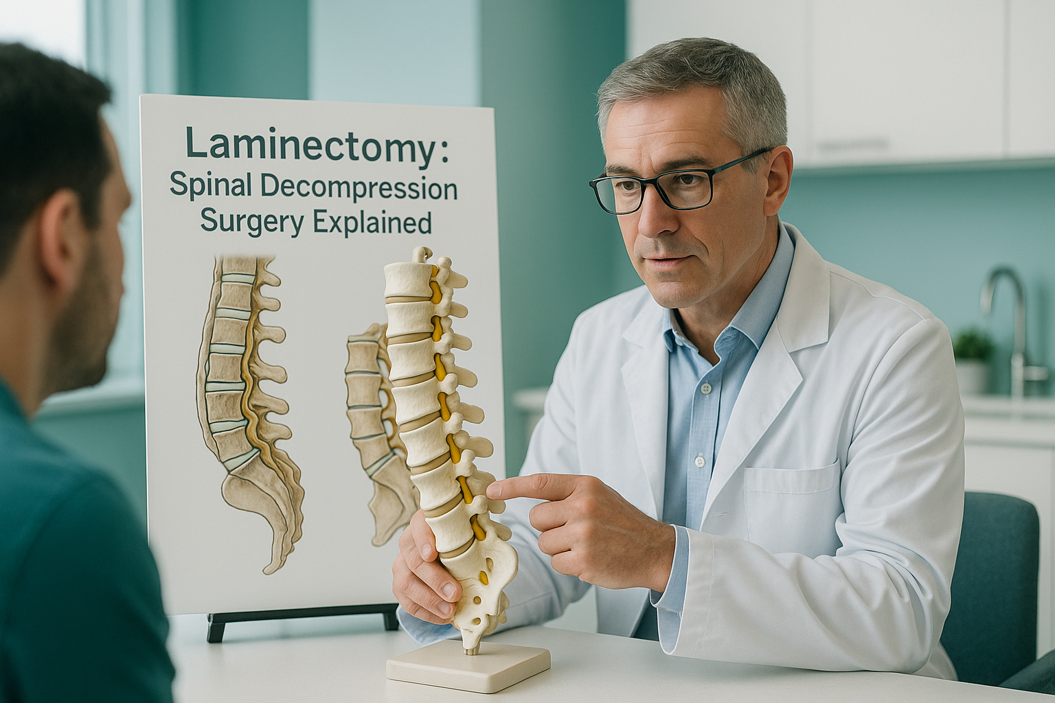
An EOS scan takes a full-length, standing image of your spine, pelvis and legs in a single pass, with about 90% less radiation than a standard X-ray, around ten times less. The image captures the true alignment and loading of every segment. Your surgeon can see exactly where narrowing is worst and how the rest of your spine compensates, giving them better information for planning the procedure.
When MRI is the better tool: EOS shows bones, joints and alignment clearly. For soft tissue such as discs, nerves and the spinal cord itself, MRI is usually the better choice. Many surgical teams use both: MRI for the soft tissue detail and a standing EOS scan for the whole-spine alignment picture.
Wondering whether a standing alignment scan could give your surgeon more information before your laminectomy?
Book a free video consultationWhat should you expect after a laminectomy?
Recovery after a laminectomy varies from person to person and depends on factors such as how many spinal levels were decompressed, whether a fusion was done at the same time, and your general health. Your surgical team will give you a personalised recovery plan.
In general terms, most people are up and walking within a day of surgery. You will typically be advised to avoid heavy lifting and twisting for several weeks. Physiotherapy usually plays an important part in rebuilding strength and flexibility. Some people notice an improvement in leg symptoms quickly, while numbness or tingling can take longer to settle. Your consultant is the best person to discuss realistic timescales with you.
Laminectomy risks
All surgery carries risks. Common ones for a laminectomy include infection, bleeding, nerve injury and spinal fluid leak. There is also a chance that symptoms do not fully improve or that the spine becomes less stable at the operated level over time. Your surgeon will explain these risks in the context of your specific situation before you give consent.
Better pre-surgical imaging can help reduce surprises. When your surgeon can see the full picture of your spinal alignment under load, they can plan more precisely, choose the right levels to decompress, and decide whether a fusion is needed. This is where a standing EOS scan adds value as a complement to your existing MRI or CT.
Frequently asked questions about laminectomy
A laminectomy removes part of the bony lamina to relieve pressure in the spinal canal. A discectomy removes part of a damaged disc that is pressing on a nerve. Sometimes both are done in the same operation.
Recovery time varies. Most people are walking within a day and can return to light daily activities within a few weeks. Full recovery, including return to more demanding activities, can take several months. Your surgical team will guide you.
A laminectomy is one type of spinal decompression surgery. Other types include laminotomy, where only a small window of bone is removed, and foraminotomy, which widens the opening where the nerve root exits the spine. Your surgeon will recommend the most appropriate approach.
A laminectomy removes the lamina. A laminoplasty hinges it open and uses spacers to keep the canal wider without removing the bone entirely. Laminoplasty is more commonly used in the cervical spine.
No. You can self-refer. Many people book after a free video consultation to discuss whether a standing alignment scan is appropriate for their situation. You can also be referred by your GP, physiotherapist or consultant.
An EOS scan uses about 90% less radiation than a standard X-ray, around ten times less. This makes it one of the lowest-dose skeletal imaging options available.
No. EOS and MRI do different things. EOS shows bones, joints and whole-body alignment while standing. MRI shows soft tissue, discs, nerves and the spinal cord. Most surgical teams use both for a complete picture.
Spinal stenosis symptoms are typically worse when standing and walking because gravity compresses the already-narrow canal. Lying-down scans remove that gravitational load, potentially making the narrowing look less severe than it actually is when you are upright.
For current pricing and what is included, please visit the FAQs and cost page.
No. ScanAlign is a diagnostic imaging clinic. We provide standing EOS scans and detailed reports that you and your medical team can use to plan treatment. We do not carry out surgery, injections or any form of treatment.
See your spine the way your surgeon needs to
A standing EOS scan can show your full spinal alignment under load, giving your surgical team the detail they need to plan precisely. Start with a free video consultation.
Book a free video consultationSources
- NHS. "Lumbar decompression surgery." nhs.uk. Available at: nhs.uk/conditions/lumbar-decompression-surgery
- American Academy of Orthopaedic Surgeons (AAOS). "Lumbar Spinal Stenosis." OrthoInfo. Available at: orthoinfo.aaos.org
- Illés T, Somoskeöy S. "The EOS imaging system and its uses in daily orthopaedic practice." International Orthopaedics. 2012;36(7):1405-1411.
Written by Abbas Dhami (Specialist Diagnostic Radiographer)
This page is general information and not a diagnosis.
ScanAlign is the trading name of CoreMed Solutions Ltd and operates under The Harley Street Hospital's CQC licence.