
Lumbar Radiculopathy: What Causes Sciatica and Nerve Root Pain in Your Lower Back?
Lumbosacral radiculopathy happens when a nerve root in the lower spine is compressed or irritated, sending pain, tingling or weakness down your leg. It is the medical name for what most people call sciatica. Standing whole-body imaging can reveal spinal alignment problems that contribute to nerve compression but do not show up when you are lying down.
Key takeaways
- Lumbar radiculopathy is a pinched or irritated nerve root in the lower back, commonly called sciatica.
- Pain, numbness or weakness can travel from the buttock down the leg to the foot.
- Lying-down scans can miss alignment shifts that compress nerve roots under your body weight.
- EOS standing imaging captures your full spine and pelvis in the position where symptoms actually occur.
- ScanAlign provides diagnostic imaging only. It does not treat the condition.
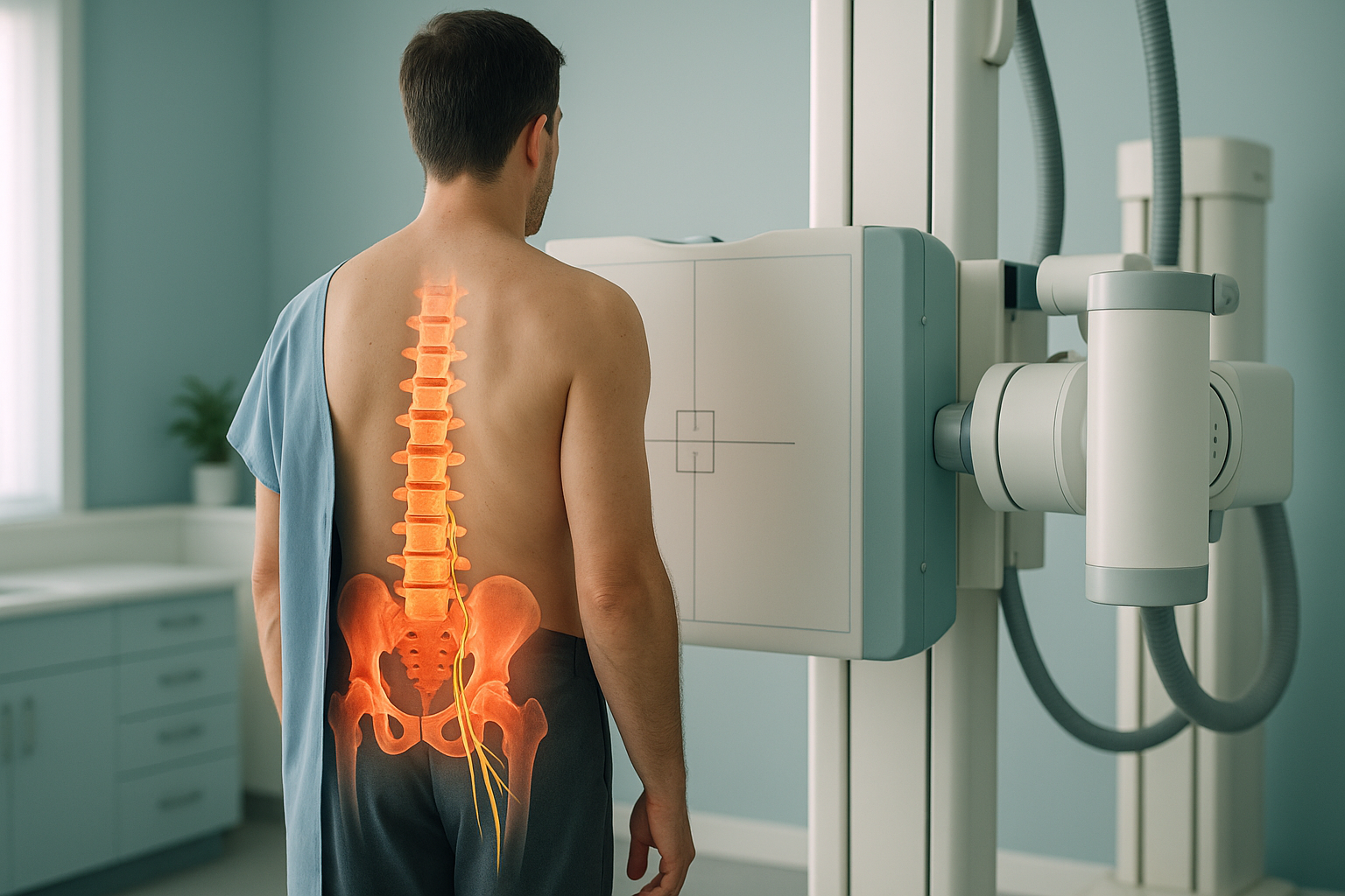
Your lower spine (the lumbar and sacral region) carries most of your body weight. Nerves branch out from the spinal cord between each vertebra, forming nerve roots that run down through your buttock and into your leg. When one of these roots is pinched, squeezed or irritated, the signals it carries get disrupted. The result is radiculopathy of the lumbosacral region, which most people simply know as sciatica.
The important thing to understand is that the pain you feel in your leg often starts with a structural problem in your spine. And that structural problem can sometimes only be seen clearly when you are standing upright, bearing your own weight.
What are the symptoms of lumbar radiculopathy?
The main symptom is pain that travels from the lower back or buttock down one leg, following the path of the affected nerve. Which part of the leg hurts depends on which nerve root is involved.
A sharp or burning pain that runs from the buttock down the back, side or front of the leg, sometimes reaching the foot.
A "pins and needles" feeling in the leg, calf or foot, or patches of reduced sensation.
Difficulty lifting the foot (foot drop), weakness when pushing off, or a leg that gives way.
Symptoms often flare when upright, coughing, sneezing or bending, then ease when lying flat.
Urgent warning. If you develop sudden loss of bladder or bowel control, numbness around the groin or saddle area, or rapidly worsening weakness in both legs, seek immediate medical help. Contact your GP, call 111, or go to A&E. These symptoms may indicate a serious condition that needs emergency care.
What causes sciatica and lumbar radiculopathy?
Lumbar radiculopathy occurs when something presses on a nerve root as it exits the spine. Several structures can be responsible, and often more than one factor is involved.
The soft centre of a spinal disc pushes outward and presses on the nearby nerve root.
The bony canal around the spinal cord or nerve roots narrows, squeezing them.
One vertebra slips forward over the one below, narrowing the space for nerve roots.
Bony growths from wear and tear can press directly on the nerve exit point.
Your body is a connected chain from your feet to your head. If your pelvis tilts, your hip sits unevenly, or your lumbar curve is too deep or too flat, the load on your lower spine changes. Over time, this uneven loading can cause disc wear, joint thickening or vertebral slippage in one area while another area compensates. This is why L4-L5 nerve root pain can be connected to an alignment problem much further along the chain.
How does a standing scan help diagnose lumbar radiculopathy?
A standing, weight-bearing scan shows your spine in the position where symptoms actually happen. When you lie down for a standard X-ray, CT or MRI, gravity is removed, muscles relax, and vertebrae can shift back into a more normal position. That means a narrowed nerve exit or a vertebral slip can look less severe, or even disappear, on a lying-down image.
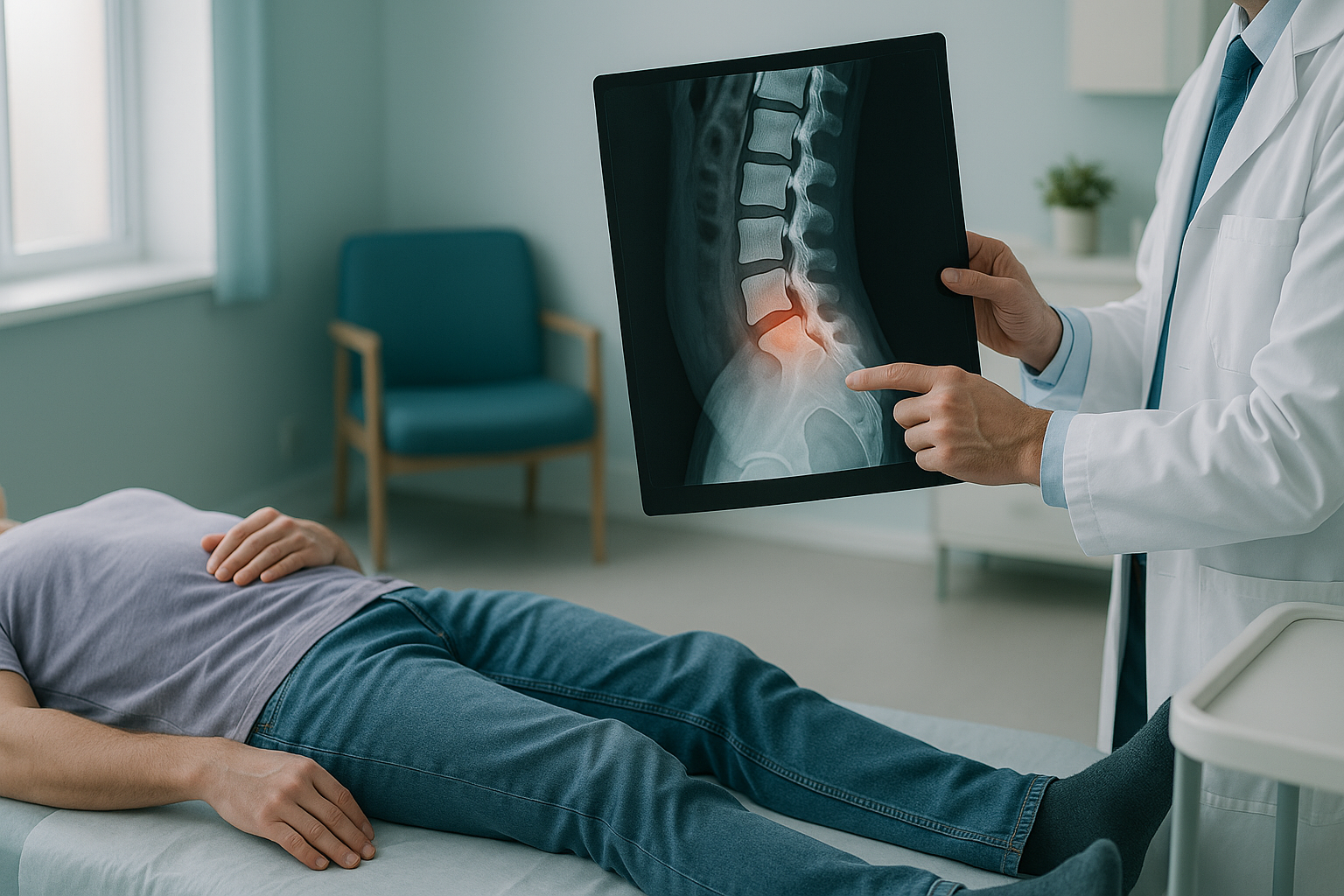
EOS whole-body standing imaging captures your full skeleton, from your head to your feet, in a single upright scan. It uses about 90% less radiation than a standard X-ray, around ten times less. This gives your specialist a complete picture of how your lumbar spine, pelvis, hips and legs interact under real-world load.
Because the image shows your entire alignment, it can reveal whether a pelvic tilt, a leg-length difference, or an altered spinal curve is placing extra stress on the nerve roots at a specific level. A conventional scan focused only on the lower back would miss these broader mechanical factors.
Want to see how your full spinal alignment could be contributing to your nerve pain?
Book a free video consultationA note on MRI. EOS excels at showing bony alignment, joint spacing and overall spinal balance in the standing position. For detailed views of soft tissue, such as discs, nerves and the spinal cord itself, MRI is usually the better tool. In many cases of lumbar radiculopathy, both types of imaging give the most complete picture. Your clinician can advise which you need.
What generally helps with lumbar radiculopathy?
Most cases of sciatica improve within weeks to months. Management usually involves a combination of approaches guided by a healthcare professional.
- Staying active. Gentle movement is generally encouraged over prolonged bed rest. Walking and light stretching can help maintain mobility.
- Physiotherapy. Specific exercises can strengthen the muscles supporting the spine and improve flexibility. Your physiotherapist can tailor these to your situation.
- Pain management. Your GP or specialist may recommend appropriate medication or other pain-relief strategies.
- Understanding the cause. Accurate imaging helps clinicians identify the structural driver behind the nerve compression, so that any intervention targets the right level and the right problem.
ScanAlign provides the diagnostic imaging that informs these decisions. It does not offer treatment. Your scan results and clinical report are shared with your chosen clinician so they can plan the best course of action for you.
Frequently asked questions about lumbar radiculopathy
They refer to the same problem. Sciatica is the everyday term for pain along the sciatic nerve. Lumbar radiculopathy, or radiculopathy of the lumbosacral region, is the clinical name. It means a nerve root in the lower spine is compressed or irritated, causing symptoms in the leg.
The L4, L5 and S1 nerve roots are the most frequently involved. Each supplies different muscles and areas of sensation in the leg. The specific pattern of pain or weakness helps clinicians narrow down the level affected.
Standard X-rays show bones but not the nerves or discs directly. However, a standing EOS image can reveal narrowing of the disc space, vertebral slippage, or alignment problems that create the conditions for nerve compression. MRI is usually needed to see the nerve and disc detail directly.
When you lie down, gravity is removed. Muscles relax and vertebrae can shift back into a more normal position. A vertebral slip or disc-space narrowing that compresses a nerve under your body weight may look less severe, or even normal, on a lying-down scan. Standing imaging captures what is happening in real life.
Yes. EOS uses about 90% less radiation than a standard X-ray, around ten times less. You can find more details on our FAQs and safety page.
No. ScanAlign accepts self-referrals. You can book a free video consultation directly without needing a letter from your GP.
ScanAlign is a private, self-pay clinic. For current pricing information, please visit our FAQs page.
Yes. Your body works as a connected chain. A tilted pelvis, uneven leg lengths, or an altered hip position can shift the load on your lower spine, increasing pressure at a specific spinal level. Whole-body standing imaging reveals these connections, whereas a scan of the lower back alone would not.
Your images are reviewed and a detailed clinical report is produced. This is shared with you and, if you wish, with your treating clinician. ScanAlign provides diagnostic imaging only and does not offer treatment.
Targeted exercises, guided by a physiotherapist, can help many people with lumbar radiculopathy by improving core support and flexibility. However, effective rehabilitation depends on understanding the structural cause. Accurate imaging gives your physiotherapist the information they need to prescribe the right exercises for your situation.
Learn more about spinal imaging
Explore how standing whole-body imaging works and how it can help with conditions related to your spine and alignment.
Understand what is behind your nerve pain
A free video consultation lets you discuss your symptoms with a specialist radiographer and find out whether standing whole-body imaging could help.
Book a free video consultationSources
- NHS. Sciatica. nhs.uk/conditions/sciatica. Accessed 2024.
- National Institute for Health and Care Excellence (NICE). Low back pain and sciatica in over 16s: assessment and management (NG59). 2016, updated 2020.
- Deschênes S, et al. Diagnostic imaging of spinal deformities: reducing patients' radiation dose with a new slot-scanning X-ray imager. Spine. 2010;35(9):989-994.
Written by Abbas Dhami (Specialist Diagnostic Radiographer)
This page is general information and not a diagnosis.
ScanAlign is the trading name of CoreMed Solutions Ltd and operates under The Harley Street Hospital's CQC licence.