

What Is Military Neck, and Does a Straight Cervical Spine Matter?
Military neck is the informal name for a loss of the normal inward curve in your neck, called cervical lordosis. On imaging the cervical spine looks unusually straight or even slightly reversed. It can cause stiffness and discomfort, but many people have a straighter neck without any symptoms at all. Context matters more than the shape alone.
Key takeaways
- Military neck means the normal gentle curve in your neck has flattened or reversed.
- It is often found incidentally on imaging and is not always a problem.
- Muscle spasm, posture habits and lying-down positioning during scans can all make the neck look straighter than it really is.
- A standing, weight-bearing image shows what your neck alignment actually looks like in daily life.
- Knowing the full picture helps you and your clinician decide whether anything needs to change.
What does military neck actually mean?
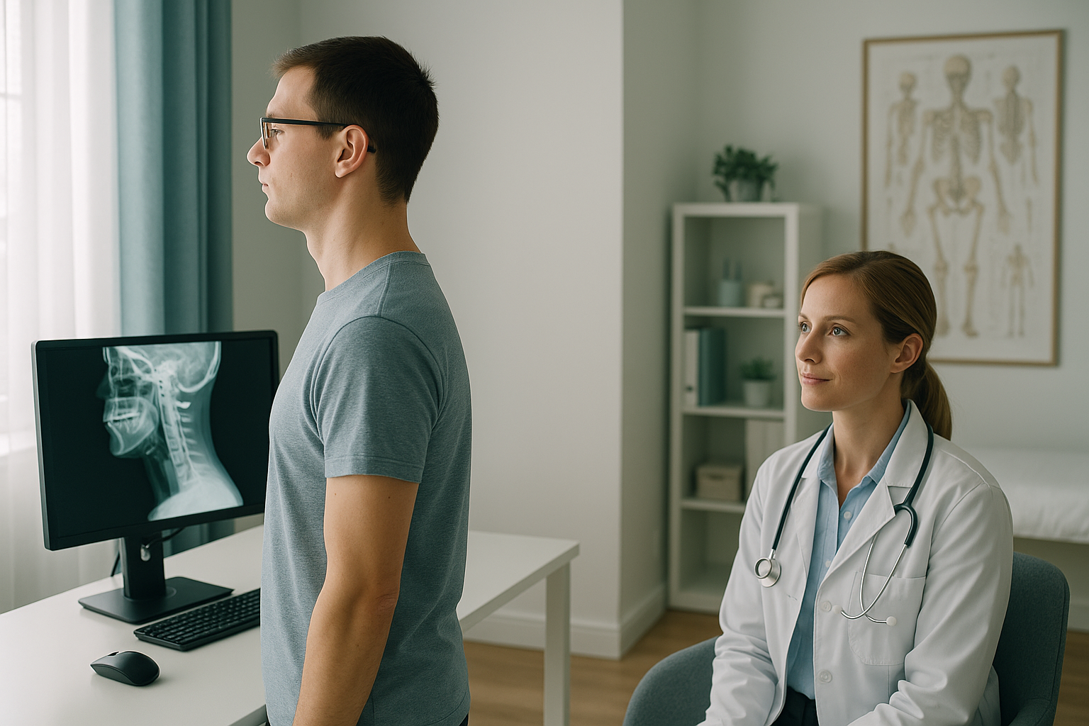
Military neck describes a cervical spine that has lost its natural lordotic curve. Normally, the seven vertebrae in your neck form a gentle inward arc. This curve helps absorb the weight of your head and allows flexible movement. When that curve flattens out, or in more pronounced cases tips slightly the wrong way (sometimes called a reversed cervical curve or cervical kyphosis), reports may use the term "straightening of the cervical spine" or "military neck."
The name comes from the upright, chin-tucked posture of a soldier on parade. It sounds dramatic, but the label itself is descriptive rather than diagnostic. It tells you the shape of the curve, not the cause or the significance.
What are the signs of military neck?
Some people notice symptoms. Others discover a loss of cervical lordosis only when a scan is done for another reason entirely. The most common things people report include:
A feeling of restricted movement when turning or tilting your head, especially first thing in the morning.
Dull pain at the base of the neck or across the upper back after sitting or screen work.
Tension-type headaches starting at the back of the skull, sometimes linked to forward head posture.
Plenty of people with a straight cervical spine feel fine. A finding on a report does not automatically mean a problem.
What causes a loss of cervical lordosis?
There is rarely one single cause. The straightening of the cervical spine usually results from a combination of factors. Some are structural, some are muscular, and some are simply about how you were positioned when the image was taken.
Hours of screen work or phone use can encourage a forward head position, gradually flattening the neck curve over time.
When neck muscles go into spasm, they can pull the spine straighter than normal. This is temporary but shows up clearly on imaging.
A whiplash injury can alter the neck's resting curve, sometimes permanently. The straightening may appear on imaging years later.
As discs lose height with age, the spacing and curve of the cervical spine can shift. This is common and not always painful.
Some people naturally have less cervical curve. Research shows a wide range of "normal" across the population.
This is an important point. A report that mentions straightening of the cervical spine is describing a shape, not necessarily a disease. How you feel, how you move and what your whole spinal alignment looks like all matter just as much as the curve itself.
Why does a standing scan show military neck more accurately?
A standing, weight-bearing image captures your neck the way it actually works, upright, supporting the weight of your head, with muscles active. This matters because most conventional imaging is done lying down.
Weight-bearing EOS imaging captures the cervical spine while you stand naturally. Gravity, muscle tone and postural habits all influence the curve. If your neck straightens because of how you hold yourself during the day, a standing image will show that. It also captures the rest of your spine in the same shot, so a clinician can see whether cervical tilt or changes lower down are contributing to the loss of curve. The EOS system uses about 90% less radiation than a standard X-ray, around ten times less.
Lying-down imaging (standard X-ray, CT, MRI) relaxes the muscles around your spine. That relaxation can temporarily change the shape of the cervical curve. Muscle spasm may ease when you lie flat, making the neck look more normal than it feels during the day. Conversely, the position of your head on a pillow or table can flatten the curve artificially, making a normal neck look abnormally straight. Positioning on the image is one of the most common reasons for a "loss of lordosis" finding.
Because your body is a connected chain from your feet to your head, a problem at one level can make another area compensate. A flattened thoracic curve, a pelvic shift or even a difference in leg length can alter how your neck sits. ScanAlign standing EOS imaging captures the full picture in a single upright image, so compensation patterns are visible rather than hidden.
Wondering whether your military neck finding tells the full story? A standing scan shows your cervical alignment as it really is.
Book a free video consultationCan military neck be improved?
That depends on what is causing it and whether it is actually producing symptoms. ScanAlign is a diagnostic imaging clinic, not a treatment provider, so we cannot prescribe a course of action. What we can say is that understanding your alignment accurately is the first step.
If muscle tension or habitual posture is the main driver, many clinicians recommend targeted exercises, ergonomic changes and physiotherapy to encourage the curve back. If degenerative changes are the cause, the approach may be different. And if the finding turns out to be positional, meaning it appeared on a lying-down scan but is not present when standing, you may not need any intervention at all.
A detailed standing alignment assessment gives you and your clinician the information to make that decision with confidence, rather than guessing.
When MRI is the better tool. EOS imaging shows bones, joints and alignment clearly. If your clinician suspects a disc bulge, nerve compression or soft-tissue problem in the neck, MRI is usually the more appropriate investigation. The two are complementary rather than competing.
Frequently asked questions about military neck
Yes. Military neck is the informal term for a loss of cervical lordosis. Both describe a cervical spine that has lost its normal inward curve and appears straight or slightly reversed on imaging.
It can. When the neck curve flattens, muscles at the base of the skull may work harder to support the head. This extra strain can trigger tension-type headaches, particularly if it is combined with forward head posture.
Not necessarily. A straight-looking neck can be caused by muscle spasm at the time of the scan, by the way your head was positioned, or simply by normal variation. Many asymptomatic people have a reduced cervical curve with no problems at all.
A straightened neck has lost its curve and appears flat. Cervical kyphosis goes a step further, meaning the curve has reversed so the neck bows slightly outward instead of inward. Kyphosis is less common and generally warrants closer clinical attention.
Yes. A whiplash injury can damage the ligaments and muscles that maintain the cervical curve. This may result in straightening that persists long after the initial injury has healed. It is one of the most frequently studied associations in the research.
No. ScanAlign accepts self-referrals. You can book a standing neck and spine scan directly. If you prefer, your GP, physiotherapist or consultant can also refer you.
An EOS scan uses about 90% less radiation than a standard X-ray, around ten times less. This makes it a very low-dose option for full standing alignment imaging.
EOS imaging shows bones, joints and overall alignment. It does not image soft tissues like discs or nerves directly. If a disc problem is suspected, MRI is usually the better investigation. The two scans complement each other well.
Pricing details are available on the ScanAlign website. You can start with a free video consultation to discuss whether the scan is right for you before committing to anything.
If the straightening is primarily driven by muscle tension and habitual posture, targeted exercises and physiotherapy may help restore some of the curve. The evidence varies depending on the underlying cause. Accurate imaging helps your clinician decide the best approach for you.
See your neck alignment as it really is
A free video consultation lets you discuss your findings and decide whether a standing scan is the right next step. No obligation, no pressure.
Book a free video consultationWritten by Abbas Dhami (Specialist Diagnostic Radiographer)
Sources
- Gore DR, Sepic SB, Gardner GM. Roentgenographic findings of the cervical spine in asymptomatic people. Spine. 1986;11(6):521-524.
- NHS. Whiplash. nhs.uk. Available at: https://www.nhs.uk/conditions/whiplash/
- Harrison DD, Harrison DE, Janik TJ et al. Modeling of the sagittal cervical spine as a method to discriminate hypolordosis. Spine. 2004;29(22):2485-2492.
This page is general information and not a diagnosis.
ScanAlign is the trading name of CoreMed Solutions Ltd and operates under The Harley Street Hospital's CQC licence.