

Cervical Tilt: What Causes a Tilted Neck Posture and How Is It Assessed?
A cervical tilt is when your head leans persistently to one side instead of sitting centred over your shoulders. It can come from muscle tightness, joint wear, spinal asymmetry or compensation for a problem lower down the body. Standing whole-body imaging helps identify the structural cause by showing your full alignment under gravity.
Key takeaways
- Cervical tilt means the head sits off-centre, leaning laterally toward one shoulder.
- The cause is often somewhere other than the neck itself, such as the mid-back, pelvis or hips.
- Lying-down scans can miss the tilt because gravity is removed and muscles relax.
- An EOS standing scan captures the full skeleton in a natural, weight-bearing position.
- Accurate imaging guides the right professional to treat the root cause, not just the symptom.
What is a cervical tilt?
A cervical tilt is a lateral lean of the head and upper neck to one side. In everyday language, it is the posture people notice when their head always seems to sit closer to one shoulder. It is different from simply turning (rotating) the neck. The tilt happens in the frontal plane, meaning the ear drops toward the shoulder rather than the chin turning left or right.
Some people develop a cervical tilt gradually over years. Others notice it suddenly after an injury or a period of pain. When the tilt is caused by sustained muscle contraction pulling the head to one side, clinicians may use the term torticollis in adults. However, a tilt is not always muscular. It can reflect bony asymmetry, disc height loss or joint degeneration in the cervical spine.
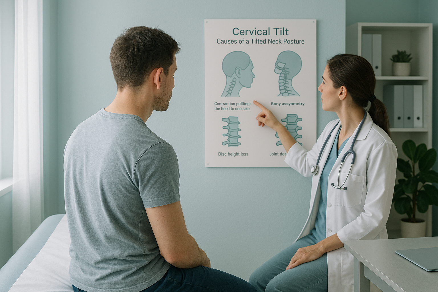
Your body is a connected chain from your feet to your head. The cervical spine sits at the very top of that chain, so even a small shift in the pelvis, a leg-length difference or a curve in the thoracic spine can travel upward and show itself as a head tilt. This is why looking at the neck alone often misses the real story.
How do you know if your head is tilted to one side?
Many people first notice a cervical tilt in photographs or when a hairdresser or optician points it out. Others feel it as persistent one-sided neck ache, shoulder tension or headaches. Below are the most common signs.
Your head consistently sits closer to one shoulder, noticeable in the mirror or in photos.
You find it harder to bend or turn your neck in one direction, with tightness that returns even after stretching.
One shoulder appears higher than the other, and shirt collars or bag straps keep slipping on one side.
Tension headaches focused on one side, often starting at the base of the skull.
A note on nerve symptoms. If your head tilt comes with numbness, tingling or weakness spreading into your arms or hands, or if you notice any loss of bladder or bowel control, please seek urgent medical attention through your GP, 111 or A&E. These symptoms need immediate assessment.
What causes a head tilted to one side in adults?
A cervical tilt in adults usually develops from one or more structural or muscular factors. The cause is not always in the neck. Your body compensates constantly, and a problem at one level can create a visible tilt at another.
Uneven disc height loss or facet joint degeneration can cause the spine to lean to one side at a specific level.
Chronic contraction of muscles on one side of the neck, particularly the sternocleidomastoid, pulls the head into a tilt.
A curve lower in the spine forces the neck to tilt the opposite way to keep the eyes level with the horizon.
If one leg is effectively shorter, the pelvis tilts, the spine curves to compensate, and the head follows.
This compensation pattern is the reason a cervical tilt is often misunderstood. Treating just the neck may ease symptoms temporarily, but if the underlying cause is in the pelvis or mid-back, the tilt usually returns.
How is a cervical tilt assessed with standing imaging?
A cervical tilt is best assessed when you are upright and weight-bearing, because that is how your body actually functions. Standard imaging methods, such as X-ray, CT or MRI, usually capture the neck while you are lying down. In that position, gravity is removed, the muscles relax, and the tilt may partly or fully disappear. The image can look reassuringly normal even though the problem is clearly there when you stand up.
An EOS scan takes a full-length standing image of your skeleton, from head to feet, in a single capture. It uses about 90% less radiation than a standard X-ray, around ten times less. Because the whole body is visible in one image, the reporting clinician can trace the alignment chain and see exactly where the tilt starts, whether that is the cervical spine itself, the thoracic curve below it, a pelvic obliquity or a difference in leg length.
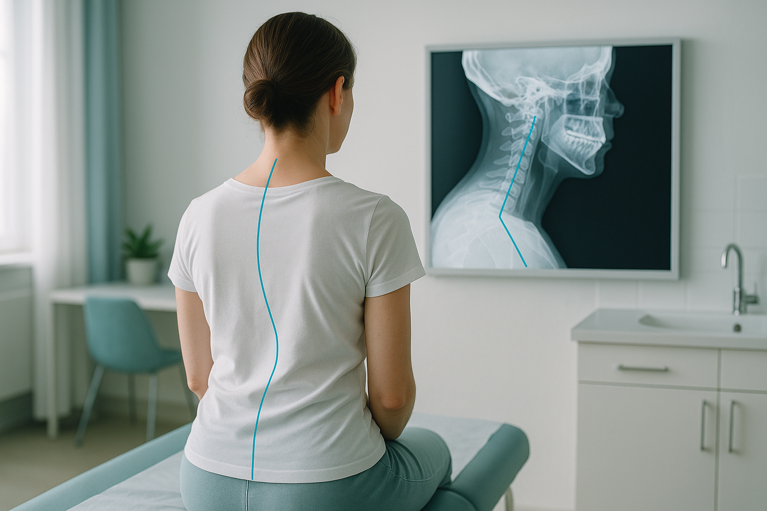
This full-picture approach is what makes standing alignment imaging different from a single-area neck scan. You get a clear map of the cause, not just the symptom.
Want to understand what is behind your neck tilt? A free video consultation lets you discuss your symptoms with a specialist radiographer before committing to a scan.
Book a free video consultationWhat helps with a cervical tilt?
The right approach depends entirely on the underlying cause. A cervical tilt driven by muscle tightness may respond to targeted physiotherapy or manual therapy. One caused by joint degeneration or structural asymmetry may require a different pathway. That is why accurate imaging comes first.
General strategies that clinicians often consider include targeted stretching and strengthening of the neck and upper back muscles, postural retraining, ergonomic adjustments at work or during sleep, and in some cases, referral to a specialist for further investigation.
ScanAlign does not provide treatment. It provides the detailed standing alignment imaging that helps your treating clinician, whether a physiotherapist, osteopath, chiropractor or consultant, understand the full picture and plan the right course of action.
When MRI is the better tool. EOS imaging excels at showing bones, joints and overall alignment. If your clinician suspects a disc bulge, nerve compression or soft-tissue problem in the neck, MRI is usually the more appropriate scan. The two are complementary, not competing.
Frequently asked questions about cervical tilt
A persistent head tilt usually means something structural is pulling or pushing the head off centre. This could be tight muscles on one side of the neck, joint wear in the cervical spine, or a compensation pattern caused by a curve or imbalance lower in the spine or pelvis.
They overlap but are not identical. Torticollis specifically refers to a muscular contraction that tilts and often rotates the head. A cervical tilt is a broader term covering any lateral lean of the head, whether muscular, bony or compensatory.
Yes. Your body is a connected chain. A pelvic tilt, a lumbar curve or a leg-length difference can cause the thoracic spine to shift, and the neck then tilts in the opposite direction to keep the eyes level. This is one of the most common and most overlooked causes.
When you lie down, gravity is removed and muscles relax. A tilt driven by posture, muscle tension or weight-bearing asymmetry may partly or fully correct itself in that position, making the image look normal even though the problem is obvious when you stand.
An EOS scan captures your entire skeleton from head to feet in one standing image, using about 90% less radiation than a standard X-ray, around ten times less. A normal X-ray typically images only one area, so the connections between your neck, spine, pelvis and legs are not visible.
No. ScanAlign is a private self-pay clinic. You can refer yourself directly. The process starts with a free video consultation to discuss your symptoms and confirm whether an EOS scan is appropriate for you.
Yes. The EOS system uses about 90% less radiation than a standard X-ray, around ten times less. You can find more details about safety, preparation and what to expect on the frequently asked questions page.
Pricing details are on the ScanAlign FAQs and cost page. The free video consultation is a good first step to understand which scan is right for you before any commitment.
Exercises can help with muscular causes, but they are unlikely to resolve a tilt caused by joint degeneration, a spinal curve or a leg-length difference. Understanding the structural cause through imaging helps ensure the right approach is used.
You speak with a specialist radiographer by video. They discuss your symptoms, review any previous imaging if you have it, and advise whether a standing EOS scan is likely to be helpful. There is no obligation to book a scan afterwards.
Ready to understand your cervical tilt?
A free video consultation with a specialist radiographer is the first step. No referral needed, no obligation.
Book a free video consultation Learn more about EOS imagingSources
- NHS. "Neck pain and stiff neck." nhs.uk, 2023.
- Illés, T. and Somoskeöy, S. "The EOS imaging system and its uses in daily orthopaedic practice." International Orthopaedics, 36(7), 2012, pp.1405–1411.
- Bao, H. et al. "Coronal alignment in adult spinal deformity: assessment and clinical implications." The Spine Journal, 19(3), 2019, pp.537–547.
Written by Abbas Dhami (Specialist Diagnostic Radiographer)
Related pages
Learn how spinal alignment issues connect to pain and posture on the spine and posture page.
This page is general information and not a diagnosis.
ScanAlign is the trading name of CoreMed Solutions Ltd and operates under The Harley Street Hospital's CQC licence.