
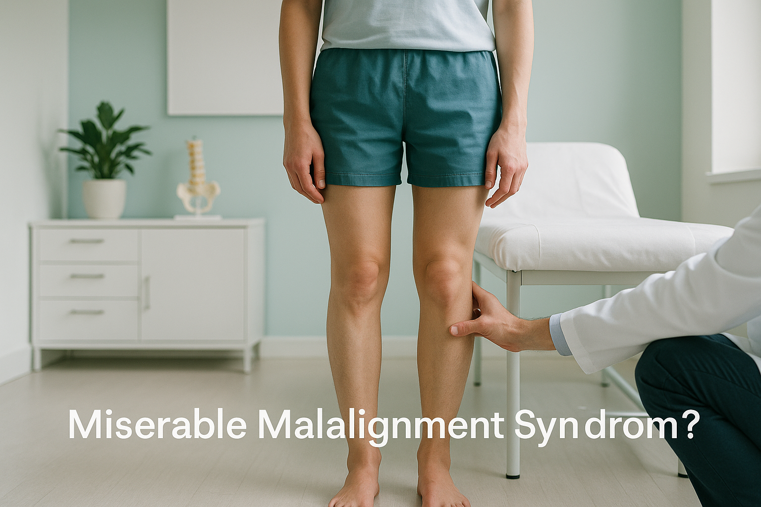
What Is Miserable Malalignment Syndrome and Why Does It Cause Knee Pain?
Miserable malalignment syndrome is a combined rotational deformity of the legs. The thighbone twists inward (femoral anteversion) while the shinbone twists outward (external tibial torsion). Together, these twists force the kneecap out of its normal tracking path and place extra stress on the knee, hip and ankle. Because the problem is rotational, it only shows clearly on imaging that captures 3D bone position while you are standing.
Key takeaways
- The syndrome combines two rotational twists, one at the hip and one at the shin, that together overload the knee.
- It is a common but under-recognised cause of persistent front-of-knee pain, especially in young women.
- "Squinting patellae" (kneecaps that point inward) is the classic visible sign.
- Standard X-rays and MRI often miss rotational problems because they do not measure twist accurately.
- A standing 3D scan can measure the rotational profile of the whole leg in one image.
What are the signs of miserable malalignment syndrome?
The most common sign is persistent pain at the front of the knee that does not respond to rest or standard physiotherapy. Because the kneecap is being pulled out of alignment by the combined twist, you may also notice that your kneecaps seem to point inward when you stand. This appearance is called "squinting patellae."
You might also notice that your feet point outward when you walk, even though your knees point inward. This contradictory pattern happens because the outward twist in the shin compensates for the inward twist at the hip. The result is a gait that looks slightly awkward and places each joint under strain it was not designed for.
What causes combined rotational deformity in adults?
The combined twist usually develops during childhood growth and persists into adulthood. In most people the thighbone gradually loses its inward twist as they grow. When it does not, the body compensates by twisting the shinbone outward so the foot can still point forward. By adulthood, this double twist is fixed in the bone and cannot be changed by exercise alone.
Because the cause is structural, people with this syndrome often spend years trying treatments aimed at muscles or soft tissue without lasting improvement. The underlying rotational mismatch, which is the real driver, goes unrecognised because standard scans are not designed to measure twist. Understanding the difference between anteversion versus retroversion helps make sense of why the direction of the twist matters so much.
Why does rotational malalignment need a standing scan?
Rotation in the leg bones is nearly invisible on a standard X-ray, because a flat image cannot show twist. CT scanning can measure rotation, but it requires high radiation and scans you lying down, which removes the effect of gravity and weight-bearing on your alignment.
A standing EOS scan captures your entire leg from hip to ankle while you bear your full body weight. It produces a 3D reconstruction of every bone in the chain, showing exactly how much each bone is twisted and where the rotation sits. The rotational profile of the femur and tibia can be measured together in one low-dose image.
Because the EOS system uses about 90% less radiation than a standard X-ray, around ten times less, it is well suited to the full-limb imaging this condition requires.
When you lie on a table for CT or MRI, gravity no longer loads your joints. Your muscles relax, and the rotational stress on the knee is reduced. The scan may show structurally normal-looking joints, even though the real problem, the twist between them, causes significant pain when you stand and move.
This is one of the reasons miserable malalignment syndrome is so often missed. The imaging that most people receive simply is not designed to see it.
At ScanAlign, 3D standing skeletal imaging, we produce a full rotational profile of both legs in a single standing scan. This gives your surgeon or physiotherapist the measurements they need to plan the right approach, whether that is targeted rehabilitation or a discussion about surgical correction.
How does the rotational twist affect the kneecap?
When the thighbone twists inward and the shinbone twists outward, the kneecap is caught between two opposing forces. The inward pull of the femur drags the kneecap medially, while the outward twist of the tibia pulls the patellar tendon laterally. This is a key mechanism behind patellar maltracking.
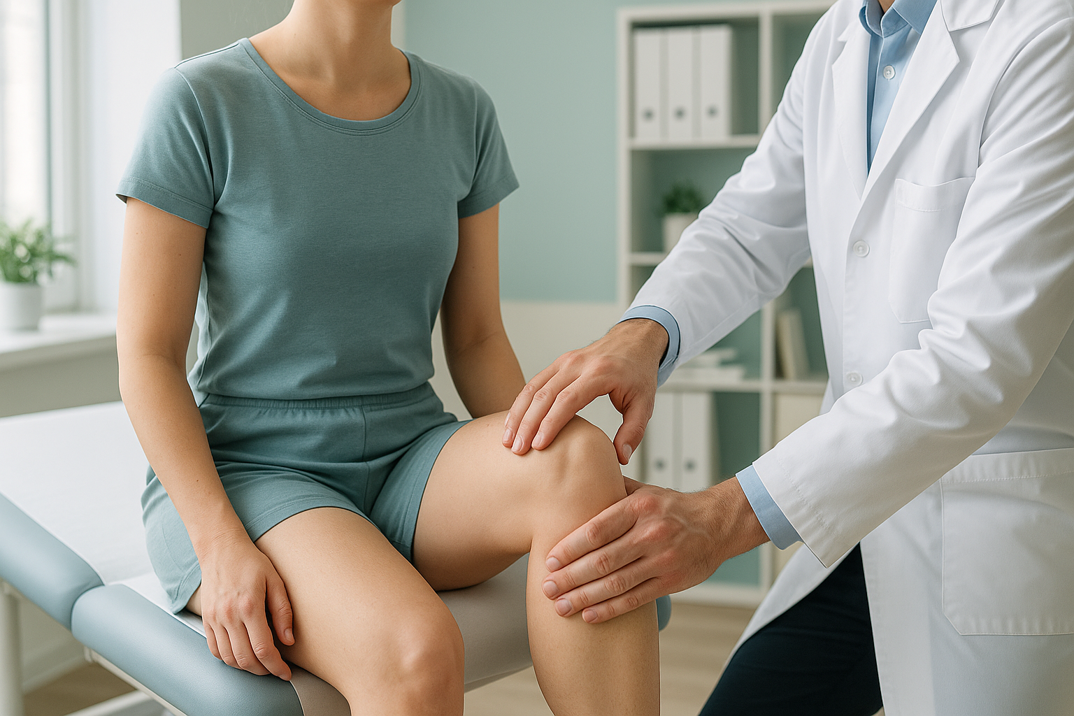
The diagram above shows the key difference. On the left, the femur and tibia rotate in opposite directions, catching the kneecap between competing forces. On the right, a normally aligned leg keeps the kneecap tracking in a straight line. This is why addressing only the kneecap, without measuring the full rotational profile, often fails to solve the pain.
What can be done about miserable malalignment syndrome?
The first step is getting an accurate measurement of the rotational deformity. Without knowing how much twist exists, and exactly where it sits, no treatment plan can be properly targeted. That measurement is what a standing 3D scan provides.
Once the rotational profile is clear, your clinician can decide between two broad paths.
- Targeted physiotherapy and load management. If the twist is mild, strengthening the muscles around the hip and knee can help control symptoms. This works best when the physiotherapist knows the precise rotational picture.
- Surgical discussion (derotation osteotomy). For more significant rotational deformity that does not respond to rehabilitation, a surgeon may discuss a procedure to correct the twist in the bone. This requires precise pre-operative measurements, which is exactly what 3D standing imaging provides.
ScanAlign does not provide treatment. We provide the diagnostic imaging that helps you and your clinician make an informed decision.
Want to understand your rotational profile? A standing 3D scan can measure the twist in both legs in a single visit.
Book a free video consultationWhen is MRI more useful? EOS shows bones, joints and alignment. If your clinician suspects cartilage damage, a torn meniscus, or nerve involvement, MRI is usually the better tool. Many people with miserable malalignment syndrome benefit from both types of imaging.
Is a standing alignment scan right for you?
This scan is most useful when the problem is structural and rotational. It is not a replacement for MRI or blood tests when soft-tissue or inflammatory conditions need to be ruled out.
- Persistent knee pain despite normal MRI results
- Squinting patellae or in-toeing gait
- Recurrent kneecap dislocations or subluxations
- Pre-surgical planning for rotational correction
- Previous scans that did not explain your symptoms
- Sudden knee swelling after an injury (MRI first)
- Suspected ligament or meniscal tear
- Pain with numbness, weakness, or bladder changes (seek urgent medical attention)
Frequently asked questions
Most people describe a deep, aching pain at the front of the knee that worsens with stairs, squatting, or sitting for a long time. You may also feel discomfort in the hip or ankle because the rotational twist stresses the joints above and below the knee.
The name reflects both the persistent nature of the pain and the frustration patients feel after multiple normal scan results. The condition was named by the orthopaedic surgeon James Staheli to highlight how disabling it can be when it goes undiagnosed.
Squinting patellae describes kneecaps that point inward rather than straight ahead when you stand with your feet together. It is a visible sign of femoral anteversion and one of the hallmarks of miserable malalignment syndrome.
The underlying twist in the bones develops during childhood growth and persists into adulthood. Adults do not develop new rotational deformity, but symptoms often appear or worsen in adulthood as activity increases or compensatory mechanisms begin to fail.
MRI is excellent for soft tissue but is not designed to measure rotational alignment across the full length of the leg. The cartilage, ligaments and menisci may look perfectly healthy, while the rotational mismatch causing the pain is simply not visible on a standard MRI.
The EOS system uses about 90% less radiation than a standard X-ray, around ten times less. It is one of the lowest-dose medical imaging technologies available for full-body skeletal imaging.
No. You can self-refer. The process starts with a free video consultation where a specialist radiographer reviews your history and confirms whether a standing alignment scan is appropriate for your situation.
You can find current pricing details on the book a rotation and alignment scan page or by getting in touch during your free consultation.
Physiotherapy cannot change the shape of the bone, but it can strengthen the muscles around the hip and knee to manage symptoms. For milder deformities, targeted rehabilitation guided by accurate rotational measurements can make a significant difference. For larger deformities, a surgical discussion may be more appropriate.
A derotation osteotomy is a surgical procedure in which the bone is cut and rotated to correct the twist. It is typically considered when the rotational deformity is significant and conservative treatment has not helped. Precise pre-operative 3D measurements are essential for planning this surgery.
Ready to see the full picture?
A standing 3D scan can measure the rotational profile of both legs in a single visit. Start with a free video consultation to find out if it is right for you.
Book a free video consultationAuthor: Abbas Dhami (Specialist Diagnostic Radiographer)
Sources
- Staheli LT. Rotational problems in children. Journal of Bone and Joint Surgery, 1993.
- Teitge RA. The power of the transverse plane rotational alignment of the lower extremity. Operative Techniques in Sports Medicine, 2007;15(2):51-60.
- Illés T, Somoskeöy S. The EOS imaging system and its uses in daily orthopaedic practice. International Orthopaedics, 2012;36(7):1405-1411.
This page is general information and not a diagnosis.
ScanAlign is the trading name of CoreMed Solutions Ltd and operates under The Harley Street Hospital's CQC licence.