
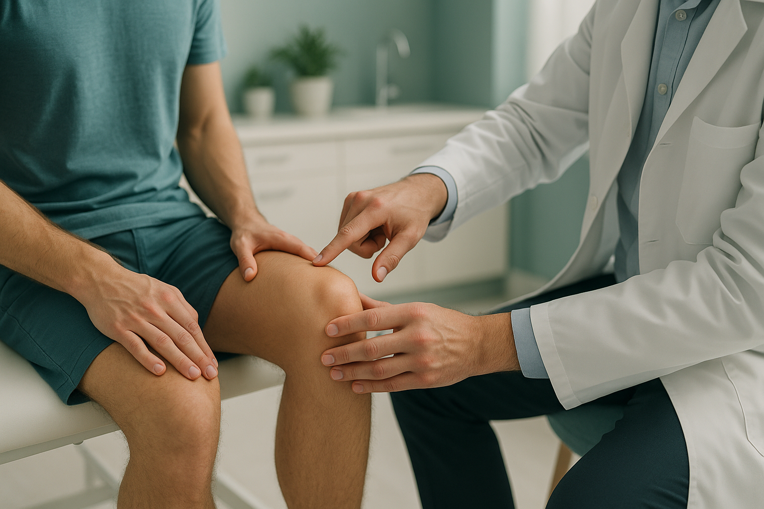
Patellar Maltracking: Why Does Your Kneecap Shift to the Side?
Patellofemoral maltracking means the kneecap does not glide centrally in the groove at the front of the thigh bone. Instead it drifts, tilts or catches, usually outward. This causes pain at the front of the knee, especially on stairs, squatting or sitting for a long time. It often involves the alignment of the whole leg, not just the knee itself.
Key takeaways
- The kneecap sits in a shallow groove and relies on muscles, ligaments and leg alignment to track straight.
- Patellofemoral maltracking is a common cause of front-of-knee pain in adults.
- Problems higher up (hip rotation, pelvic tilt) or lower down (flat feet, shin twist) can pull the kneecap off course.
- Lying-down scans relax the muscles, so the kneecap may sit normally. Standing, weight-bearing imaging can reveal the real tracking problem.
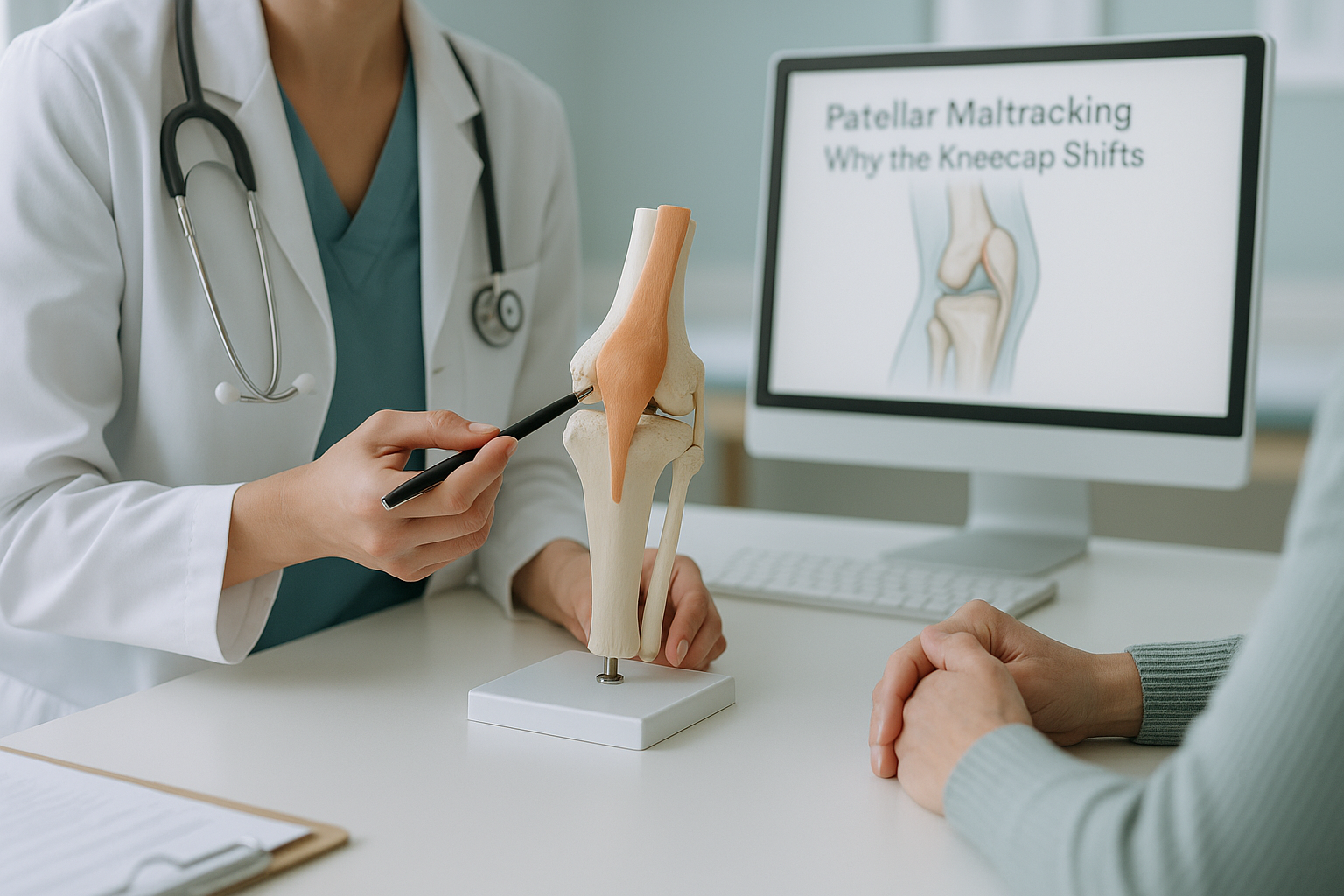
Your kneecap (patella) is the small, rounded bone at the front of the knee. It sits inside the tendon of your quadriceps muscle and slides up and down in a groove on the thigh bone (femur) every time you bend or straighten your leg. When everything lines up well, you barely notice it working.
With a patellar tracking disorder, the kneecap pulls to one side, usually the outer (lateral) side. It might tilt, shift slightly out of its groove (subluxation), or catch and grind as you move. Over time this can wear down the cartilage on the underside of the kneecap and cause persistent pain.
What does patellar maltracking feel like?
Most people notice a dull, aching pain around or behind the kneecap that gets worse with activity. The pain usually builds gradually rather than starting with a single injury.
Walking downstairs often hurts more than going up, because the kneecap presses harder into the groove.
A grating sensation or audible click when bending the knee, sometimes called crepitus.
Pain or stiffness after sitting with knees bent for a long time, sometimes called "cinema sign".
The knee may feel like it is about to give way, especially on uneven ground or when turning.
Important. If your knee locks completely, gives way suddenly, or you notice significant swelling after an injury, seek medical attention promptly through your GP, 111, or A&E. These may indicate a different, more urgent problem.
What causes the kneecap to track incorrectly?
There is rarely a single cause. Patellofemoral maltracking usually results from a combination of structural shape and muscle or alignment imbalances that pull the kneecap off centre.
A kneecap that sits higher than usual has less contact with the groove and is more likely to shift.
Some people are born with a flatter trochlear groove, giving the kneecap less of a channel to follow.
Weak inner quadriceps or tight outer structures can pull the kneecap laterally.
Knock knees, a twisted shin bone, or flat feet change the angle of pull on the kneecap.
Can hip or foot problems cause kneecap pain?
Yes. Your body is a connected chain from your feet to your spine. When one link is out of position, other areas compensate, and the knee is often the place that pays the price.
If your hip rotates inward too far, the thigh bone turns with it. That changes the angle between your thigh muscle and the kneecap tendon, pulling the kneecap outward. Flat feet or excessive ankle rolling can have a similar effect from below. A tilted pelvis can change the load on one leg compared to the other.
This is why treating kneecap pain in isolation sometimes fails. The kneecap is just the point where the forces meet. Unless you see how the whole leg lines up under load, you may miss the real driver.
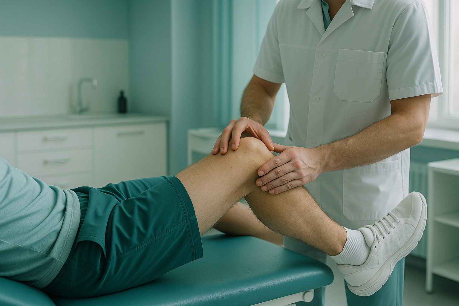
How is patellofemoral maltracking assessed when standing?
Standing, weight-bearing imaging shows the kneecap under real-life load, which is when tracking problems actually happen. Lying-down scans relax the muscles and take gravity out of the equation, so the kneecap may sit perfectly in its groove even when it does not in daily life.
An EOS scan captures the full length of both legs while you stand naturally. It shows the kneecap position, leg rotation, joint angles and overall alignment from hip to ankle in a single, low-dose image. The scan uses about 90% less radiation than a standard X-ray, around ten times less.
Because your muscles are active and gravity is acting on your joints, the image reflects how your body actually works. If the kneecap drifts laterally under load, it will show up.
Standard X-rays, CT and MRI are usually taken lying down. Without body weight compressing the joints and without your muscles firing, the kneecap often centres itself in the groove. The result can look normal even when the patient has clear symptoms while standing or walking.
These scans are still valuable for checking bone shape, cartilage (MRI) and ruling out other problems. But they may not catch a tracking issue that only appears under load.
At ScanAlign, the EOS system captures your whole leg alignment in one standing image. Your specialist or physiotherapist can then see exactly where the chain breaks down, whether it is at the hip, the knee, the shin or the foot. Learn more about how standing imaging helps with knee problems.
When MRI is the better tool. EOS shows bones, joints and alignment clearly. For soft-tissue detail such as cartilage damage, ligament tears or swelling inside the joint, MRI is usually the better choice. In many cases, both types of imaging together give the fullest picture.
What can be done about patellar maltracking?
Treatment depends on the cause and severity, and is always guided by a clinician, not an imaging clinic. ScanAlign provides the diagnostic imaging that informs those decisions. Common approaches include the following.
- Physiotherapy. Strengthening the inner quadriceps and stretching tight outer structures is often the first step.
- Orthotics or footwear changes. If flat feet or excessive foot roll contribute, correcting foot position can change how forces reach the kneecap.
- Taping or bracing. Patellar taping can guide the kneecap during exercise and rehabilitation.
- Surgery. In persistent cases where the groove is very shallow, the kneecap sits too high (patella alta), or non-surgical options have not helped, procedures such as lateral release or tibial tubercle transfer may be considered.
What all of these have in common is that they work best when the clinician understands the full alignment picture, not just what the kneecap looks like in isolation.
Not sure whether standing imaging would help your kneecap problem? Speak to us first, at no cost.
Book a free video consultationFrequently asked questions about patellar maltracking
It is a condition where the kneecap does not glide centrally in the groove on the front of the thigh bone. Instead it shifts, tilts or catches, usually toward the outer side. This causes pain at the front of the knee, particularly during activities like climbing stairs, squatting or sitting for a long time.
They are related but not identical. Maltracking means the kneecap does not follow its groove properly. Subluxation means it partially slips out of the groove. Subluxation is a more severe form of maltracking. Both can cause pain and instability.
Common reasons include muscle imbalance (weak inner quadriceps, tight outer structures), a shallow groove on the thigh bone, a kneecap that sits too high (patella alta), or alignment issues in the hip, shin or foot that change the angle of pull on the kneecap.
Yes. When the foot rolls inward excessively (overpronation), the shin bone can rotate, changing the forces that act on the kneecap. This can contribute to lateral maltracking.
When you lie down, gravity no longer compresses your joints and your muscles relax. The kneecap may settle back into its groove, making the image look normal. Standing, weight-bearing imaging captures the kneecap under real-life load, when tracking problems actually occur.
An EOS scan captures the full length of both legs while you stand. It shows kneecap position, leg rotation, joint angles and overall alignment from hip to ankle. This helps identify whether a problem elsewhere in the chain is contributing to the kneecap issue.
Yes. The EOS system uses about 90% less radiation than a standard X-ray, around ten times less. You can find more details on our safety and FAQs page.
No. ScanAlign is a private, self-pay clinic. You can refer yourself. We offer a free video consultation first so we can check whether standing imaging is right for your situation.
Pricing details are on our FAQs and cost page.
Many people improve with physiotherapy, taping and footwear changes. The right approach depends on the cause. Accurate imaging helps your clinician decide what is most likely to work for you. ScanAlign provides the imaging, not the treatment itself.
See the full picture, standing up
A free video consultation lets us understand your kneecap problem and explain whether standing imaging could help. No referral needed, no obligation.
Book a free video consultationSources
- NHS. "Patellofemoral pain syndrome." nhs.uk, National Health Service.
- Post WR, Teitge R, Amis A. "Patellofemoral malalignment: looking beyond the viewbox." Clinics in Sports Medicine, 2002; 21(3): 521-546.
- Defined Diagnostic. "Standing weight-bearing imaging in lower limb alignment assessment." European Journal of Radiology, EOS imaging clinical literature.
Written by Abbas Dhami (Specialist Diagnostic Radiographer)
This page is general information and not a diagnosis.
ScanAlign is the trading name of CoreMed Solutions Ltd and operates under The Harley Street Hospital's CQC licence.