
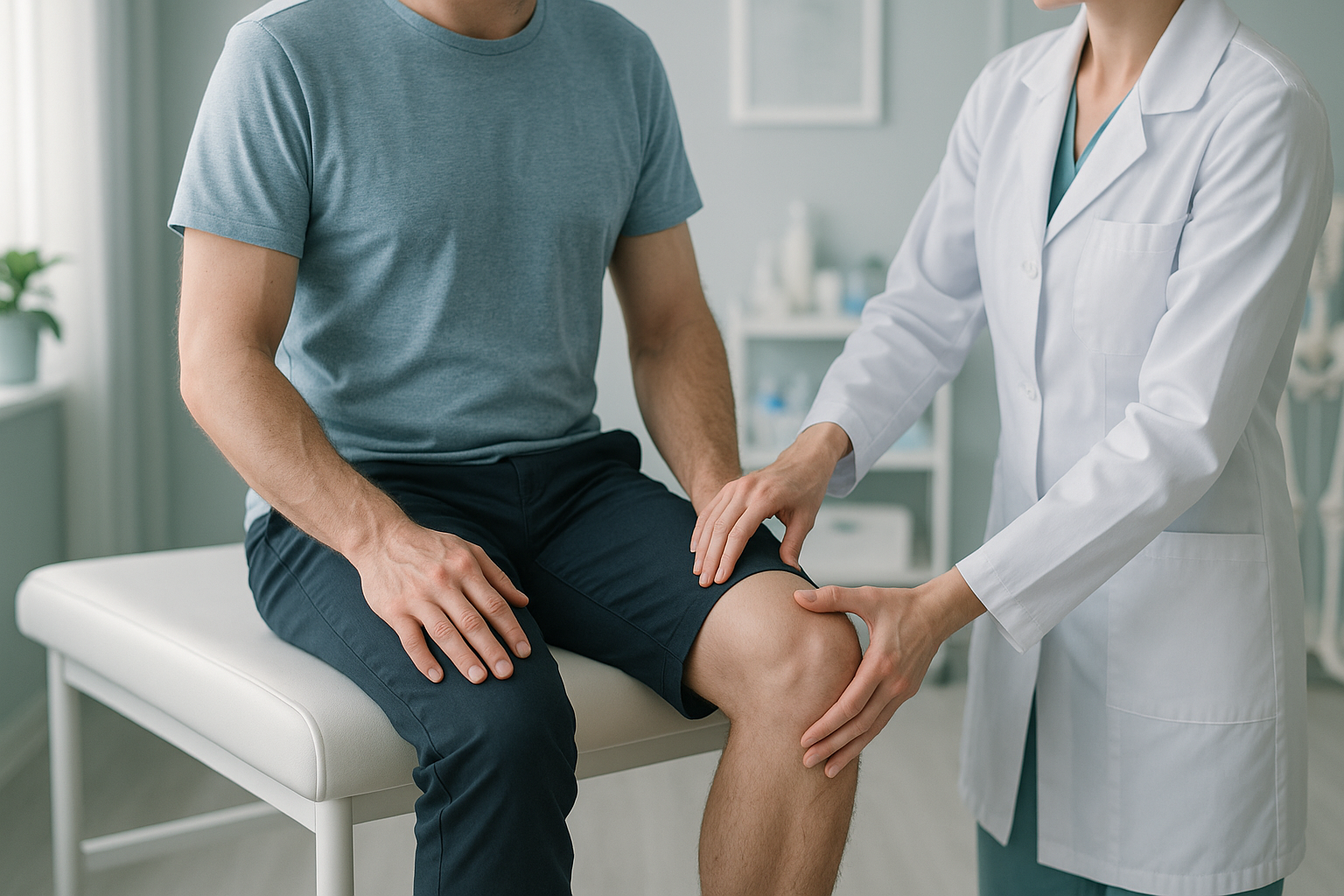
Femoral Anteversion and Retroversion: What Are They and Could They Be Causing Your Pain?
Femoral anteversion and femoral retroversion describe how the thighbone (femur) is twisted along its length. Too much forward twist is anteversion. Too little, or a backward twist, is retroversion. Both can change how your hip, knee and foot line up when you stand, leading to pain, stiffness or an unusual gait that is only visible under weight-bearing imaging.
Key takeaways
- Femoral version is the natural twist in your thighbone. Everyone has some, but too much or too little can cause problems.
- Anteversion tends to make you walk with your toes pointing inward. Retroversion tends to make you walk with your toes pointing outward.
- Pain often appears at the hip, knee or lower back rather than where the twist actually is.
- Standard lying-down scans can miss the real alignment picture because your muscles relax and gravity is removed.
- A standing whole-body EOS scan shows the full chain, from your feet to your spine, under natural load.
What is femoral version and why does it matter?
Femoral version is the amount of twist along the shaft of your thighbone. Picture the femur as a long rod. At the top it angles into the hip socket, and at the bottom it meets the knee. The natural twist between these two ends determines how your leg is oriented when you stand and walk.
Everyone has a small amount of forward twist. This is normal anteversion. When there is too much forward twist, the condition is called excessive femoral anteversion. When there is too little twist, or the bone twists backward, it is called femoral retroversion.
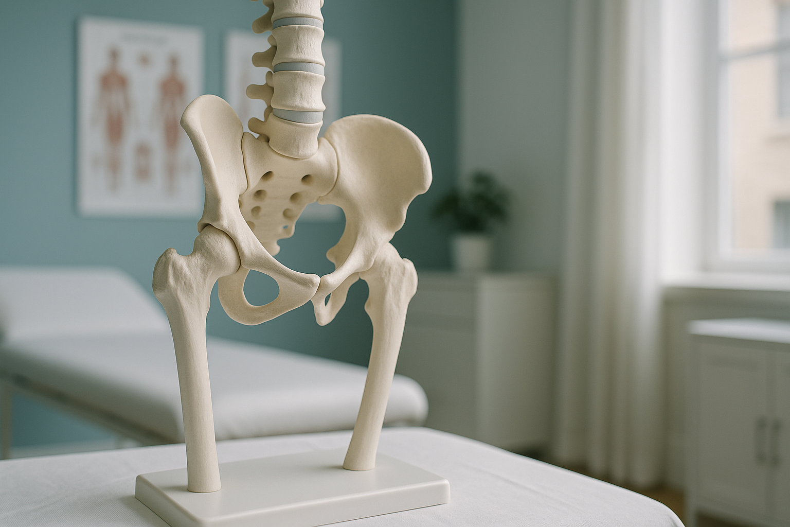
Femoral anteversion vs retroversion
With excessive anteversion, the femoral head points more forward than it should. To keep the hip joint seated properly, your whole leg tends to rotate inward. This is why in-toeing gait is one of the clearest everyday signs.
With femoral retroversion, the opposite happens. The femoral head is oriented further back, and the leg naturally rotates outward. You may notice an out-toeing gait, a feeling of tightness at the front of the hip, or difficulty sitting cross-legged.
Both conditions alter the mechanical loading through your hip joint. Over time, this can contribute to cartilage wear, labral stress, and early hip arthritis.
What are the signs of a femoral version abnormality?
Many adults with abnormal femoral version do not realise the twist in their thighbone is behind their symptoms. The signs below are common, though none of them on their own confirms the diagnosis.
Your feet point noticeably inward or outward when you walk, even though you are not doing it deliberately.
Aching at the front or side of the hip, especially after standing, walking or exercise.
The altered rotation changes loading at the knee, sometimes causing patella tracking issues or medial knee strain.
Your spine compensates for the twisted leg alignment, creating strain further up the chain.
Because these symptoms overlap with so many other conditions, a femoral version problem is often missed when imaging focuses on just one area.
What causes femoral anteversion or retroversion in adults?
In most adults, abnormal femoral version is a developmental variant. It means the natural twist that is present from childhood did not fully correct during growth. Less commonly, it can follow a healed fracture, hip surgery, or a condition affecting bone development.
The thighbone twist present at birth did not fully reduce during growth.
Bone healing or implant placement can alter the rotational profile of the femur.
Years of adapting your gait can mask the underlying twist but cause pain elsewhere.
Your body is a connected chain from your feet to your head. A twist at the thighbone forces the knee, ankle and even the lower back to compensate. That is why pain often appears far from the actual cause, and why whole-body imaging is so valuable.
How is femoral version assessed when you are standing?
Femoral version is best assessed when your body is weight-bearing, because that is when your muscles, joints and alignment are working as they do in real life. Standard imaging taken lying down removes gravity, relaxes the muscles, and can mask the way a twisted femur affects the rest of your skeleton.
An EOS scan captures a full-length, standing image of your skeleton from head to feet in a single pass. It uses about 90% less radiation than a standard X-ray, around ten times less. The image lets a clinician see how your femoral twist relates to your hip socket, your knee alignment and your spine, all on one picture.
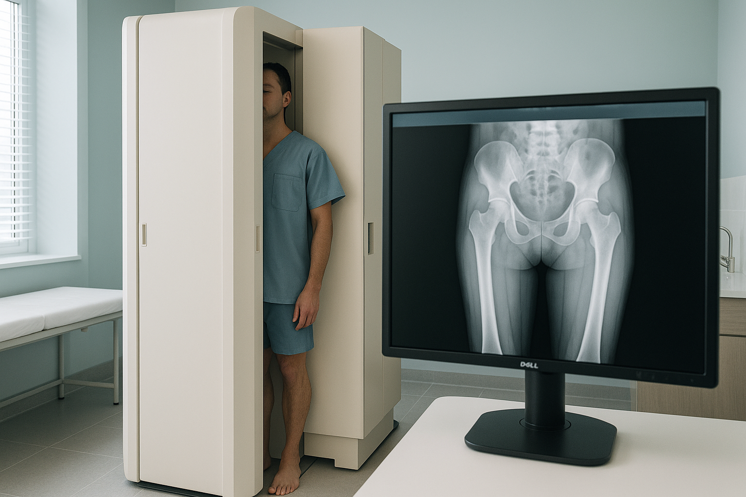
This matters for femoral version because the rotational twist at the thighbone changes the way your knee tracks, the way your pelvis tilts, and the way your spine curves above it. A scan of just the hip, taken lying down, cannot reveal those connected effects.
If you have hip, knee or gait concerns and want to understand how your alignment looks under real weight-bearing conditions, a standing EOS scan can show the full picture.
Book a free video consultationWhat can be done about femoral version problems?
The right approach depends entirely on how severe the twist is, how it affects your alignment, and what symptoms it is causing. ScanAlign is a diagnostic imaging clinic, not a treatment centre, so the goal is to give you and your clinician the clearest possible picture of what is happening.
In many cases, targeted physiotherapy focusing on hip and core strengthening, gait retraining, and flexibility can reduce symptoms. For adults with significant femoral version abnormalities that cause ongoing pain or joint damage, orthopaedic specialists may discuss a procedure called femoral derotation osteotomy, which corrects the twist surgically. Accurate imaging of the full lower limb is essential before any such decision.
Whatever path is right for you, it starts with knowing exactly what your alignment looks like when you are upright and weight-bearing.
When is MRI the better choice? EOS shows bones, joints and alignment clearly. For soft-tissue problems such as labral tears, cartilage damage or nerve issues, MRI is usually the better tool. In many cases, both types of imaging complement each other.
Frequently asked questions about femoral version
Anteversion means the thighbone twists forward more than normal, which tends to make the leg and foot turn inward. Retroversion means it twists less than normal or even backward, which tends to make the leg and foot turn outward. Both change the way your hip joint and the rest of your leg are loaded.
Yes. Excessive anteversion changes the position of the femoral head inside the hip socket, which can create abnormal contact, impingement and increased wear. This may cause deep groin or front-of-hip pain, especially with activity.
It can. When the thighbone is twisted differently, the knee joint has to compensate. This can lead to abnormal patella tracking, medial or lateral compartment overload, and long-term wear. That is why whole-body imaging is important, it shows how the hip twist affects the knee below it.
Lying down removes gravity and relaxes the muscles. Your leg may naturally rotate into a comfortable position that hides the effect of the twist. A standing scan shows how your bones actually line up when bearing your full weight, which is when the twist has a real impact.
An EOS scan is a standing, full-length X-ray that captures your entire skeleton from head to feet. It uses about 90% less radiation than a standard X-ray, around ten times less. Because it images you upright, it reveals the real-world effect of femoral twist on your hip, knee and spine alignment. You can learn more about how an EOS scan works.
Yes. It uses about 90% less radiation than a standard X-ray, around ten times less. You can find more details about safety and what to expect on the ScanAlign FAQs page.
No. ScanAlign is a private, self-pay clinic. You can refer yourself. The process starts with a free video consultation to discuss your symptoms and confirm that a scan is appropriate for you.
Pricing details are available on the ScanAlign FAQs page.
It is a surgical procedure where an orthopaedic surgeon cuts and rotates the thighbone to correct the abnormal twist. It is generally considered only when conservative measures have not helped and the version abnormality is causing significant symptoms or joint damage. ScanAlign does not perform surgery, but the imaging can help your surgeon plan if this option is being discussed.
In many cases, physiotherapy, gait retraining and strengthening work can improve how you walk and reduce related pain. The first step is understanding whether the gait pattern is being driven by the twist in the thighbone, and that requires accurate whole-body alignment imaging.
Understand your alignment, standing up
If hip pain, knee pain or an unusual gait has not been explained by previous scans, a standing whole-body EOS image may reveal the missing piece. Start with a free video consultation.
Book a free video consultationLearn more about hip alignment and how EOS imaging can help.
Sources
- Tönnis D, Heinecke A. Acetabular and femoral anteversion: relationship with osteoarthritis of the hip. Journal of Bone and Joint Surgery (American). 1999;81(12):1747-1770.
- Sangeux M, et al. Femoral anteversion and tibial torsion in adult populations. Journal of Orthopaedic Research. 2015;33(9):1381-1387.
- National Institute for Health and Care Excellence (NICE). Osteoarthritis: care and management. Clinical guideline CG177. 2014 (updated 2022).
Written by Abbas Dhami (Specialist Diagnostic Radiographer)
This page is general information and not a diagnosis.
ScanAlign is the trading name of CoreMed Solutions Ltd and operates under The Harley Street Hospital's CQC licence.