

My Scans Are Normal but I Am Still in Pain. What Now?
A normal scan does not mean there is nothing wrong. Standard imaging is done lying down and usually looks at one body area. It can miss alignment and loading problems that only appear when you are standing and weight-bearing. The answer is often not a better scan, but a different question.
Key takeaways
- Lying-down scans relax muscles and joints, so alignment problems can disappear from the image.
- Pain in one area is often caused by a problem somewhere else in the chain, from feet to head.
- A standing, whole-body assessment can reveal what single-region lying-down imaging misses.
- A "normal" result does not mean your pain is imagined. It may mean the imaging asked the wrong question.
- You do not need a GP referral to book a standing assessment.
Why is my scan normal when I am in pain?
There are several genuine reasons a scan can come back clear while you still feel real pain. None of them mean the pain is in your head.
Lying flat removes gravity. Joints decompress, muscles relax, and alignment shifts that cause your pain can vanish from the image.
A standard scan focuses on one area. If the cause sits above or below the painful spot, the scan simply does not include it.
Your body is a connected chain. A tilt or twist at the pelvis can overload a knee, or a stiff hip can strain the lower back. Pain often shows up far from its true source.
There is also a well-recognised phenomenon called central sensitisation, where the nervous system amplifies pain signals over time. In these cases, imaging may genuinely appear normal because the problem is in how the body processes pain, not in a structural injury visible on a scan. This is real and valid, but it is a clinical diagnosis best made by your treating clinician, not by a scan report alone.
What is the difference between lying-down and standing imaging?
Most imaging, including MRI, CT and standard X-ray, is performed with you lying flat on a table. This removes the very force that creates your symptoms: gravity and your own body weight. Read more about weight-bearing versus lying imaging.
What happens: You lie flat. Gravity is removed. The spine decompresses and the pelvis settles into a neutral position. Muscles switch off.
What the scan shows: The anatomy at rest. A disc bulge or arthritic change may be visible, but subtle tilts, shifts and compensations are hidden. Many disc bulges seen on MRI cause no symptoms at all, which can make the report confusing.
What happens: You stand naturally, bearing your own weight. Gravity compresses joints and reveals how your body actually loads itself day to day.
What the scan shows: True alignment from head to ankle in a single image. A pelvic tilt, a leg-length difference, or a sideways shift in the spine becomes visible because the body is under its normal load.
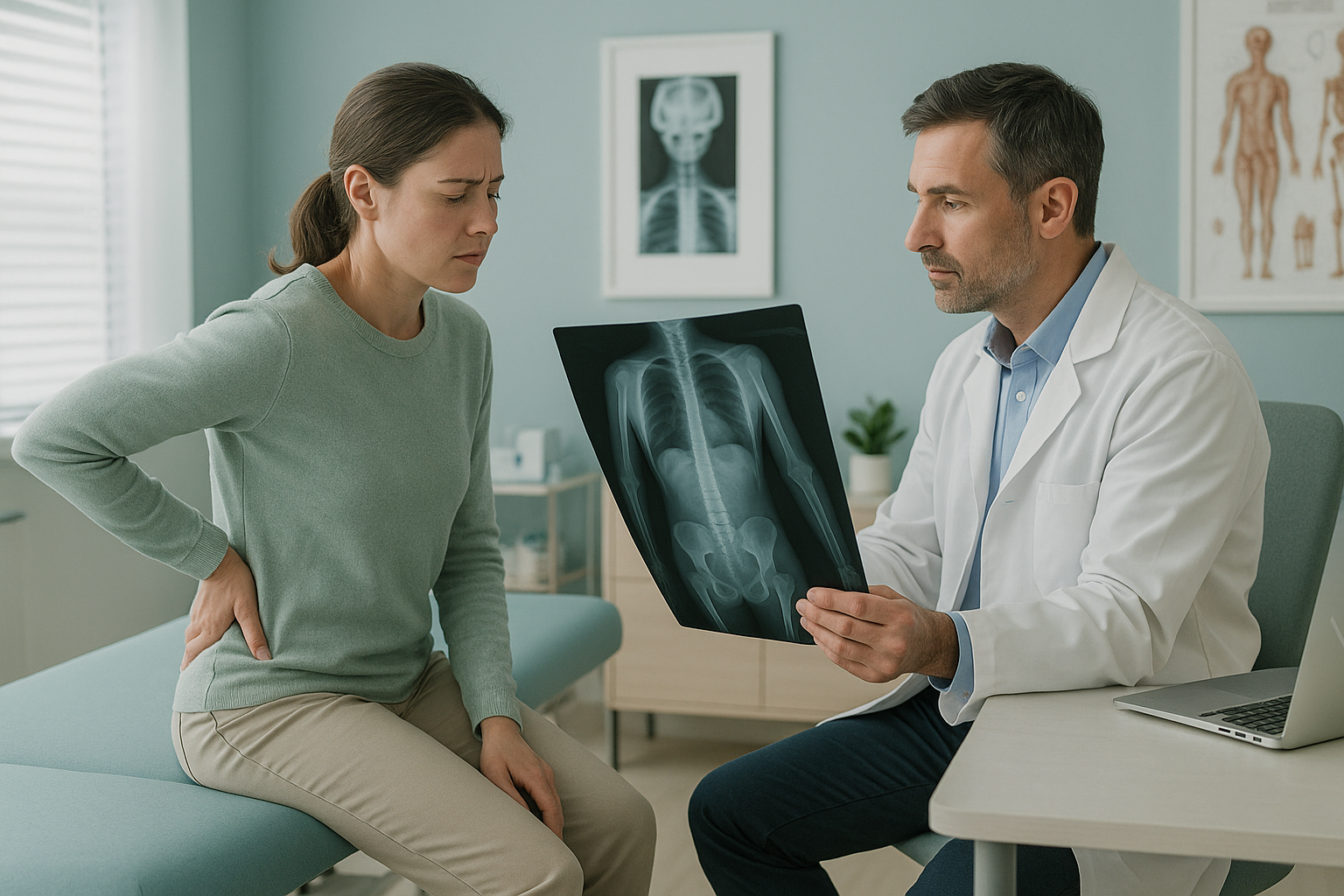
This is why a standing, whole-body approach can answer the question that lying-down imaging never asked. At ScanAlign, whole-body standing imaging captures you from head to ankle in a single exposure, with about 90% less radiation than a standard X-ray, around ten times less.
What are the signs that my pain has a postural component?
Pain with a postural or alignment component often follows a recognisable pattern. If several of these sound familiar, a standing assessment may be worth considering.
Pain increases with time on your feet and eases when you sit or lie down.
The pain is mostly on one side, or it moves between your back, hip and knee at different times.
You have had X-rays, MRIs, or both, and every report says "no significant findings."
Physiotherapy or injections have given only short-term relief because the underlying cause has not been identified.
A note on MRI. EOS standing imaging shows bones, joints and alignment under load. For soft tissue, discs or nerves, MRI is usually the better tool. The two are complementary. If your MRI was normal but the pain remains, the issue may not be soft tissue at all, it may be how your skeleton loads itself when you stand. Learn more about when an MRI shows nothing but the pain remains.
Important. If you have numbness, weakness in your legs, or any loss of bladder or bowel control, please seek urgent medical attention. Contact your GP, call 111, or go to A&E. These symptoms need immediate assessment, not a routine scan booking.
What should I do if my scans are clear but I still have pain?
The most productive next step is usually not another scan of the same type. It is asking a different question. Instead of "what is wrong with my knee?" you might ask "how does my whole body align when I stand, and is something else causing my knee to overwork?"
A standing, whole-body assessment captures everything from your head to your ankles in one image while you bear your own weight. It shows how each part of the chain relates to the next. A tilt in your pelvis, a twist in your spine, or a difference in leg length can become visible for the first time, even after years of normal lying-down results.
This is a diagnostic step, not a treatment. The images and measurements give your treating clinician, whether that is a physiotherapist, osteopath, or orthopaedic surgeon, new information to guide what happens next. Visit our back pain clinic page for more on how this fits into your care.
Not sure if a standing scan is right for you? You can talk it through first, free of charge, with no obligation.
Book a free video consultationFrequently asked questions
MRI is excellent at showing soft tissue, discs and nerves. But it is done lying down, so it removes gravity. Alignment problems that only appear when you stand and bear weight will not show on a lying-down MRI. Your pain may have a postural or loading cause that MRI simply was not designed to detect.
Yes. A standard X-ray typically images one area, such as a single knee or a section of spine, and is often taken lying down. It may miss problems above or below the painful area, and it cannot show how your whole skeleton aligns under load.
A standing whole-body scan captures your skeleton from head to ankle in one image while you bear your own weight. It reveals alignment, pelvic tilt, leg-length differences and compensatory shifts that disappear when you lie down for conventional imaging.
No. A normal scan means that particular type of imaging, in that position, did not find a visible cause. It does not mean there is nothing wrong. Many real, physical causes of pain, especially those related to alignment and loading, are simply not captured by standard lying-down imaging.
No. ScanAlign is a self-pay clinic and you can book directly without a referral. The process starts with a free video consultation to check that a standing scan is appropriate for your situation.
Yes. The EOS system uses about 90% less radiation than a standard X-ray, around ten times less. The scan takes only a few seconds and you simply stand still inside the machine.
ScanAlign is a private self-pay service. For current pricing and what is included, please visit the FAQs and cost page.
Yes, this is common. Your body is a connected chain. A tilt at the pelvis can overload a knee. A stiff hip can strain the lower back. Pain frequently appears in the area that is compensating, not the area where the original problem sits.
You receive a detailed report showing your alignment from head to ankle. This report can be shared with your GP, physiotherapist, osteopath, or surgeon to guide the next steps in your care. ScanAlign provides the imaging, not treatment.
ScanAlign is based at 19 Harley Street, London. It operates under The Harley Street Hospital's CQC licence.
Ready to ask a different question?
A free video consultation lets you talk through your history, your scans, and whether a standing assessment could show what has been missed.
Book a free video consultationAuthor: Abbas Dhami (Specialist Diagnostic Radiographer)
Sources
- Brinjikji W, Luetmer PH, Comstock B, et al. Systematic literature review of imaging features of spinal degeneration in asymptomatic populations. AJNR Am J Neuroradiol. 2015;36(4):811-816.
- Illés T, Somoskeöy S. The EOS imaging system and its uses in daily orthopaedic practice. Int Orthop. 2012;36(7):1405-1411.
- National Institute for Health and Care Excellence (NICE). Low back pain and sciatica in over 16s: assessment and management (NG59). 2016, updated 2020.
This page is general information and not a diagnosis.
ScanAlign is the trading name of CoreMed Solutions Ltd and operates under The Harley Street Hospital's CQC licence.