
Weight-Bearing vs Lying-Down Imaging: Why Does Standing Matter?
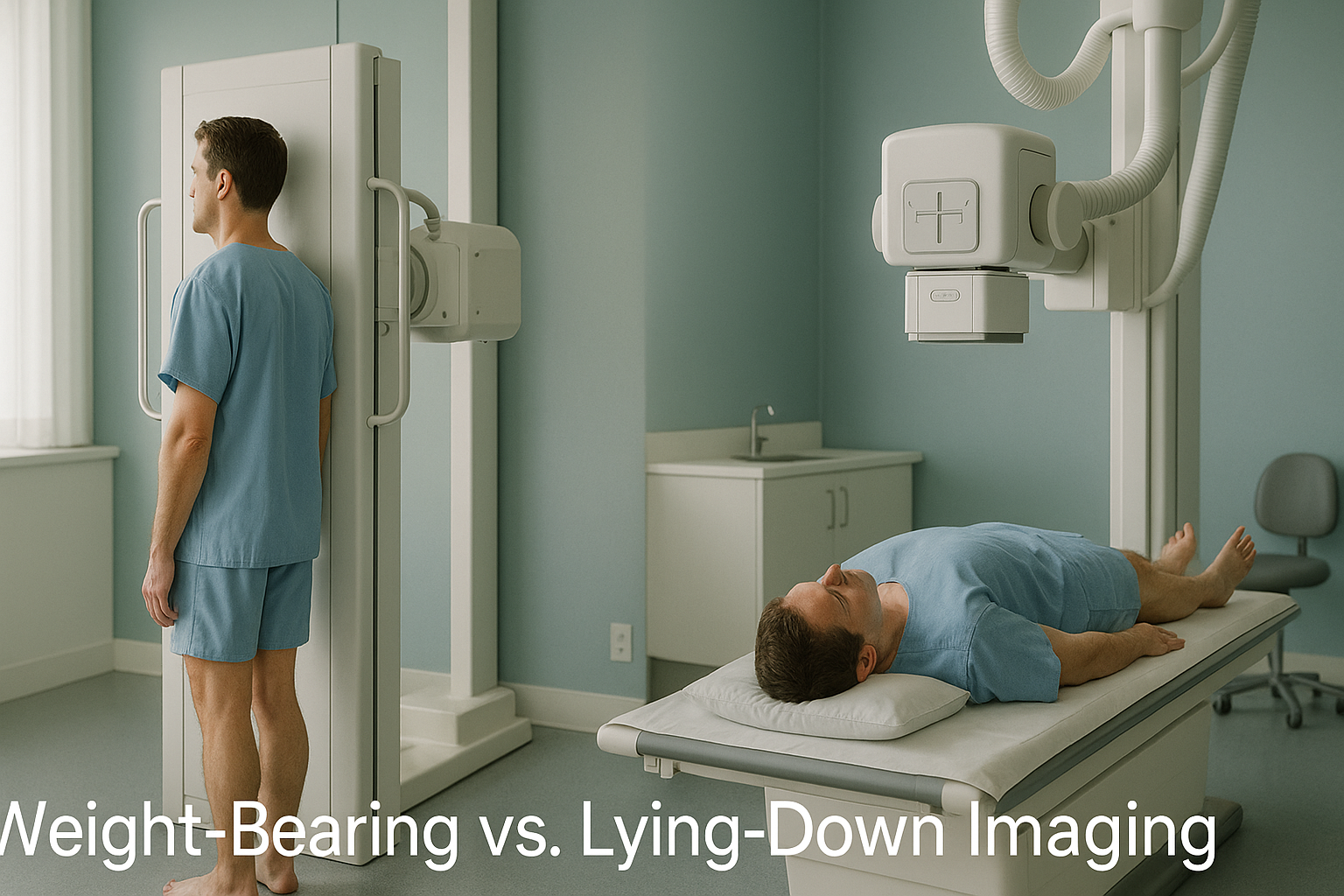
When you lie down for a scan, gravity stops compressing your joints and your muscles relax. This can hide the alignment problems that cause your pain during daily life. Weight-bearing imaging captures your skeleton as it actually works, standing under load, so your clinician can see what a lying-down scan may miss.
Key takeaways
- Lying down removes gravity from the picture, which can mask joint narrowing, curves and tilts.
- Standing weight-bearing images show how your bones, joints and spine behave under real-world load.
- Your body is a connected chain. A problem at one level often causes pain somewhere else.
- EOS imaging captures your full body standing, from head to feet, in one low-dose scan.
- EOS is not a replacement for MRI when soft tissue, disc or nerve detail is needed.
What changes between standing up and lying down?
Your alignment changes the moment you stand. Gravity compresses your discs, loads your joints and engages muscles that stay relaxed when you are flat on a table. A spine that looks well-aligned on a lying-down MRI may show a noticeable curve or tilt once body weight presses through it. A knee joint that appears fine on a standard X-ray may narrow significantly when you stand on it.
Research shows that spinal curves, pelvic tilt and leg-length differences can all measure differently depending on whether the patient is standing or lying. If the image does not match the position you are in when you feel pain, the image may not tell the full story.
How do you know a lying-down scan may have missed something?
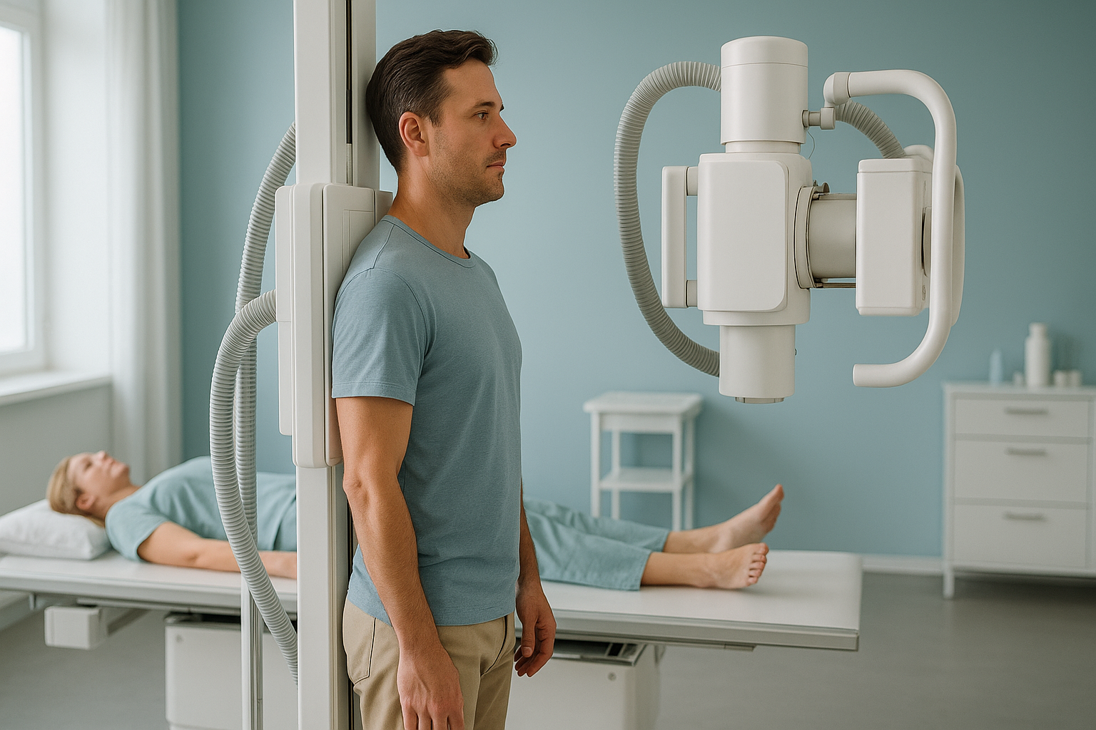
If you have had imaging that came back normal but you are still in pain, a weight-bearing scan may reveal what was hidden. Common signs include the following.
MRI or X-ray results look fine, yet your back, hip or knee still hurts when you walk or stand.
Symptoms increase when you are upright, walking or carrying weight, but ease when you sit or lie down.
You lean to one side, one shoulder sits higher, or you feel off-balance when standing still.
Pain moves around, affecting your back one day and your knee the next, suggesting a linked problem.
Why does a problem in one joint cause pain in another?
Your body is a connected chain from your feet up through your knees, hips, pelvis, spine and head. When one part of that chain is out of position, the areas above and below compensate to keep you balanced. Over time, that compensation creates strain, and pain often appears far from the original cause.
For example, a slight pelvic tilt can change the way your lower spine curves, leading to pain in your mid-back or even your neck. A knee that angles inward may start as an alignment issue at the hip. Single-area scans only look at one part of the chain. They cannot show how the rest of the body is adapting.
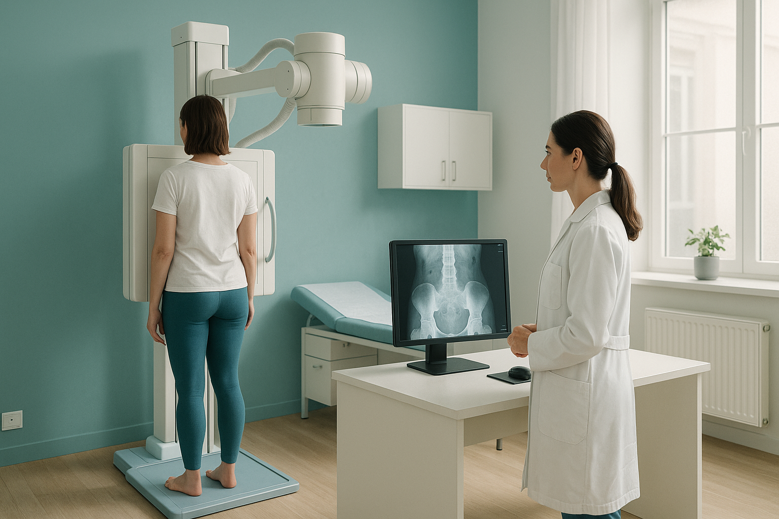
This is exactly why weight-bearing, whole-body imaging is so valuable. It captures every link in the chain at once, so your clinician can trace the cause rather than just treat the symptom.
How does EOS imaging capture your body standing up?
EOS is a specialist imaging system that scans you while you stand naturally. It captures your full skeleton, from head to feet, in a single upright exposure. That means the image shows your bones, joints and alignment exactly as they are when you are on your feet and gravity is acting on them.
Because EOS captures such a wide view, your clinician can measure how each part of the body relates to the others. They can see whether a hip sits higher on one side, whether the spine is compensating, and how the knees and ankles line up below. It uses about 90% less radiation than a standard X-ray, around ten times less, which makes it a practical choice for the detailed imaging alignment assessment requires.
Want to understand more about how the EOS scan works? Read the full overview of EOS imaging at ScanAlign.
When MRI is the better choice: EOS shows bones, joints and alignment beautifully. For soft tissue detail, such as discs, nerves, ligaments or cartilage, MRI is usually the better tool. In many cases, both types of imaging together give the fullest picture.
What can you do if lying-down scans have not explained your pain?
If previous imaging has not given you clear answers, a standing weight-bearing scan can be a sensible next step. It does not replace the scans you have already had. It adds a different perspective, one that may reveal the alignment or loading pattern your clinician needs to see.
ScanAlign is a diagnostic imaging clinic. It does not provide treatment. The detailed alignment data from your scan goes to your clinician, whether that is a surgeon, physiotherapist or osteopath, so they can plan the right approach for you. You do not need a GP referral to book.
- Your pain is worse when standing or walking
- Lying-down scans came back normal but you are still in pain
- You are being assessed for spinal alignment or posture
- You need a full picture before hip or knee surgery planning
- You need soft tissue, disc or nerve detail (MRI is better)
- You have acute trauma that needs urgent assessment
- You cannot stand unsupported for around 20 seconds
Have questions about the scan, safety, what to expect or cost? Visit the frequently asked questions page.
Frequently asked questions
When you stand, gravity compresses your joints and engages your muscles. This changes spinal curves, joint spacing and pelvic position compared with lying flat. A standing scan captures these real-world forces so your clinician sees what is actually happening when you are upright and in pain.
Weight-bearing imaging means the scan is taken while your body carries its own weight, usually standing. This shows how your skeleton behaves under load, which lying-down scans cannot replicate.
Yes. MRI is excellent for soft tissue, discs and nerves, but the patient lies flat. Curves, tilts and joint narrowing that appear under gravity may flatten out in a lying position, so they can go undetected.
EOS is a standing imaging system that captures your whole body, from head to feet, in one upright scan. Unlike a standard X-ray, it uses about 90% less radiation, around ten times less, and produces a full-length image that shows how every joint relates to the others.
No. ScanAlign accepts self-referrals. You can book a free video consultation directly without needing a letter from your GP.
Yes. EOS uses about 90% less radiation than a standard X-ray, around ten times less. It is designed to give detailed skeletal images at a very low dose.
Pricing details are available on the ScanAlign FAQs page.
Yes. Surgeons planning joint replacements benefit from seeing how the joint loads under body weight. A full standing scan shows the alignment of the whole leg and pelvis, giving the surgeon precise data for implant positioning.
Wear comfortable, loose-fitting clothing without metal zips, buttons or underwired bras. You may be asked to change into a gown for certain scan areas. Full details are on the what to expect page.
ScanAlign is based at 19 Harley Street, London. It operates under The Harley Street Hospital's CQC licence.
See what lying-down scans may have missed
A free video consultation lets you discuss your symptoms, ask questions and find out whether a standing EOS scan is the right next step for you.
Book a free video consultationSources
- Lazennec, J.-Y., et al. "Pelvis and total hip arthroplasty acetabular component orientations in sitting and standing positions." Orthopaedics & Traumatology: Surgery & Research, 2011.
- Illés, T. & Somoskeöy, S. "The EOS imaging system and its uses in daily orthopaedic practice." International Orthopaedics, 2012.
- NHS. "Back pain: diagnosis." NHS.uk. Available at: nhs.uk/conditions/back-pain.
Written by Abbas Dhami (Specialist Diagnostic Radiographer)
This page is general information and not a diagnosis.
ScanAlign is the trading name of CoreMed Solutions Ltd and operates under The Harley Street Hospital's CQC licence.