
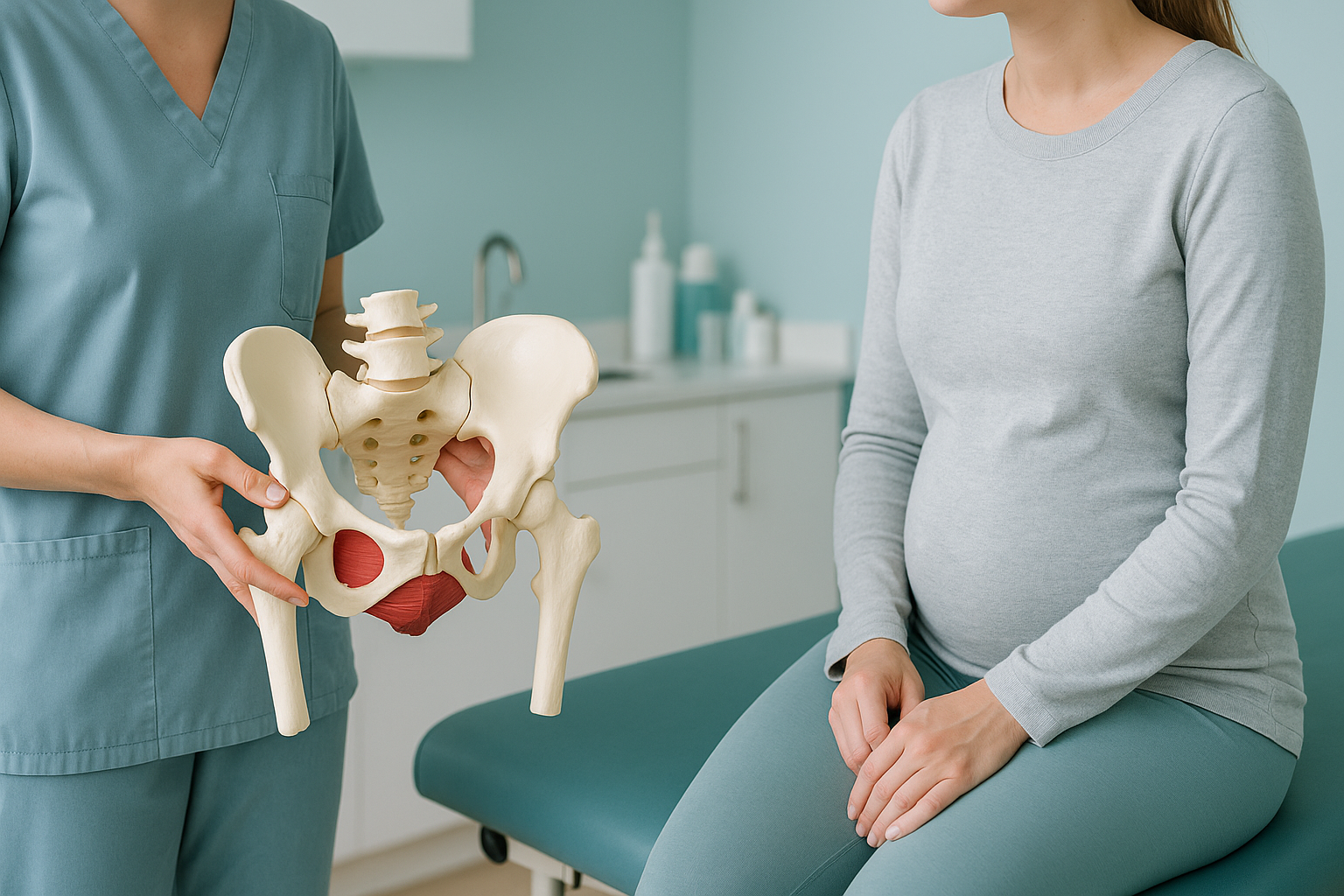
How Does Your Pelvis Recover After Giving Birth?
Your pelvis widens during pregnancy and birth, then gradually returns towards its pre-pregnancy position over the following months as the hormone relaxin clears and ligaments tighten. For some women, the pelvis does not fully realign. A standing whole-body image can show whether persistent pain is linked to a remaining shift, tilt or asymmetry.
Key takeaways
- Relaxin softens pelvic ligaments during pregnancy. It can take three to six months (or longer) after birth for them to firm up again.
- Pelvic girdle pain, postnatal back pain and postpartum posture changes are common and usually improve, but not always fully.
- Lying-down scans can miss alignment problems that only appear when you are standing and weight-bearing.
- A standing EOS image shows the whole skeleton in one shot, revealing whether your pelvis has stayed shifted, tilted or rotated.
- Most women can be scanned from around three months after a vaginal birth, or around four months after a caesarean section. Your clinician will advise on timing.
What happens to your pelvis during pregnancy and birth?
Your body releases the hormone relaxin to loosen the ligaments around the pelvis, making room for your baby to pass through. The two halves of the pelvis at the front (the pubic symphysis) and the sacroiliac joints at the back all become more mobile. This is normal and necessary.
After delivery, relaxin levels drop and your ligaments slowly tighten. For most women, the pelvis returns close to its original position within a few months. But for some, the joint surfaces do not settle evenly. One side may sit slightly higher, or the pelvis may stay rotated. You might not notice this at all, or you might feel it as ongoing pain in your lower back, hips or groin.
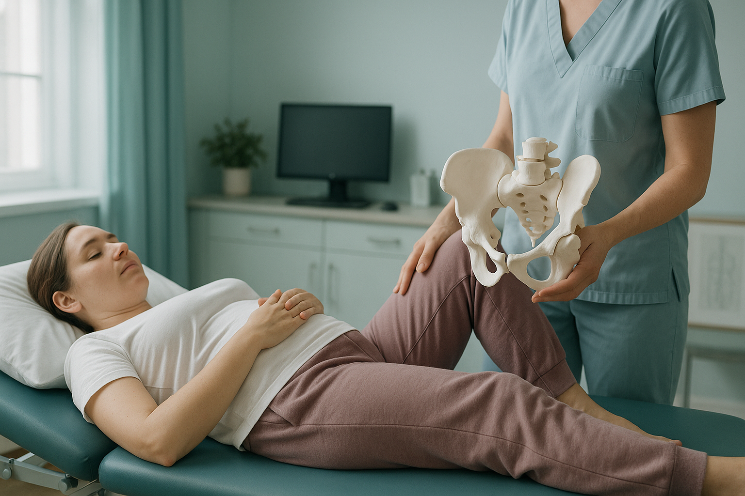
Diastasis recti, a separation of the abdominal muscles, often accompanies these pelvic changes. When the deep core muscles are weakened, your spine and pelvis lose some of their usual support. This can contribute to postnatal back pain and altered postpartum posture.
What are the signs your pelvis hasn't fully recovered?
Ongoing pelvic girdle pain after birth is the most common sign. But the effects of a pelvis that has not fully realigned can show up in several ways.
A sharp or aching sensation at the front of the pelvis, especially when walking, climbing stairs or turning in bed.
Persistent aching across the lower back or sacroiliac joints that does not settle with rest or simple exercises.
A feeling that one hip sits higher, or a limp or waddle that lingers well after delivery.
A forward-leaning stance, increased lower back curve, or feeling that your balance has shifted since pregnancy.
If you also experience sudden numbness, weakness in your legs, or any loss of bladder or bowel control, please seek urgent medical attention through your GP, 111 or A&E. These symptoms need immediate assessment.
Why does postnatal pelvic pain continue for some women?
Several factors can slow or prevent full pelvic recovery. Often more than one is at play.
Breastfeeding can keep relaxin levels elevated, prolonging ligament laxity for months after birth.
Separated abdominal muscles reduce core support, putting extra load on the pelvis and lower back.
Holding a baby on one hip or feeding in a twisted position loads the pelvis unevenly, day after day.
A pelvic tilt or lateral pelvic tilt that was present before pregnancy may become more pronounced after birth.
The body is a connected chain from your feet to your head. When the pelvis shifts, your spine, hips and knees compensate. That is why postnatal back pain can be felt far from the pelvis itself, and why a rotated pelvis can affect your gait and knee comfort on one side.
How is postnatal pelvic alignment assessed standing up?
A standing, weight-bearing image is the most reliable way to see how the pelvis sits under real load. When you lie down for a standard X-ray or MRI, your muscles relax and gravity is removed. A tilt, rotation or leg-length difference that causes pain when you stand may flatten out and appear normal on a lying-down scan.
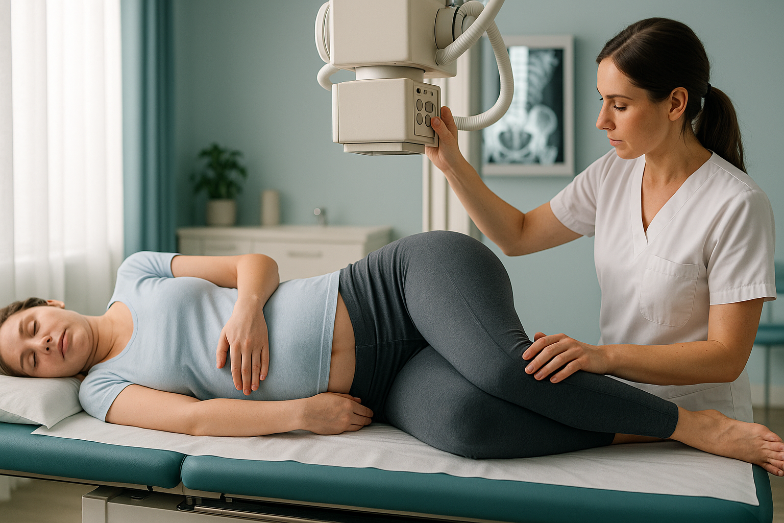
An EOS scan captures your entire skeleton, from your head to your feet, in a single standing image. It uses about 90% less radiation than a standard X-ray, around ten times less. The scan itself takes only a few seconds and you simply stand still inside the machine.
The resulting image lets your clinician measure the exact position of the pelvis relative to your spine, hips and legs. If one side is higher, or the pelvis has stayed rotated, it will show clearly. This level of whole-body context is what makes ScanAlign standing EOS imaging particularly useful for postnatal alignment concerns.
Still have questions about timing or what the scan involves?
A short video call with our team can help you decide if an alignment assessment is right for you at this stage.
Book a free video consultationWhat helps postnatal pelvic recovery?
Recovery is usually a combination of time, gradual loading and, in some cases, targeted physiotherapy. Here are the general approaches most commonly recommended.
- Pelvic floor rehabilitation. Restoring pelvic floor strength helps support the joints from below. A specialist physiotherapist can guide you through safe progressions.
- Core restoration. Gentle deep-core work, particularly for diastasis recti, helps stabilise the pelvis and reduce postnatal back pain. Avoid high-impact exercise until your core can handle the load.
- Carrying and feeding habits. Switching the hip you carry your baby on, and setting up supported feeding positions, reduces repetitive one-sided loading.
- Gradual return to exercise. Building load slowly gives the ligaments time to firm up. Jumping straight back into running or heavy lifting while relaxin is still present can prolong symptoms.
- Alignment assessment. If symptoms persist beyond the early months, a standing whole-body image gives your physiotherapist or consultant the structural detail they need to refine your plan.
When is MRI more appropriate?
EOS excels at showing bones, joints and whole-body alignment. If your clinician suspects a disc problem, nerve compression, labral tear or other soft-tissue issue, MRI is usually the better tool. The two scans answer different questions and sometimes both are needed.
How soon after birth can you have a pelvic alignment scan?
There is no single rule, but as a general guide, most women can be scanned from around three months after a vaginal birth, or around four months after a caesarean section. By this point, early healing is well underway and any lingering alignment issues are more likely to represent the pelvis's settled position rather than temporary post-delivery swelling.
If you are still breastfeeding, relaxin may remain elevated, so your alignment could continue to change. That does not mean a scan is pointless. It means the result is a snapshot of where you are now, which your clinician can use to guide rehabilitation. A follow-up image later can show whether things have shifted further as hormones normalise.
Your six-week postnatal check is a good time to discuss persistent symptoms with your GP. If pain continues beyond that point, an alignment assessment becomes more relevant.
Who benefits from a postnatal alignment assessment?
- Pelvic girdle pain or SPD that has not settled months after birth
- Postnatal back pain that physiotherapy alone has not resolved
- A feeling of unevenness, one hip higher or a persistent limp
- Planning to return to sport and wanting a structural baseline
- Diastasis recti with ongoing pelvic or lower back symptoms
- Still in the first few weeks after birth (too early for a settled reading)
- Symptoms that are clearly improving with physiotherapy alone
- Suspected disc, nerve or soft-tissue injury (MRI is usually better)
- Sudden numbness, weakness or bladder/bowel changes (seek urgent medical help)
Frequently asked questions
For most women, symphysis pubis dysfunction (SPD) improves within a few weeks to a few months after birth. In some cases, discomfort persists for six months or longer, especially if the pelvis has not fully realigned or if ligament laxity continues due to breastfeeding.
Yes. When the abdominal muscles separate, the deep core loses some of its ability to support the spine and pelvis. This can transfer extra load to the lower back, contributing to postnatal back pain. Targeted core rehabilitation usually helps, but persistent pain may benefit from an alignment assessment to check for pelvic asymmetry as well.
Mild back discomfort in the first weeks after birth is very common. If it persists beyond three to four months, or gets worse rather than better, it is worth investigating. Raise it at your six-week check and consider further assessment if simple measures are not helping.
As a general guide, from around three months after a vaginal birth or four months after a caesarean section. By this point, early healing has progressed enough for the image to represent your settled alignment rather than temporary swelling. Your clinician will advise on the best timing for you.
Breastfeeding can keep relaxin levels elevated, which means ligaments may remain looser for longer. This does not mean you should stop breastfeeding, but it is useful context. Your pelvis may continue to settle after you finish.
A standard X-ray usually images one area while you lie down. An EOS scan captures your whole skeleton, from head to feet, while you stand. It uses about 90% less radiation than a standard X-ray, around ten times less, and shows how the pelvis relates to your spine, hips and legs under real weight-bearing conditions.
No. ScanAlign accepts self-referrals. You can book a postnatal alignment assessment directly. The process starts with a free video consultation to discuss your symptoms and confirm that the scan is appropriate for you.
Pricing details are available on the ScanAlign website. The free video consultation is a good first step to understand what is included and whether a scan is the right next move for you.
In most cases, a pregnancy-related pelvic tilt resolves as ligaments tighten and muscles strengthen. However, if the pelvis settles in a shifted position and remains there for months, it can become the body's new normal. Identifying a persistent tilt early means rehabilitation can be targeted more effectively.
Yes. The EOS scan uses about 90% less radiation than a standard X-ray. This very low dose does not affect breast milk or breastfeeding. You can feed as normal before and after the scan.
Still uncomfortable months after birth?
A short, free video call can help you understand whether a standing alignment scan is the right next step for your recovery.
Book a free video consultationWritten by Abbas Dhami (Specialist Diagnostic Radiographer)
Sources
- Vleeming A, Albert HB, Östgaard HC, Sturesson B, Stuge B. European guidelines for the diagnosis and treatment of pelvic girdle pain. European Spine Journal. 2008;17(6):794–819.
- NHS. Pelvic pain in pregnancy (SPD). NHS.uk. Available at: https://www.nhs.uk/pregnancy/related-conditions/common-symptoms/pelvic-pain/
- Sperstad JB, Tennfjord MK, Hilde G, Ellström-Engh M, Bø K. Diastasis recti abdominis during pregnancy and 12 months after childbirth. British Journal of Sports Medicine. 2016;50(17):1092–1096.
This page is general information and not a diagnosis.
ScanAlign is the trading name of CoreMed Solutions Ltd and operates under The Harley Street Hospital's CQC licence.