
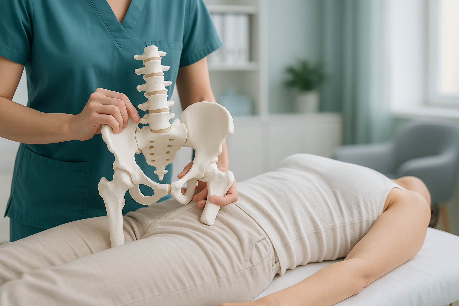
Rotated Pelvis: What Causes It and How Do You Know You Have One?
A rotated pelvis means one side of your pelvis has twisted forward or backward relative to the other. This shifts the way your body bears weight, often causing pain in your back, hip or knee rather than in the pelvis itself. Standing, weight-bearing imaging is usually the clearest way to see the rotation and measure it accurately.
Key takeaways
- A rotated pelvis, sometimes called pelvic torsion, is when one side of the pelvis sits further forward or back than the other.
- Symptoms often appear away from the pelvis because the body compensates through the spine, hips and legs.
- Lying-down scans can miss the rotation because gravity relaxes the muscles that hold the twist in place.
- An EOS standing scan captures your whole alignment from head to feet in a single image, using about 90% less radiation than a standard X-ray.
What is a rotated pelvis?
A rotated pelvis is a positional shift where one half of your pelvis turns forward while the other turns backward. Think of your pelvis as a bowl. When it sits level, your weight spreads evenly through both hips, both legs and your spine. When it twists, everything above and below has to adjust.
This is different from a pelvic tilt, where the whole pelvis tips forward or backward. With pelvic torsion, the two sides move in opposite directions. It can be subtle, but even a small rotation changes the way your joints line up under load.

What are the symptoms of a twisted pelvis?
Twisted pelvis symptoms vary widely because the rotation forces other joints to compensate. You may never feel pain in the pelvis itself. Instead, pain often shows up in places that are absorbing the extra load.
The spine curves to counterbalance the rotation, overloading muscles and joints on one side of the lower back.
One hip socket is pushed into a slightly different position, which can pinch or stretch the surrounding soft tissue.
Rotation changes the angle of the thigh bone, which can shift stress onto the knee or make one leg feel functionally shorter.
You may notice your shoes wear unevenly, or that walking feels slightly lopsided.
Important: If you experience sudden numbness, weakness in your legs, or loss of bladder or bowel control, seek urgent medical attention. Contact your GP, call 111, or go to A&E. These symptoms need immediate assessment, not an imaging booking.
What causes a rotated pelvis?
Pelvic torsion usually develops gradually. It is rarely caused by a single event. Several factors can pull or hold the pelvis into a twisted position over time.
Tight hip flexors or a weak glute on one side can gradually pull the pelvis out of alignment.
Sitting cross-legged, always leaning on one hip, or standing with weight on one leg for long periods.
Even a small difference in leg length, whether structural or functional, can tilt and twist the pelvis over time.
A past hip, back or leg injury can leave scar tissue or guarding patterns that hold the pelvis in a rotated position.
Why does lying-down imaging miss a rotated pelvis?
Lying-down imaging, including standard X-rays, CT and MRI, removes the effect of gravity and relaxes your muscles. When you lie flat, a mild pelvic rotation can temporarily correct itself. The twist only shows up consistently when you are standing and your body is bearing its own weight.
Your body works as a connected chain from your feet to your head. A rotation in the pelvis does not stay local. It causes the spine to curve, one hip to load differently, and the knees and ankles to adjust. A single-area scan of the painful spot, such as a knee MRI, might look normal. It does not capture the upstream cause.
Shows the real picture. When you stand, gravity pulls on your skeleton and your muscles engage. A pelvic rotation is visible because it is actively happening. A full-length standing image captures how the rotation affects your spine, hips, knees and ankles all at once.
Can mask the problem. Lying flat lets your muscles relax and your joints settle into a neutral position. A mild or moderate pelvic rotation can appear to correct itself on the table, leading to images that look normal despite ongoing symptoms.
How does an EOS scan assess a rotated pelvis?
An EOS scan captures a full-length, weight-bearing image of your skeleton from head to feet in a single pass while you stand naturally. This gives a clear view of how the pelvis sits relative to the spine above and the legs below, exactly as it is in everyday life.
The scan uses about 90% less radiation than a standard X-ray, around ten times less. It takes under a minute, requires no injections or contrast dye, and you stay fully clothed in loose-fitting clothing.
The resulting image allows your clinician to see the pelvic rotation in context. Rather than looking at the pelvis in isolation, they can trace the chain of compensation all the way through your body and understand where the loading problems begin.
Want to find out if standing imaging could explain your symptoms? Talk to our team at no cost.
Book a free video consultationWhen MRI is the better tool: EOS excels at showing bones, joints and whole-body alignment. If your clinician suspects a soft-tissue problem, such as a labral tear, disc issue or nerve compression, MRI is usually the more appropriate scan. The two are complementary, not competing.
How is a rotated pelvis typically managed?
Management depends entirely on the cause, the severity and how much it is affecting your daily life. ScanAlign is a diagnostic imaging clinic, not a treatment provider, so we do not prescribe exercises or therapies. What we can do is give you and your treating clinician a clear, accurate picture of what is happening in your body under load.
Common approaches that clinicians and physiotherapists use for pelvic torsion include targeted muscle strengthening, manual therapy, gait retraining and, in some cases, orthotics. The right approach depends on having the right diagnosis first. That is where accurate, standing imaging fits in.
You can learn more about how hip alignment is assessed at ScanAlign, or read about how the spine compensates for pelvic problems.
Frequently asked questions about a rotated pelvis
Yes. When the pelvis rotates, the spine has to curve or twist to compensate. This puts uneven load on the muscles, discs and joints of the lower back, often on one side more than the other.
Common signs include one-sided back or hip pain, a feeling that one leg is shorter, uneven shoe wear, or difficulty sitting comfortably for long periods. A standing, weight-bearing scan is the most reliable way to confirm the rotation.
No. A pelvic tilt means the whole pelvis tips forward or backward. A rotated pelvis, or pelvic torsion, means one side has twisted forward while the other sits further back. Both can occur together.
Many people see improvement with the right combination of physiotherapy, strengthening and manual therapy. The first step is getting an accurate diagnosis so your clinician knows exactly what they are working with.
No referral is needed. ScanAlign is a private, self-pay clinic. You can book directly. We recommend starting with a free video consultation so our team can confirm the scan is appropriate for you.
An EOS scan uses about 90% less radiation than a standard X-ray, around ten times less. It is one of the lowest-dose imaging methods available for skeletal assessment.
Most standard X-rays are taken lying down, which removes gravity and allows the muscles to relax. A mild rotation can temporarily correct itself on the table, making the image appear normal even though the problem exists when standing.
It can. Pelvic rotation changes the angle of the thigh bone, which alters how the kneecap tracks and how weight is distributed through the knee joint. Some people are treated for a knee problem when the root cause is in the pelvis.
You can find current pricing and what is included on our FAQs and cost page.
You stand still inside the scanner for under a minute while the machine captures a full-length image. There is no tunnel, no noise and no injections. You wear loose-fitting clothing with no metal. The whole visit is straightforward and quick. More details are on our what to expect page.
Wondering if a pelvic rotation is behind your pain?
A free video consultation with our team can help you understand whether a standing EOS scan is the right next step.
Book a free video consultationSources
- Dubousset, J. et al. "EOS: A new imaging concept." European Spine Journal, 2010. Overview of EOS low-dose biplanar imaging for full-body skeletal assessment.
- NHS. "Back pain: causes, treatment and prevention." nhs.uk. General guidance on musculoskeletal back pain and when to seek imaging.
- Legaye, J. et al. "Pelvic incidence: a fundamental pelvic parameter for three-dimensional regulation of spinal sagittal curves." European Spine Journal, 1998. Research on the relationship between pelvic position and spinal alignment.
Written by Abbas Dhami (Specialist Diagnostic Radiographer), ScanAlign, 19 Harley Street, London.
This page is general information and not a diagnosis.
ScanAlign is the trading name of CoreMed Solutions Ltd and operates under The Harley Street Hospital's CQC licence.