

Lateral Pelvic Tilt: Why Does One Hip Sit Higher Than the Other?
A lateral pelvic tilt means your pelvis is not level when you stand. One hip sits higher than the other, shifting your weight unevenly. This can strain muscles and joints from your lower back down to your knees. Standing whole-body imaging shows the tilt under real load, helping identify where the imbalance starts.
Key takeaways
- Lateral pelvic tilt is when one side of the pelvis is higher than the other while standing.
- It often causes pain away from the pelvis itself, in the lower back, hip or knee.
- Lying-down scans can miss it because gravity and relaxed muscles let the pelvis level out.
- A standing whole-body image captures the tilt under your full body weight.
- Accurate imaging helps clinicians find the root cause rather than chasing symptoms.
If you have ever been told one hip is higher than the other, or noticed your waistband sits unevenly, you may have a lateral pelvic tilt. It is surprisingly common and often goes unnoticed for years. The pelvis is the bridge between your spine and legs. When it tilts to one side, everything above and below has to adapt.
What are the signs of a lateral pelvic tilt?
The most obvious sign is uneven hips when you stand in front of a mirror. But many people feel the effects long before they see them.
One hip bone appears higher when you stand relaxed. Trousers or a belt may sit at an angle.
The spine curves to compensate for the tilt, overloading muscles on one side of your back.
The lower hip bears more weight, which can stress the hip joint or the knee below it.
You may scuff one shoe more than the other or feel that one leg has to "reach" further when walking.
What causes a lateral pelvic tilt?
A lateral pelvic tilt happens when muscles, joints or bones on one side of the body pull or push the pelvis out of level. There is rarely a single cause. Common contributors include the following.
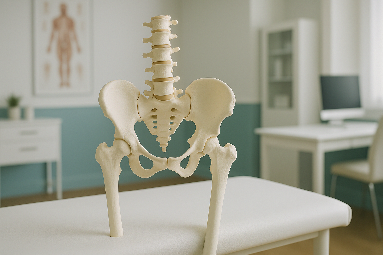
Standing on one leg, crossing the same leg, or carrying a bag on one shoulder repeatedly.
Even a small difference in leg length can tilt the pelvis when you stand or walk.
Tight or weak muscles on one side of the hip, such as the quadratus lumborum or hip abductors.
Scoliosis, hip arthritis or previous injury can shift the pelvis structurally.
How does a lateral pelvic tilt affect the rest of your body?
Your body is a connected chain from your feet to your head. When the pelvis tilts sideways, the spine curves to keep your head centred over your feet. This is called compensation. The muscles on one side of your back work harder. Your hip joints bear uneven load. Your knees and ankles adjust too.
This is why lateral pelvic tilt often causes pain somewhere other than the pelvis. You might feel it in your lower back, your outer hip, or even your knee. Treating the painful area alone may not help if the underlying tilt is the real driver.
Pain that keeps coming back? A standing whole-body image can show whether pelvic tilt is part of the picture.
Book a free video consultationWhy does a standing scan show lateral pelvic tilt better than lying down?
A standing scan captures your skeleton under the same gravity and muscle forces you experience every day. When you lie down for a conventional X-ray, CT or MRI, your muscles relax and gravity pulls differently. The pelvis can settle into a more level position, hiding the very tilt that causes your symptoms.
An EOS scan takes a full-length image of your body while you stand naturally. It shows the pelvis, spine and legs in a single view, so the relationship between all three is clear. If one hip is higher, the image shows it. If the spine is curving to compensate, that appears too. And it uses about 90% less radiation than a standard X-ray, around ten times less.
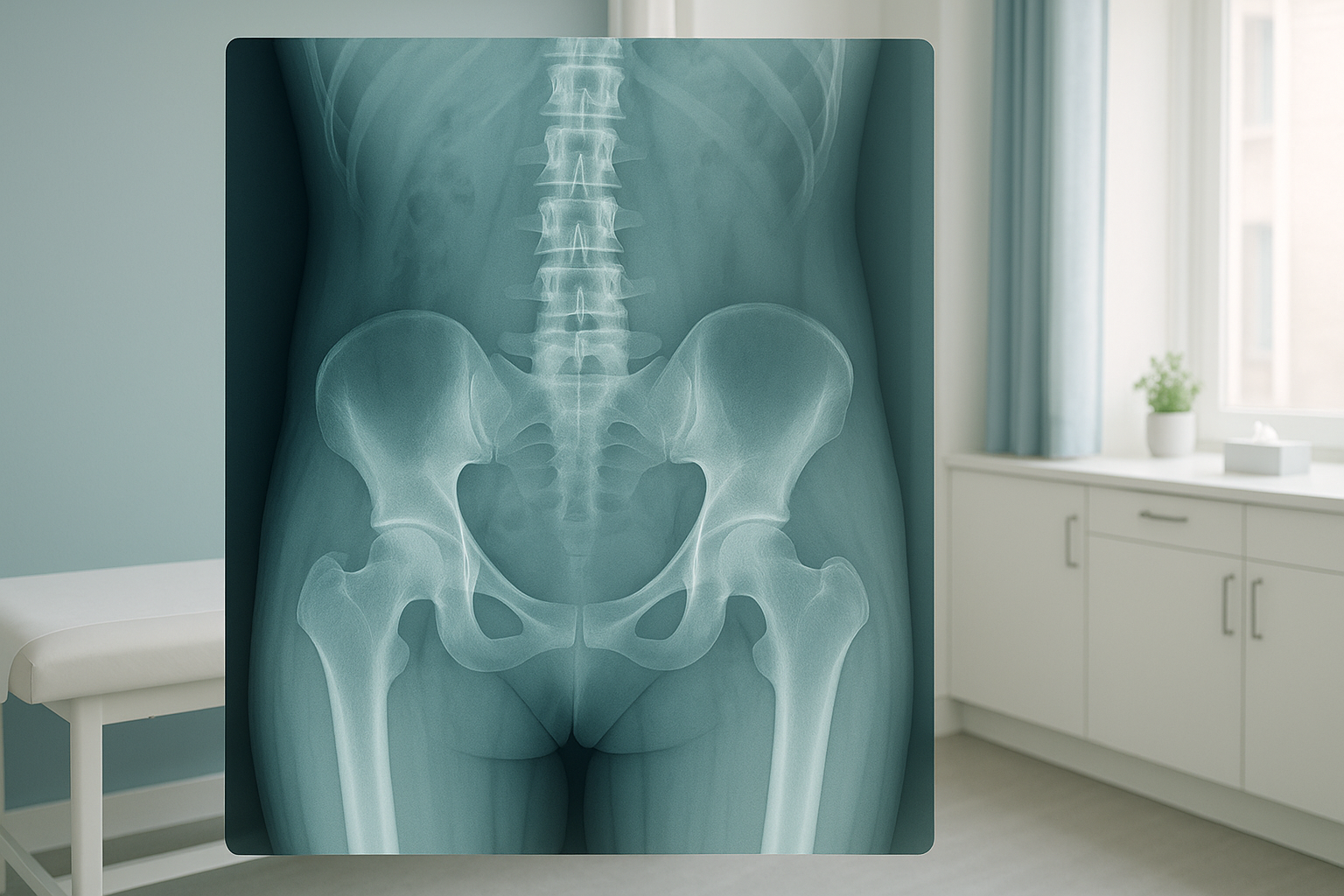
This whole-body, weight-bearing view gives your clinician the information they need to plan the right next step, whether that is physiotherapy, orthotics, or further investigation.
What helps with a lateral pelvic tilt?
The right approach depends on the cause. Accurate imaging is the starting point because it tells your clinician whether the tilt is driven by a structural issue, like a leg length difference or joint change, or by a functional pattern, like muscle tightness.
Common approaches recommended by clinicians include targeted physiotherapy to rebalance muscles around the hip and pelvis, heel lifts or orthotics if leg length plays a role, and manual therapy to address stiffness. In some cases, further investigation may be needed.
ScanAlign does not provide treatment. It provides the diagnostic imaging that helps the right clinician make the right plan for you.
When is MRI the better choice? EOS shows bones, joints and alignment in a standing position. If your clinician suspects soft-tissue problems such as a disc issue, a labral tear, or nerve involvement, MRI is usually the better tool. Both types of imaging can work together to give a complete picture.
Frequently asked questions about lateral pelvic tilt
It means your pelvis is tilted sideways so one hip sits higher than the other when you stand. This can affect how your spine, hips and knees share your body weight.
Common causes include a difference in leg length, muscle imbalance around the hips, habitual posture patterns, scoliosis, or hip joint changes such as arthritis.
You can stand in front of a mirror and place your hands on the top of your hip bones. If one hand is noticeably higher, a tilt may be present. However, this is not a substitute for proper imaging, which measures the tilt accurately and shows the cause.
It can. When the pelvis is not level, the spine curves to compensate. This puts extra strain on muscles and joints on one side of the back, which can lead to persistent one-sided lower back pain.
When you lie flat, gravity pulls evenly on both sides and your muscles relax. The pelvis can settle into a more level position, hiding the tilt that appears when you stand and bear your full weight.
An EOS scan is a standing, full-body X-ray that captures your skeleton from head to feet in a single image. It shows your pelvis, spine and legs under real weight-bearing conditions, making it well suited to assessing alignment and pelvic tilt. It uses about 90% less radiation than a standard X-ray, around ten times less.
No. ScanAlign is a private self-pay clinic, and you can self-refer. You start with a free video consultation so the team can make sure the scan is appropriate for your concern.
Pricing details are available on the ScanAlign FAQs and cost page.
Yes, they describe the same thing. Pelvic obliquity is the clinical term often used by specialists. Both mean the pelvis is tilted to one side so the hips are not level.
Many cases improve with the right approach, but the first step is understanding the cause. Imaging helps determine whether the tilt is structural or functional, which guides the plan your clinician recommends. ScanAlign provides the imaging, not the treatment itself.
Wondering if pelvic tilt is behind your pain?
A free video consultation lets you discuss your symptoms and find out whether a standing whole-body scan is the right next step.
Book a free video consultationLearn more about how EOS whole-body imaging works on the ScanAlign EOS scan overview.
Sources
- Khamis, S. and Yizhar, Z. (2007). Effect of feet hyperpronation on pelvic alignment in a standing position. Gait & Posture, 25(1), pp.127-134.
- Illés, T. and Somoskeöy, S. (2012). The EOS imaging system and its uses in daily orthopaedic practice. International Orthopaedics, 36(7), pp.1315-1323.
- NHS (2023). Back pain: causes, treatment and prevention. NHS.uk.
Written by Abbas Dhami (Specialist Diagnostic Radiographer)
This page is general information and not a diagnosis.
ScanAlign is the trading name of CoreMed Solutions Ltd and operates under The Harley Street Hospital's CQC licence.