
Pelvic Tilt: What Is It and How Do You Know You Have One?
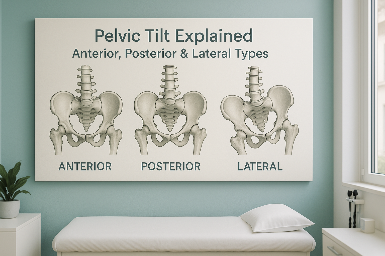
A pelvic tilt is a shift in the position of your pelvis away from its neutral alignment. It can tilt forward (anterior), backward (posterior) or drop to one side (lateral). Because the pelvis connects your spine to your legs, even a small tilt can change how your whole body carries weight and may cause pain in your back, hips or knees.
Key takeaways
- There are three main types of pelvic tilt: anterior, posterior and lateral.
- Pain often appears away from the pelvis itself, in the lower back, hip or knee.
- Lying-down scans can relax muscles and hide a tilt that only shows when you stand.
- An EOS standing scan captures your full alignment from head to feet on one image.
- Identifying the tilt is a diagnostic step, not a treatment. It helps guide the right care.
Your pelvis is the central hub of your skeleton. It sits between your spine and your legs and acts as a platform for your upper body. When it is level and balanced, your weight spreads evenly. When it tilts, every joint above and below has to adjust. That adjustment is called compensation, and it is often the reason people feel pain in a place that seems unrelated to the actual problem.
What are the different types of pelvic tilt?
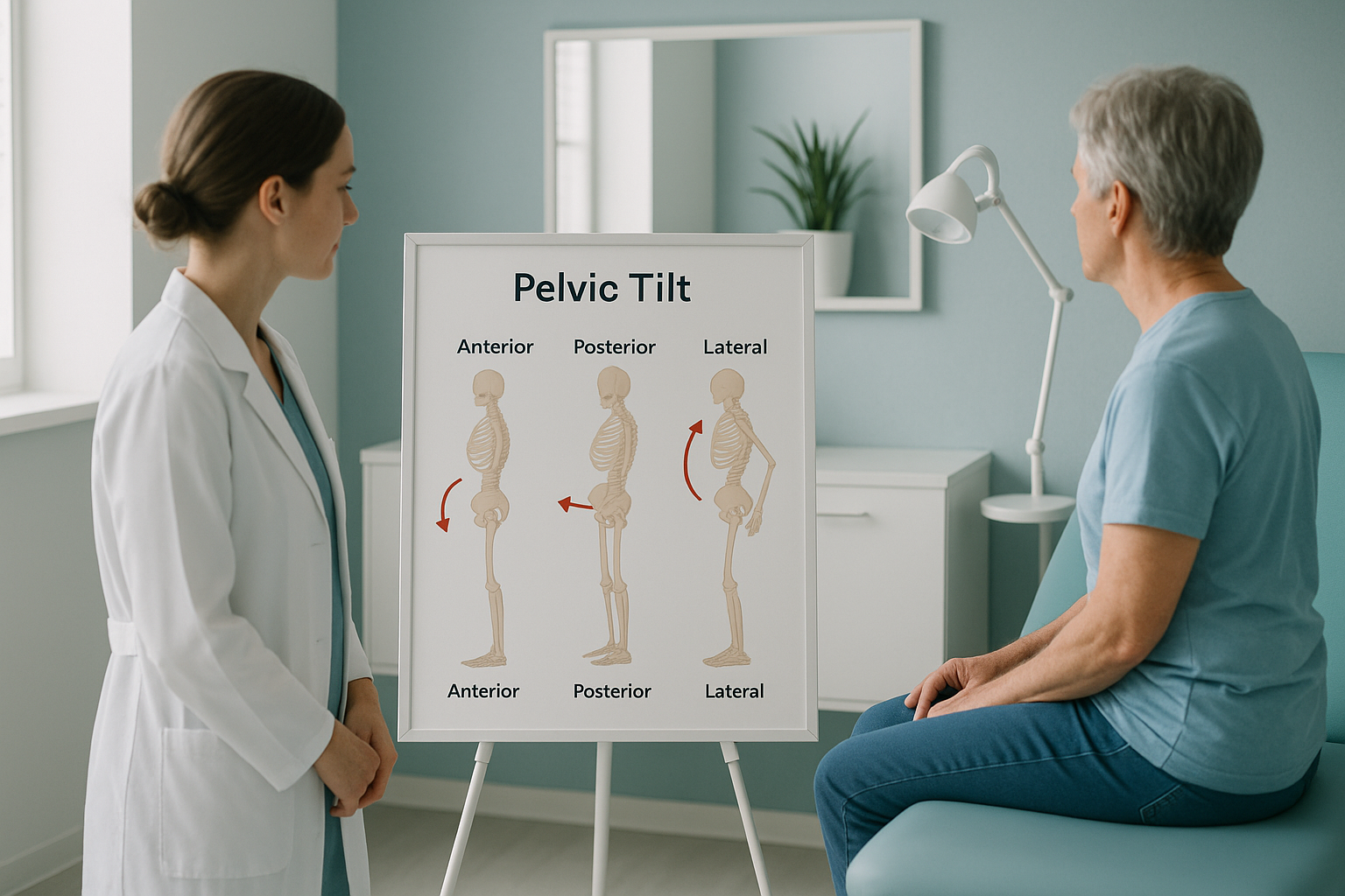
There are three main types: anterior (forward), posterior (backward) and lateral (side to side). Each one shifts the pelvis in a different direction and creates a different pattern of strain through the body.
The front of the pelvis drops forward and down. This increases the curve of the lower back and often pushes the stomach forward. It is the most common type.
The pelvis tucks under, flattening the lower back curve. This can create a slouched posture and put extra load through the lumbar discs.
One side of the pelvis sits higher than the other. This can make one leg feel shorter and changes how you walk, often straining the hip or knee on the lower side.
Many people have a combination of two types at once, for example an anterior tilt with a lateral drop. That is why looking at the pelvis in isolation rarely tells the whole story. You need to see how it relates to the spine above and the legs below.
How do you know if you have a pelvic tilt?
You may notice visual clues in the mirror or feel recurring aches that do not match a clear injury. Common signs include the following.
- An exaggerated lower back curve or a visibly flat lower back.
- One hip appearing higher than the other when you stand in front of a mirror.
- Trousers or a belt sitting unevenly around the waist.
- Recurring lower back, hip or knee pain that does not improve with rest.
- Tightness in the hip flexors or hamstrings on one or both sides.
- Feeling off-balance or favouring one leg when you stand still.
These signs can overlap with other conditions, so accurate imaging is important to confirm whether a tilt is present and how far it extends through your alignment.
What causes a pelvic tilt?
A pelvic tilt develops when the muscles and joints around the pelvis are loaded unevenly over time. Common contributing factors include prolonged sitting, previous injury, muscle imbalance and natural variation in leg length or joint shape.
Hours at a desk shorten the hip flexors and weaken the glutes, pulling the pelvis forward.
Even a small difference in leg length can tilt the pelvis to one side when you stand and walk.
A fracture, surgery or sprain on one side can change how you load your pelvis as you recover.
Tight or weak muscles on one side of the body pull the pelvis out of its neutral position over time.
Your body is a connected chain from your feet to your head. When the pelvis tilts, the spine curves to compensate, the shoulders may shift, and the knees or ankles take extra load. That is why the pain you feel might be in your knee or lower back, but the underlying cause could be a pelvic tilt that nobody has measured.
Why does a pelvic tilt need to be assessed standing up?
A pelvic tilt is a weight-bearing problem. It only shows its true effect when you are upright and gravity is acting on your skeleton. When you lie down for a standard X-ray, CT or MRI, your muscles relax, your joints decompress, and the tilt can partially or fully disappear. The scan may come back looking normal, even though you feel anything but normal when you stand and walk.
An EOS scan takes a full-length image of your body while you stand naturally. It captures your pelvis, spine, hips, knees and ankles in one exposure, so a specialist can see how one area affects every other. EOS also uses about 90% less radiation than a standard X-ray, around ten times less, which makes it a practical choice for whole-body imaging.
Not sure whether your symptoms could be related to pelvic alignment? Talk it through with a specialist radiographer at no cost.
Book a free video consultationWhat helps once a pelvic tilt is identified?
Identifying the type and extent of a pelvic tilt is the diagnostic step. ScanAlign provides the imaging and clinical report, not treatment. Once you have a clear picture of your alignment, your clinician can use it to plan the most appropriate next step.
Depending on the findings, that might include physiotherapy to address muscle imbalances, a referral to an orthopaedic specialist, footwear adjustments for a leg length difference, or further investigation. Having a standing whole-body image gives any treating clinician a measurable baseline they can refer back to over time.
When MRI is the better tool. EOS shows bones, joints and alignment clearly. If your clinician suspects a soft tissue, disc or nerve problem, MRI is usually the better choice. The two types of imaging complement each other well.
You can learn more about hip alignment assessment or read about spinal alignment and posture on the ScanAlign site.
Frequently asked questions about pelvic tilt
A pelvic tilt is a shift in the position of your pelvis away from neutral. It can tip forward (anterior), backward (posterior) or drop to one side (lateral). It affects how your spine, hips and legs share your body weight.
An anterior tilt means the front of the pelvis drops forward, increasing the lower back curve. A posterior tilt means the pelvis tucks under, flattening the lower back. Both change how forces travel through the spine and legs.
Yes. A tilted pelvis changes the curve of the lower spine, which can overload certain joints and muscles. Many people with unexplained lower back pain discover a pelvic tilt is a contributing factor once it is properly assessed.
A clinician may suspect a tilt from a physical examination. Imaging confirms and measures it. A standing full-body scan, such as EOS, is particularly useful because it captures the pelvis under natural weight-bearing conditions, unlike lying-down scans.
When you lie down, gravity is removed and your muscles relax. This can allow the pelvis to return closer to neutral, hiding the tilt that causes your symptoms when you are upright. A standing scan shows what is actually happening under load.
EOS uses about 90% less radiation than a standard X-ray, around ten times less. It is a very low-dose system designed for full-body imaging. You can find more details on the ScanAlign FAQs page.
No. ScanAlign accepts self-referrals. You can book a free video consultation directly and discuss your symptoms with a specialist radiographer before deciding whether to proceed with a scan.
Pricing details are available on the ScanAlign FAQs page. The free video consultation will also confirm exactly what is included.
Yes. Because the pelvis connects the spine to the legs, a tilt can change the loading on the hip joints, alter leg alignment and put extra stress on the knees. Pain in the knee or hip can sometimes be traced back to pelvic alignment.
You receive a detailed report showing your alignment from head to feet. This report can be shared with your GP, physiotherapist or consultant to guide the next steps. ScanAlign provides diagnostic imaging, not treatment, so your treating clinician uses the findings to plan your care.
Ready to see the full picture?
Talk to a specialist radiographer about your symptoms, at no cost and with no obligation, before deciding whether a standing alignment scan is right for you.
Book a free video consultationAuthor: Abbas Dhami (Specialist Diagnostic Radiographer)
Sources
- Herring JA. Tachdjian's Pediatric Orthopaedics, 6th ed. Elsevier, 2022. (Chapters on pelvic and spinal alignment biomechanics.)
- Legaye J, Duval-Beaupère G. "Sagittal plane alignment of the spine and gravity." European Spine Journal, 2005;14(7):654-658.
- NHS. "Back pain: causes, treatment and prevention." nhs.uk/conditions/back-pain.
This page is general information and not a diagnosis.
ScanAlign is the trading name of CoreMed Solutions Ltd and operates under The Harley Street Hospital's CQC licence.