

Anterior Pelvic Tilt: What Causes It and How Do You Know You Have One?
Anterior pelvic tilt is when the front of your pelvis drops forward and the back rises up, creating an exaggerated curve in your lower back. It is usually caused by muscle imbalances from prolonged sitting and can lead to lower back pain, hip tightness and poor posture. A standing whole-body scan reveals the tilt under real weight-bearing conditions.
Key takeaways
- Anterior pelvic tilt tips the pelvis forward, increasing the arch in your lower back.
- Common causes include tight hip flexors, weak glutes and long hours sitting.
- Pain often shows up in the lower back or hips, not the pelvis itself.
- Lying-down scans can miss alignment problems that only appear when you are standing.
- A standing EOS scan captures the whole body in one image, showing how the pelvis affects everything above and below it.
What is anterior pelvic tilt?
Anterior pelvic tilt is a postural pattern where the pelvis rotates forward around the hip joints. Picture your pelvis as a bowl of water. In a neutral position, the water stays level. With an anterior tilt, the bowl tips forward and water would spill out of the front.
When this happens, the lower spine curves inward more than it should. This is sometimes called an increased lumbar lordosis. The knock-on effect travels both upward and downward. Your body is a connected chain from your feet to your head, so the pelvis sitting out of position can change the way your knees track, how your mid-back rounds and even how your neck sits.
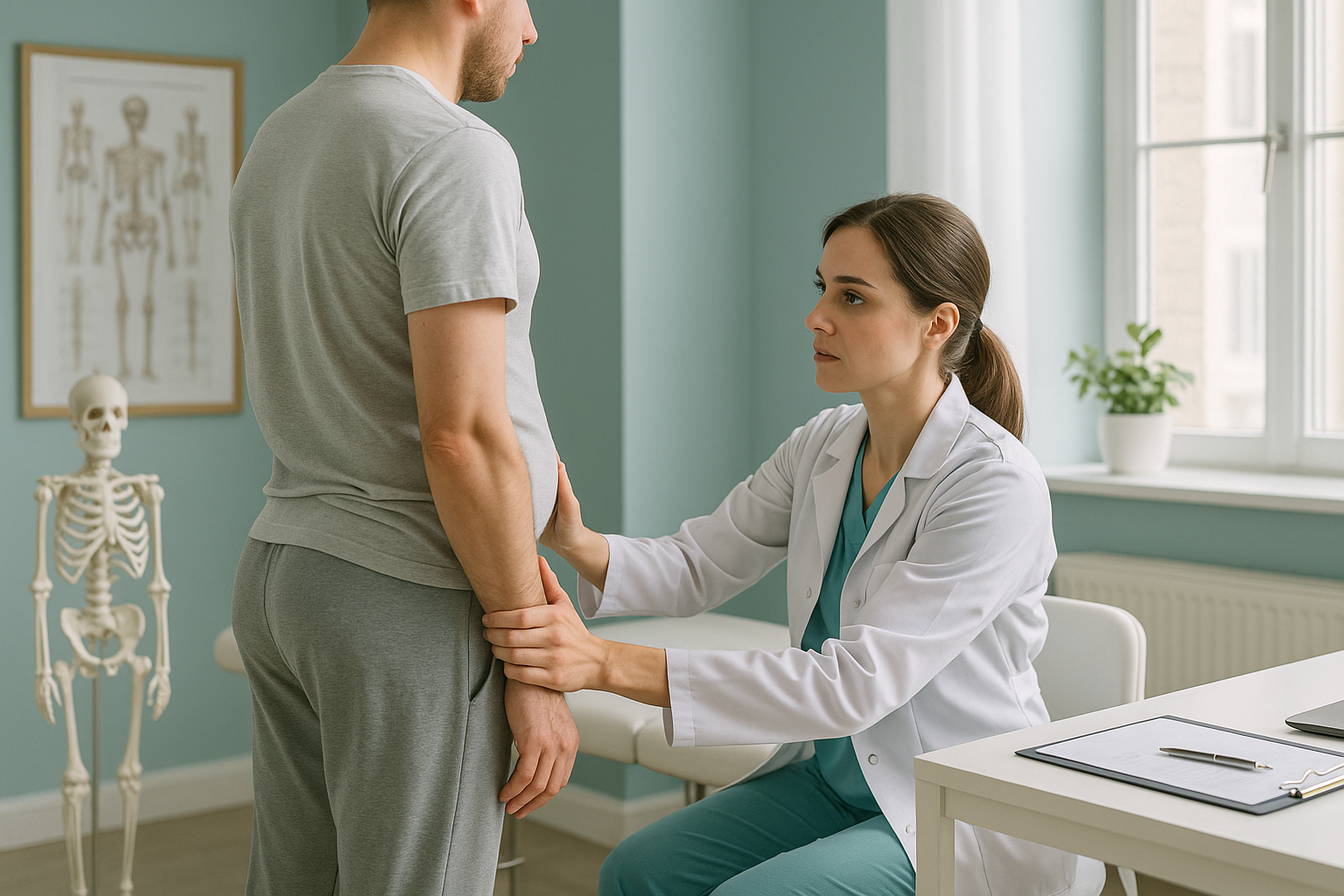
A small degree of forward tilt is normal. The pelvis naturally sits with a slight anterior angle. It only becomes a problem when the tilt is excessive enough to overload the lower back, change your gait or cause pain elsewhere.
What are the symptoms of anterior pelvic tilt?
The most common symptom is a dull, persistent ache in the lower back, especially after standing or walking for a long time. Because the tilt changes your entire posture chain, you may also feel pain or tightness in areas that seem unrelated to the pelvis.
A tight, achy feeling across the lower back that worsens with prolonged standing.
A pulling sensation at the front of the hip, particularly when you try to stand up straight.
Your belly pushes forward even at a healthy weight, because the pelvis tips the torso forward.
The changed leg alignment can stress the outside of the knee or the thigh.
Many people with anterior pelvic tilt also notice their buttocks appear to stick out more than usual, giving the impression of an exaggerated "duck" posture. If you experience sudden numbness, weakness in your legs, or loss of bladder or bowel control, seek urgent medical attention through your GP, 111 or A&E. These are not typical of a simple pelvic tilt and need immediate assessment.
What causes anterior pelvic tilt?
Anterior pelvic tilt is most commonly caused by a combination of muscle tightness and weakness that develops over time. The pelvis is held in position by several muscle groups pulling in different directions. When some become too tight and others too weak, the balance shifts.
Hours at a desk shorten the hip flexors and switch off the glutes.
Without strong posterior muscles, nothing resists the forward pull.
Short, stiff muscles at the front of the hip actively pull the pelvis forward.
Hip joint shape, leg length differences or spinal curvatures can all contribute.
The important point is that causes are rarely limited to one area. A difference in leg length, for example, shifts the pelvis, which increases the lumbar curve, which tightens the hip flexors. Each part compensates for another. This is why looking at the pelvis alone, without seeing the full chain, can miss the underlying cause.
How is anterior pelvic tilt assessed when you are standing up?
The most accurate way to assess pelvic tilt is with imaging taken while you are upright and weight-bearing. When you lie down for a conventional X-ray, CT or MRI, gravity is no longer acting on your spine and pelvis in the same way. Your muscles relax, and the tilt can flatten out, making everything look more normal than it actually is.
An EOS scan captures a full-length image of your skeleton while you stand naturally. It uses about 90% less radiation than a standard X-ray, around ten times less. Because it shows you from head to foot in a single image, your clinician can see exactly how the pelvic tilt is affecting your lumbar spine above and your knees and feet below.
This matters because pain from an anterior tilt often appears away from its true source. You might have a knee problem that started with a pelvic shift, or lower back pain driven by a subtle leg length difference. A standing whole-body image connects the dots that single-area scans cannot.
Want to understand how your pelvis sits when you are standing? A free video consultation can help you decide whether a standing scan is the right next step.
Book a free video consultationWhen is MRI the better option? EOS shows bones, joints and alignment clearly. If your clinician suspects a disc problem, nerve compression or soft-tissue injury, MRI is usually the better tool. The two scans answer different questions and sometimes work best together.
What helps with correcting anterior pelvic tilt?
Correcting anterior pelvic tilt generally involves addressing the muscle imbalances that hold the pelvis in that position. Stretching the hip flexors and strengthening the glutes and abdominal muscles are commonly recommended by physiotherapists and exercise professionals. Improving sitting habits and taking regular breaks from prolonged desk work can also help over time.
However, targeted exercises work best when you know exactly what is going on structurally. If the tilt is partly driven by a leg length difference or a joint shape issue, stretching alone will not resolve it. That is where accurate diagnostic imaging fits in. It gives the professional guiding your rehab a clear, measurable picture of your alignment so they can tailor their approach.
ScanAlign is a diagnostic imaging clinic. We do not provide treatment, but the imaging we produce gives your physiotherapist, osteopath or surgeon the detailed alignment data they need to plan effectively. Learn more about how hip and pelvic alignment is assessed.
Frequently asked questions about anterior pelvic tilt
Many people improve their tilt with consistent stretching and strengthening exercises. But if there is a structural factor such as a leg length difference or joint issue, exercise alone may not be enough. A clear image of your alignment helps you know what you are working with.
Common signs include a noticeable arch in the lower back, a belly that pushes forward, tight hip flexors and lower back ache after standing. A standing whole-body scan provides a definitive assessment of the tilt and its effects on the rest of your body.
Yes, it is one of the more common contributors to chronic lower back pain. The increased lumbar curve compresses the joints and muscles of the lower spine, especially during prolonged standing or walking.
It can. When the pelvis tilts forward, it changes how the legs are loaded. This can place extra stress on the knees and the band of tissue running down the outside of the thigh. A full-length standing image shows the connection between pelvic position and knee alignment.
Anterior tilt means the front of the pelvis drops forward, increasing the lower back curve. Posterior tilt is the opposite, where the pelvis tucks under and the lower back flattens. Both can cause problems, and both are clearly visible on a standing EOS scan.
No. ScanAlign accepts self-referrals. You can book a free video consultation directly, and if a scan is appropriate, you can arrange it without needing a GP letter first.
Yes. EOS uses about 90% less radiation than a standard X-ray, around ten times less. You can find more detail on the safety and FAQs page.
Pricing information is available on the ScanAlign FAQs page. The initial video consultation is free and carries no obligation.
When you lie down, gravity no longer loads your spine and pelvis the way it does when you stand. Muscles relax, the tilt reduces, and the image can make things look more normal than they are in daily life. A standing scan captures your skeleton as it actually functions.
ScanAlign is based at 19 Harley Street, London. It is a private self-pay clinic. You can learn more about what to expect on the ScanAlign homepage.
Ready to see the full picture?
A free video consultation takes a few minutes. We will help you decide if a standing whole-body scan is the right next step for your pelvic tilt or alignment concern.
Book a free video consultationSources
- Levine, D. and Whittle, M.W. (1996). The effects of pelvic movement on lumbar lordosis in the standing position. Journal of Orthopaedic & Sports Physical Therapy, 24(3), pp.130-135.
- Illés, T. and Somoskeöy, S. (2012). The EOS imaging system and its uses in daily orthopaedic practice. International Orthopaedics, 36(7), pp.1325-1331.
- NHS (2023). Back pain: causes, treatment and prevention. nhs.uk. Available at: nhs.uk/conditions/back-pain.
Author: Abbas Dhami (Specialist Diagnostic Radiographer)
This page is general information and not a diagnosis.
ScanAlign is the trading name of CoreMed Solutions Ltd and operates under The Harley Street Hospital's CQC licence.