
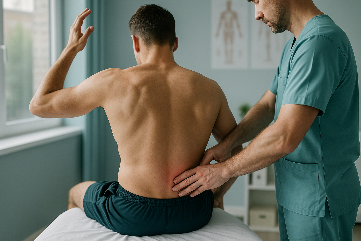
Why Do Fast Bowlers Get Lower Back Stress Injuries?
Fast bowling places enormous rotational and compressive forces on the lower spine with every delivery. Over time, these repeated loads can cause a stress reaction or fracture in a small bony bridge called the pars interarticularis. A mixed bowling action, high workload and incomplete recovery are the biggest risk factors.
Key takeaways
- The pars interarticularis is the most common site of fast bowling back injury.
- A mixed bowling action, where the shoulders and hips counter-rotate, increases risk significantly.
- MRI is the first-line test for a suspected acute stress reaction.
- Once the acute phase is over, standing imaging reveals alignment issues that lying-down scans can miss.
- Workload monitoring and technique coaching are key to reducing recurrence.
What happens to the spine when you bowl fast?
Every fast delivery compresses the lower spine under forces several times your body weight. At the point of front-foot contact, your trunk extends backwards, bends to the side and rotates, all in a fraction of a second. This combination targets one small area of the vertebra called the pars interarticularis, a thin bony bridge connecting the upper and lower joints of each spinal level.
Bowl after bowl, season after season, that bridge absorbs more load than it can repair. First, the bone develops a stress reaction, where the internal structure weakens but has not yet cracked. If bowling continues without rest, the stress reaction becomes a stress fracture, known as spondylolysis. If the fracture fails to heal and the vertebra shifts forwards, it becomes a spondylolisthesis.
What causes a fast bowling back injury?
A fast bowling back injury is almost never caused by a single delivery. It is the result of repeated stress combined with one or more risk factors.
Shoulders and hips rotate in opposite directions, multiplying the twisting force through the pars.
Bowling too many overs in a short window, or sudden spikes in training volume, outpaces the bone's ability to recover.
Excessive lateral bending at the point of delivery concentrates load on the non-bowling-arm side of the spine.
Young adult bone is still maturing and less resistant to repeated stress, making athletes in their late teens and early twenties especially vulnerable.
Pre-existing alignment issues can also raise risk. A rotated pelvis, for example, can shift the axis of trunk rotation and place uneven load across the lower spine with every delivery.
What are the signs of a lumbar stress fracture in cricket?
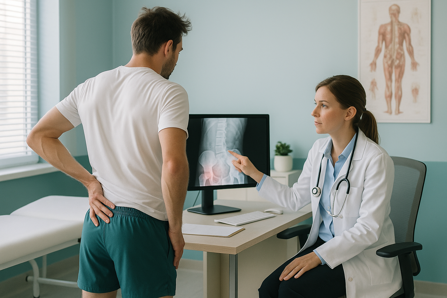
The most common sign is a gradual onset of lower back pain on the non-bowling-arm side, meaning the left side for a right-arm bowler. Pain usually worsens during bowling and eases with rest, at least at first.
Localised to the non-bowling-arm side, typically at the L4 or L5 level.
Leaning back and twisting reproduces the pain, mirroring the bowling action itself.
Starts as a dull ache during spells, then becomes painful in daily activities like sitting in a car.
The muscles on either side of the spine may tighten as the body tries to protect the injured area.
Seek urgent help if you develop numbness or tingling in your legs, weakness in a foot, or any loss of bladder or bowel control. Contact your GP, call 111, or go to A&E.
How is a fast bowler's back stress injury diagnosed?
If a stress fracture is suspected, MRI is the first-line investigation. It can detect early bone stress reactions before a crack is visible on a standard X-ray, and it does so without any radiation. MRI also shows soft tissue, including discs and nerves, which helps rule out other causes of the pain.
A note on MRI. EOS is excellent at showing bones, joints and alignment. For soft tissue, discs or nerves, and for detecting an acute stress reaction, MRI is usually the better tool. Both types of imaging play different, complementary roles in a fast bowler's assessment.
Standard lying-down X-rays may also be taken, but they can miss early stress reactions and they do not show the spine under load. A full spine x-ray taken while you are standing offers more information about alignment, but it uses a higher dose of radiation than EOS.
Why does a standing scan matter after a bowling back injury?
Once the acute phase has settled and MRI has confirmed the diagnosis, the next question is what your spine looks like under real-world load. This is where standing imaging adds value.
When you lie down for a standard X-ray or MRI, gravity is removed. Your muscles relax, your pelvis settles, and any alignment imbalance may flatten out. Standing up reintroduces the forces your spine actually deals with when you bowl, walk and stand. Problems like a mild vertebral slip, a pelvic tilt or a subtle spinal curve only become visible when the body is weight-bearing.
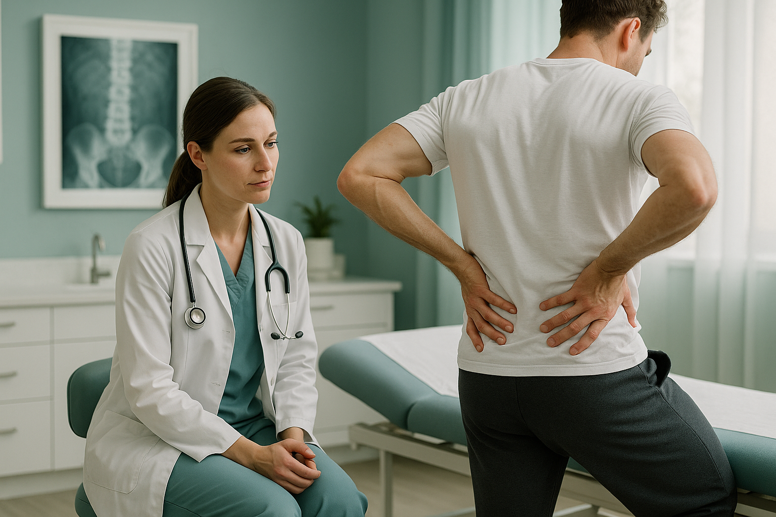
ScanAlign standing EOS imaging captures a full-length image of your spine, pelvis and legs in a single standing exposure with about 90% less radiation than a standard X-ray, around ten times less. This gives your clinician a complete picture of your alignment under load, which helps guide decisions about return to bowling, strengthening programmes and any ongoing monitoring.
Ready to see how your spine is aligned after a bowling injury? A free video consultation can help you decide if a standing scan is the right next step.
Book a free video consultationWhat helps fast bowlers manage and reduce recurrence?
Management is led by your sports medicine doctor, physiotherapist and coaching team. ScanAlign does not provide treatment, but the alignment data from a standing scan can inform the following approaches.
- Rest and graduated return. Most stress fractures require a period away from bowling, followed by a phased return guided by imaging and symptoms.
- Workload monitoring. Tracking overs, deliveries per session and rest days helps prevent the sudden spikes in volume that overload healing bone.
- Technique review. A biomechanics assessment of your bowling action, particularly the degree of counter-rotation between shoulders and hips, is one of the most important preventive steps.
- Core and trunk conditioning. Strengthening the muscles that control spinal extension and rotation can reduce the stress transferred to the bone.
- Alignment awareness. Understanding your baseline spinal alignment and pelvic balance helps your team tailor conditioning and monitor any changes over time.
Frequently asked questions
Each delivery combines spinal extension, side flexion and rotation at high speed, all while landing on a braced front leg. These forces are concentrated on a small bony bridge in the lumbar spine called the pars interarticularis.
A pars defect is a break or weakness in the pars interarticularis. It can range from a stress reaction, where bone is weakened but not yet cracked, to a complete fracture. In cricket, it is commonly called spondylolysis.
A mixed bowling action is one where the shoulders and hips rotate in opposite directions during delivery. This counter-rotation dramatically increases the twisting force through the lower spine and is considered the single biggest biomechanical risk factor for a pars stress injury.
MRI is the first-line test for a suspected acute stress injury because it can detect early bone oedema before a fracture line appears on X-ray. Once the acute phase has passed, a standing X-ray or EOS scan provides valuable alignment information that MRI cannot show in the same way.
An EOS scan captures a full-length image of your skeleton while you stand, using about 90% less radiation than a standard X-ray, around ten times less. It shows your spine, pelvis and legs in their true weight-bearing position, which can reveal alignment issues that lying-down imaging misses.
No. ScanAlign is a private self-pay clinic and you can self-refer. You start with a free video consultation where a specialist radiographer reviews your situation and advises whether a standing scan is appropriate.
Pricing details are available on the book a spine assessment page and the clinic's FAQ section. The free video consultation will also confirm what is included.
Yes. The alignment data from a standing scan gives your sports medicine team objective information about spinal position and pelvic balance under load. This can help guide the timing and conditions of your return to bowling.
EOS uses about 90% less radiation than a standard X-ray, around ten times less. It is one of the lowest-dose full-body skeletal imaging methods available.
They can contribute. A pelvic tilt, a leg-length difference or a subtle spinal curve may alter the way forces travel through the spine during bowling. Identifying these factors gives your coaching and medical team useful information for injury prevention.
Understand your alignment after a bowling injury
A free video consultation with a specialist radiographer can help you decide if a standing scan is the right next step for your back.
Book a free video consultationWritten by Abbas Dhami (Specialist Diagnostic Radiographer)
Sources
- Ranson C, Burnett A, King M, et al. "The relationship between bowling action classification and three-dimensional lower trunk motion in fast bowlers in cricket." Journal of Sports Sciences, 2008;26(3):267-276.
- Elliott B. "Back injuries and the fast bowler in cricket." Journal of Sports Sciences, 2000;18(12):983-991.
- England and Wales Cricket Board (ECB). Fast bowling directives and workload guidelines for age-group cricket. ECB, current edition.
This page is general information and not a diagnosis.
ScanAlign is the trading name of CoreMed Solutions Ltd and operates under The Harley Street Hospital's CQC licence.