
Spondylolysis (Pars Defect): What Causes It and How Is It Found?
Spondylolysis is a stress fracture or defect in a small bridge of bone called the pars interarticularis, usually in the lower back. It is often caused by repeated extension and rotation of the spine. Symptoms include a deep, one-sided ache that worsens with activity. Standing, weight-bearing imaging can reveal how the defect behaves under load, which lying-down scans may miss.
Key Takeaways
- Spondylolysis is a crack or break in the pars interarticularis, a small bony bridge connecting the joints at the back of a vertebra.
- It most often affects the lowest lumbar vertebra (L5) and is linked to sports involving repeated arching of the back.
- If the fracture allows a vertebra to slip forward, it becomes spondylolisthesis, a related but separate condition.
- Standing whole-body imaging shows the spine under gravity, revealing instability that lying-down scans can hide.
- ScanAlign provides diagnostic imaging only. Treatment is guided by your consultant or physiotherapist.
What is spondylolysis?
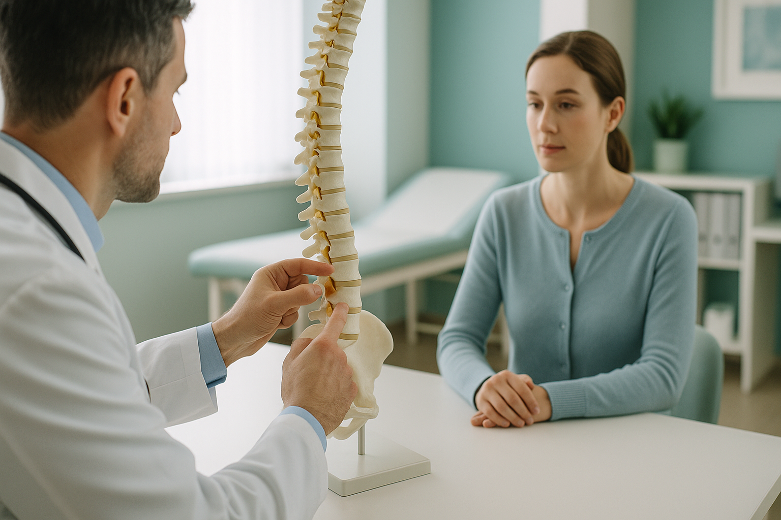
Spondylolysis is a stress fracture, or sometimes a complete break, in a part of the vertebra called the pars interarticularis. This is a thin bridge of bone that links the upper and lower facet joints at the back of each vertebra. Think of the pars as a bony strut that holds the vertebra steady. When it cracks, the vertebra loses some of its stability.
It most commonly happens at L5, the lowest lumbar vertebra, because this area bears a great deal of load and is subject to the most movement during extension (arching backwards). The defect can occur on one side (unilateral) or both sides (bilateral) of the vertebra.
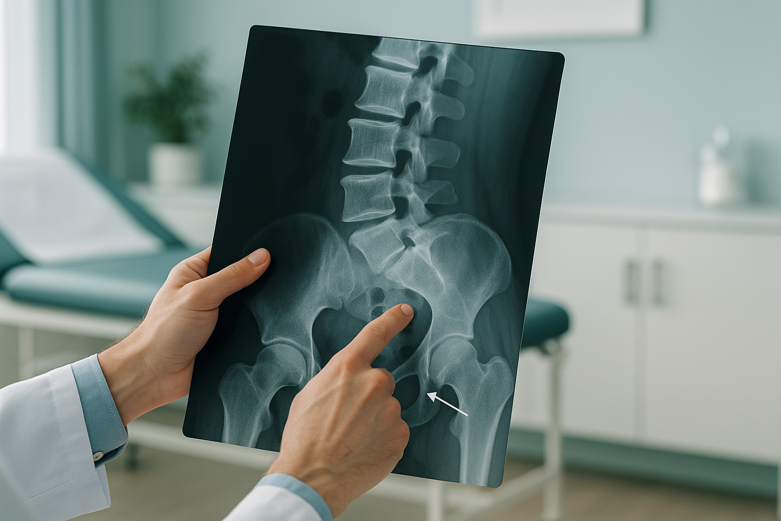
Many people have a pars defect without knowing it. In some cases the fracture heals on its own or never causes trouble. In others, it leads to persistent lower back pain, especially during or after physical activity.
What does spondylolysis feel like?
The main symptom is a dull, aching pain in the lower back, usually on one side. It tends to get worse when you arch your back, twist, or stand for a long time. Here are the most common signs.
A deep, nagging pain that usually sits to one side of the spine, just above the pelvis.
Extension movements load the pars, so bending backwards or looking up at a ceiling often makes it worse.
The lower back may feel tight and stiff after sitting or sleeping, loosening once you start to move.
Unusually tight hamstrings are a common companion. The body tightens them to limit spinal movement and protect the area.
When to seek urgent help. If you develop numbness, weakness in your legs, or any change in bladder or bowel control, contact your GP, call 111, or go to A&E straight away. These symptoms need immediate medical attention, not a routine scan.
What causes a pars fracture?
Spondylolysis is caused by repeated stress on the pars interarticularis. It is not usually the result of a single injury. Instead, the bone develops tiny cracks over time that eventually become a visible defect. The main contributing factors include the following.
Sports or activities that involve arching the back repeatedly, such as gymnastics, fast bowling, weightlifting, or ballet.
The pars is thinner and more vulnerable during adolescent growth, making younger adults more susceptible.
Some people have a naturally thinner pars bone, which can make stress fractures more likely even with moderate activity.
Because the fracture develops gradually, many people first notice it only when pain becomes persistent enough to seek medical advice. By that point, the defect may have been present for months or longer.
What is the difference between spondylolysis and spondylolisthesis?
Spondylolysis is the crack in the pars bone. Spondylolisthesis is what can happen next: if both sides of the pars are broken, the vertebra above may slip forward on the one below. Put simply, spondylolysis is the fracture, and spondylolisthesis is the slip that the fracture can allow.
Not every pars defect leads to a slip. Many people live with a bilateral pars fracture and never develop spondylolisthesis. However, knowing whether any slip is present, and whether it changes when you are standing under gravity, is important for planning the right course of action.
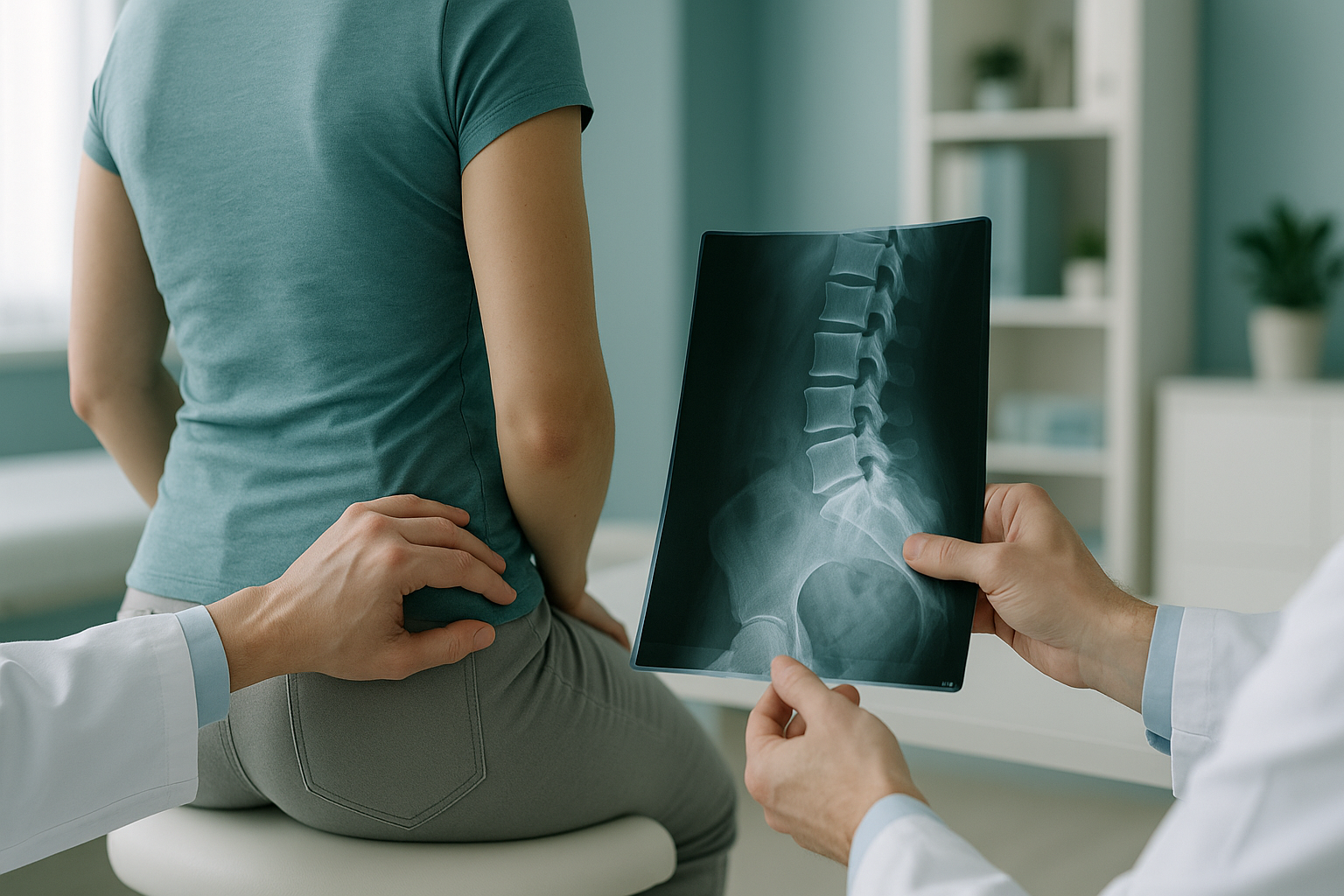
This is where standing imaging becomes especially valuable. A vertebra may look stable when you are lying down, because gravity is no longer pulling it forward. When you stand, that same vertebra might slip noticeably. Understanding whether a slip is stable or changes with posture helps your clinician decide between conservative management and further intervention.
How does standing imaging help assess spondylolysis?
Standing, weight-bearing imaging shows the spine the way it actually works, under the full load of gravity and your body weight. This is important for spondylolysis because the defect in the pars may only reveal its true impact when the spine is loaded.
Most conventional imaging, such as a standard X-ray, CT or MRI, is done while you lie down. In that position, muscles relax and gravity no longer compresses the spine. A subtle slip or instability that causes pain in everyday life may not show up at all.
An EOS whole-body standing scan captures the full skeleton from head to feet in a single upright image, using about 90% less radiation than a standard X-ray, around ten times less. Because it images you standing, it shows your true spinal alignment, any slip under load, and how the rest of your body, including your pelvis, hips and legs, may be compensating for the defect.
Your body is a connected chain. A pars defect in the lower back can change the curve of the spine above it, tilt the pelvis, and affect the way you load your hips and knees. A whole-body standing image lets your clinician see all of these connections in one view, rather than guessing from a single-area lying-down scan.
A note about MRI. EOS shows bones, joints and alignment clearly. If your clinician needs to look at soft tissue, discs, nerves, or the bone marrow itself to assess whether a fracture is still active, MRI is usually the better tool. The two types of imaging complement each other well.
How is spondylolysis usually treated?
Treatment depends on the severity of the defect, whether a slip is present, and how much pain it causes. ScanAlign provides the diagnostic imaging that informs these decisions but does not deliver treatment itself. Your consultant, physiotherapist or GP will guide the treatment plan. Common approaches include the following.
- Rest and activity modification. Reducing or stopping the aggravating activity, especially extension-heavy sports, to allow the bone to heal.
- Physiotherapy. A structured programme to strengthen the core muscles that support the lower back and improve flexibility.
- Bracing. In some cases, a brace may be recommended to limit spinal movement while the pars heals.
- Surgical referral. If the defect is unstable, causes a significant slip, or does not respond to conservative management, surgery may be discussed. Accurate standing imaging helps surgeons plan the approach.
Accurate imaging is the first step. Understanding how your spine behaves under load helps your clinician choose the right path for you.
Book a free video consultationFrequently asked questions about spondylolysis
A pars defect is a crack or break in the pars interarticularis, a thin bridge of bone at the back of a vertebra that connects the upper and lower joints. It is another name for spondylolysis.
Healing time varies depending on the individual, the severity of the fracture, and the treatment approach. Some defects heal with rest and physiotherapy over several months. Others may not fully close but can become pain-free with the right management. Your clinician can advise based on your imaging.
Spondylolysis is the fracture in the pars bone. Spondylolisthesis is the forward slip of one vertebra on another that can result from that fracture. The fracture does not always lead to a slip.
A pars defect can sometimes be seen on a standard X-ray, but it is not always clear, especially in the early stages. A standing EOS scan has the added advantage of showing whether the defect is causing any slip or instability under weight-bearing conditions.
When you lie down for a scan, gravity is removed and muscles relax. A vertebra that slips when you stand may look perfectly stable lying down. Standing imaging shows your spine under real-world loading, giving your clinician a more accurate picture.
Yes. An EOS scan uses about 90% less radiation than a standard X-ray, around ten times less. You can find more details about safety on the FAQs page.
No. ScanAlign is a self-pay clinic. You can book directly without a GP or consultant referral. You start with a free video consultation so the team can confirm the scan is appropriate for you.
Current pricing information is available on the ScanAlign FAQs page.
Yes. A pars defect can change the way your spine curves, which may tilt your pelvis and alter how you load your hips, knees and legs. Pain often appears away from the original problem because the body compensates along the whole chain.
It depends on the severity of the defect and your symptoms. Many people return to activity with appropriate physiotherapy. Your clinician or physiotherapist will advise on which exercises are safe and which to avoid during recovery.
See how your spine really behaves under load
A free video consultation lets you discuss your symptoms and find out whether a standing EOS scan is the right next step.
Book a free video consultationWritten by Abbas Dhami (Specialist Diagnostic Radiographer)
Sources
- NHS. "Back pain." nhs.uk. Available at: https://www.nhs.uk/conditions/back-pain/
- Standaert CJ, Herring SA. "Spondylolysis: a critical review." British Journal of Sports Medicine, 2000;34(6):415-422.
- The Royal College of Radiologists. "iRefer: Making the best use of clinical radiology." Available at: https://www.rcr.ac.uk/
This page is general information and not a diagnosis.
ScanAlign is the trading name of CoreMed Solutions Ltd and operates under The Harley Street Hospital's CQC licence.