
Spondylolisthesis: When a Vertebra Slips Forward, What Is Really Going On?
Spondylolisthesis happens when one vertebra slides forward over the one below it. It most often affects the lower back. Many people have no symptoms at all, while others get persistent back or leg pain. A standing, weight-bearing image is the clearest way to see how far the slip goes when your body is upright and under load.
Key takeaways
- Spondylolisthesis means a vertebra has slipped forward. It is graded from mild to severe.
- Degenerative wear and tear is the most common cause in adults.
- Pain often appears in the lower back, buttocks or legs, but some people feel nothing.
- Lying-down scans can underestimate the slip because gravity is removed.
- EOS standing imaging shows the slip under real weight-bearing conditions.
The word spondylolisthesis comes from Greek. "Spondylo" means vertebra, and "listhesis" means to slip. In plain terms, one of the bones in your spine has moved forward out of its normal position. This usually happens in the lower back, at the base of the spine.
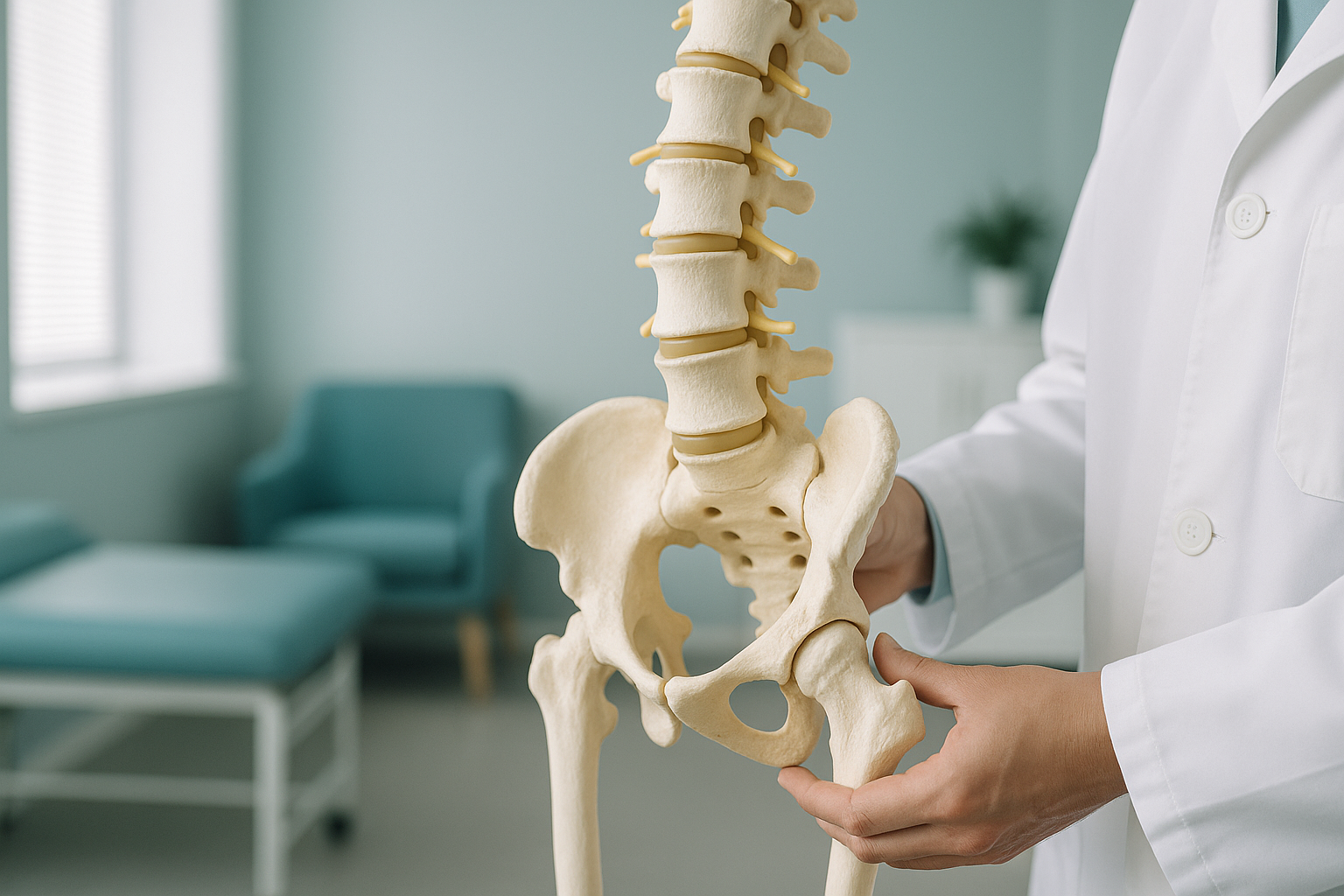
Spondylolisthesis is surprisingly common. Many people discover it by chance on an X-ray taken for something else. The key question is not just whether a slip exists, but how much it moves when you are standing upright and your spine is carrying your full body weight.
What are the symptoms of spondylolisthesis?
The most common symptom is a deep, aching pain in the lower back that gets worse when you stand, walk or arch backwards. Some people also feel pain, tingling or heaviness in one or both legs. Others have no symptoms at all.
A deep ache across the lower back, often worse after standing or walking for a while.
Pain, numbness or a "heavy" feeling that travels into the buttocks or down the legs.
Tightness in the hamstrings and a feeling that your back does not bend as easily as it should.
Some people have a slip but feel nothing. It may only show up on imaging done for another reason.
Important: If you experience sudden leg weakness, loss of bladder or bowel control, or numbness around the groin area, seek urgent medical attention. Contact your GP, call 111, or go to A&E. These symptoms need immediate assessment.
What causes spondylolisthesis in adults?
In adults, the most common cause is degenerative wear and tear. Over years, the discs and joints that hold each vertebra in place gradually weaken, allowing one bone to shift forward. Other causes include a stress fracture in the vertebra (called a pars defect) or, less often, changes from previous surgery or other bone conditions.
Age-related wear on discs and facet joints. The most common type in adults over 50.
A small crack in the back part of the vertebra, often from repetitive strain during younger years.
Previous spinal surgery, bone disease, or trauma can occasionally lead to instability and slippage.
Regardless of the cause, spondylolisthesis is graded from mild to severe based on how far the vertebra has moved. Even a mild slip can cause meaningful symptoms if it narrows the space around the spinal nerves. Conversely, a larger slip may cause no pain at all.
Is spondylolisthesis serious?
Most cases of spondylolisthesis are mild and manageable. Many people live with a small slip and experience little or no disruption to daily life. A slip becomes more of a concern when it presses on nerves, causes persistent pain, or progresses over time. That is why accurate imaging matters. Understanding the exact position of the slip, and whether it changes when you stand, helps your clinician decide whether you need active management or simple monitoring.
Why does standing imaging show spondylolisthesis more accurately?
Standing, weight-bearing imaging captures the slip under real-life conditions, with gravity pulling on the spine and your muscles working to hold you upright. When you lie down for a conventional scan, the muscles relax and the vertebra may settle back toward its normal position. This can make a slip look smaller than it really is, or hide it altogether.
An EOS scan takes a full-length, weight-bearing image of your spine and pelvis in one pass while you stand naturally. It shows the true extent of any slip, plus how your pelvis, hips and legs are compensating. The radiation dose is about 90% less than a standard X-ray, around ten times less.
Standard X-rays, CT and MRI are usually done lying down. Without the load of gravity, the vertebra can shift back into place. This may underestimate the grade of the slip and miss compensatory changes in your posture.
Your body is a connected chain from your feet to your head. When one vertebra slips forward, the rest of the spine, pelvis and legs adjust to keep you balanced. A standing, whole-body image captures all of those compensations in a single view. That context is what single-area, lying-down imaging misses.
When is MRI the better choice? EOS shows bones, joints and alignment clearly. For soft tissue, discs or nerves, MRI is usually the better tool. If your clinician suspects nerve compression from the slip, they may recommend MRI alongside or instead of EOS imaging.
Wondering whether a standing scan would add useful information about your spondylolisthesis? We can talk it through.
Book a free video consultationWhat generally helps manage spondylolisthesis?
Management depends on the severity of the slip and the symptoms it causes. Most people with a mild slip respond well to conservative approaches recommended by their clinician. These typically include physiotherapy to strengthen the core and stabilise the spine, activity modification to avoid movements that worsen symptoms, and pain management guided by your GP or specialist.
Accurate diagnostic imaging plays a key role in guiding those decisions. A clear, weight-bearing view of the slip, combined with measurements of your overall spinal and pelvic alignment, gives your treating clinician the detail they need to plan the right approach for you.
ScanAlign is a diagnostic imaging clinic. We do not provide treatment. We provide the detailed imaging that helps the right clinician make the right plan. You can learn more about what an EOS scan involves and how it works.
Frequently asked questions about spondylolisthesis
Most people describe a deep ache in the lower back that worsens with standing, walking or arching backwards. Some also feel pain, tingling or heaviness in one or both legs. Others feel nothing at all.
Spondylolisthesis is graded on a scale from 1 to 4 based on how far the vertebra has slipped. Grade 1 is the mildest, meaning only a small portion of the bone has moved forward. Many people with a grade 1 slip manage well with physiotherapy and monitoring.
It can, particularly the degenerative type, as the joints and discs continue to wear. That is one reason periodic imaging can be valuable, to track whether the slip is stable or progressing.
Movements that extend the lower back, such as deep backbends, heavy overhead lifting and high-impact activities, are generally best avoided. Your physiotherapist or specialist can give you a tailored list based on your specific slip and symptoms.
Yes. Degenerative spondylolisthesis is caused by age-related wear and tear on the facet joints and discs. It typically affects adults over 50 and most often occurs at the L4-L5 level. The isthmic type results from a stress fracture in the vertebra itself and tends to develop earlier in life.
When you lie flat, gravity is removed and your muscles relax. The slipped vertebra can settle back closer to its normal position, making the slip appear smaller than it is in real life, or hiding it entirely.
Yes. An EOS scan uses about 90% less radiation than a standard X-ray, around ten times less. You can find more detail on the FAQs and safety page.
No. ScanAlign accepts self-referrals. You start with a free video consultation where a specialist radiographer discusses your situation and confirms whether EOS imaging is appropriate for you.
Pricing details are on the FAQs and cost page. The initial video consultation is free and there is no obligation to proceed.
EOS imaging gives your surgeon or specialist a detailed, weight-bearing view of the slip and your overall spinal alignment. That information helps them decide whether conservative management is enough or whether surgery should be considered. ScanAlign does not provide treatment or surgical advice.
See the full picture of your spondylolisthesis
A free video consultation with a specialist radiographer. No referral needed. No obligation.
Book a free video consultationWritten by Abbas Dhami (Specialist Diagnostic Radiographer)
Sources
- NHS. "Spondylolisthesis." nhs.uk. Accessed 2024.
- Kalichman L, et al. "Spondylolysis and spondylolisthesis: prevalence and association with low back pain in the adult community-based population." Spine, 2009; 34(2): 199-205.
- Illés T, Somoskeöy S. "The EOS imaging system and its uses in daily orthopaedic practice." International Orthopaedics, 2012; 36(7): 1405-1411.
Learn more about spinal conditions and EOS imaging.
This page is general information and not a diagnosis.
ScanAlign is the trading name of CoreMed Solutions Ltd and operates under The Harley Street Hospital's CQC licence.