

Piriformis Syndrome or Sciatica: How Do You Tell the Difference?
Both piriformis syndrome and sciatica cause buttock pain radiating down the leg, but the source is different. Sciatica usually starts from a compressed nerve root in the lower spine. Piriformis syndrome happens when the piriformis muscle in the buttock irritates the sciatic nerve locally. Telling them apart matters because the right treatment depends on the right diagnosis.
Key takeaways
- Piriformis syndrome is a muscular problem in the buttock. Sciatica is usually a spinal nerve root problem.
- The symptoms overlap heavily, which is why many people are misdiagnosed.
- Lying-down scans can miss alignment issues that contribute to both conditions.
- A standing, weight-bearing image of the spine and pelvis shows how your body actually loads when upright.
- Accurate imaging helps your clinician choose the right path forward.
A note on MRI. EOS imaging excels at showing bone alignment, joint position and pelvic balance under load. For soft-tissue detail of the piriformis muscle, discs or nerves themselves, MRI is usually the better tool. The two scans are complementary, not competing.
Important. If you experience sudden numbness in both legs, loss of bladder or bowel control, or weakness that is getting rapidly worse, seek urgent medical attention. Contact your GP, call 111, or go to A&E.
What is piriformis syndrome?
Piriformis syndrome is a neuromuscular condition where the piriformis muscle, a small flat muscle deep in the buttock, presses on or irritates the sciatic nerve as it passes through the deep gluteal space. It is sometimes grouped under the broader term "deep gluteal syndrome," which covers any structure in that area that can trap the nerve.
The piriformis muscle runs from the front of the sacrum (the triangular bone at the base of your spine) across to the top of the thigh bone. Its job is to help rotate your hip outward. When it becomes tight, swollen or goes into spasm, it can squeeze the sciatic nerve that runs just beneath it, or in some people, right through it.

Sciatica, by contrast, is a symptom rather than a standalone diagnosis. It describes pain along the path of the sciatic nerve, most commonly caused by a compressed or irritated nerve root in the lumbar spine. Common spinal causes include disc bulges, narrowing of the spinal canal, or bony changes that press on the nerve root where it exits the spine. You can read more about this in our guide to sciatica or a spinal alignment problem.
What are the symptoms of piriformis syndrome?
The main symptom is a deep, aching pain in the buttock that can radiate down the back of the thigh. It often feels very similar to sciatica, which is exactly why the two are so often confused.
A constant ache or sharp pain deep in one buttock, often hard to pinpoint exactly.
Sitting on hard surfaces compresses the piriformis, making pain noticeably worse after a few minutes.
Buttock pain radiating down the leg, sometimes reaching the calf or foot, mimicking true sciatica.
Rotating the hip inward or crossing the affected leg over the other knee often reproduces the pain.
You may also notice tenderness when pressing into the mid-buttock area, or discomfort when climbing stairs. Some people find the pain eases with walking but flares when they stop and sit down.
How do you tell the difference between piriformis syndrome and sciatica?
The key difference is where the sciatic nerve is being irritated. In spinal sciatica (often called lumbar radiculopathy), the nerve root is compressed as it leaves the lumbar spine. In piriformis syndrome, the nerve is squeezed further down, in the buttock itself.
Where the pain starts: Deep in the buttock, often one-sided.
What makes it worse: Prolonged sitting, crossing legs, internal hip rotation.
Back pain: Usually absent or minimal.
Clinical clue: Tenderness over the mid-buttock. Pain reproduced by the FAIR test (flexion, adduction, internal rotation of the hip).
Imaging findings: Spine imaging often looks normal. The problem is muscular, not spinal.
Where the pain starts: Lower back, radiating into the buttock and down the leg.
What makes it worse: Bending forward, coughing, sneezing, prolonged standing.
Back pain: Usually present alongside the leg pain.
Clinical clue: Positive straight-leg raise test. Specific nerve-root pattern of numbness or weakness.
Imaging findings: Disc bulge, spinal canal narrowing, or bony changes visible at the nerve root exit.
There is a third possibility worth knowing about. The pain may relate to the sacroiliac joint, which sits right next to the piriformis. SI joint dysfunction can produce very similar buttock and thigh pain, adding another layer of complexity. And sitting-related buttock pain can also come from ischial bursitis, an inflamed bursa under the sitting bone.
Because the symptoms overlap so much, a clinical examination alone can be inconclusive. That is where imaging plays a crucial role.
What causes piriformis syndrome?
The piriformis can become tight, inflamed or go into spasm for several reasons. Often it is not a single event but a combination of factors, many of which relate to how your body is aligned under load.
Desk work or long drives compress the piriformis against the sciatic nerve for hours at a time.
A tilted or rotated pelvis forces one piriformis to work harder than the other, leading to overload.
Running, climbing, or a direct fall onto the buttock can inflame the muscle and trigger spasm.
In some people, the sciatic nerve passes through the piriformis rather than beneath it, increasing vulnerability.
The compensation narrative is important here. Your body is a connected chain from your feet up to your head. If your pelvis sits unevenly, perhaps from a subtle leg length difference or a rotated sacrum, the piriformis on one side works overtime to stabilise you. Over weeks and months, that overworked muscle tightens, and the sciatic nerve running beneath it starts to suffer. The pain you feel in your buttock or leg may actually be driven by an alignment issue somewhere else entirely.
How does a standing scan help diagnose piriformis syndrome?
A standing, weight-bearing scan shows your skeleton the way it actually works, upright and under gravity. This is the key advantage over lying-down imaging.
When you lie down for a conventional X-ray, CT or MRI, your muscles relax and your pelvis settles into a neutral position. A mild pelvic tilt or rotation that loads one piriformis more than the other can simply disappear from the image. You get a scan that looks reassuringly normal, while the mechanical cause of your pain goes undetected.
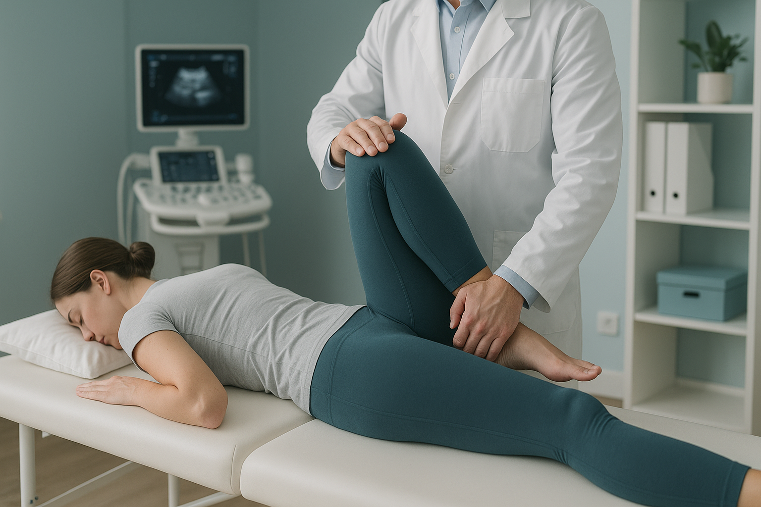
At ScanAlign, whole-body standing imaging, an EOS scan captures your full skeleton from head to ankles in a single upright image, using about 90% less radiation than a standard X-ray, around ten times less. That means your radiographer and clinician can see your spinal curves, pelvic rotation, hip alignment and leg length all at once, in the position that actually reproduces your symptoms.
If the scan reveals a level pelvis and a normal lumbar spine, your clinician can focus confidently on the piriformis itself and direct you toward the right soft-tissue investigation or treatment. If it reveals a tilt, a rotation, or a spinal cause for nerve compression, that changes the plan entirely. Either way, you leave with a clearer picture.
Wondering whether your buttock pain is coming from your spine, your pelvis, or the piriformis muscle? A standing alignment scan can help narrow it down.
Book a free video consultationWhat helps manage piriformis syndrome?
Management depends entirely on confirming that the piriformis is the real source of the problem, not the spine or the SI joint. Once the diagnosis is clear, a range of approaches may be considered by your treating clinician.
- Targeted stretching. A piriformis stretch, where you gently pull the knee of the affected side across towards the opposite shoulder, is commonly recommended to ease tightness. Your physiotherapist can tailor a programme.
- Activity modification. Avoiding prolonged sitting, using a cushion to reduce direct pressure, and taking regular movement breaks can all help reduce flare-ups.
- Physiotherapy. Strengthening the hip stabilisers and improving pelvic control may reduce the load on the piriformis over time.
- Addressing alignment. If a standing scan reveals a pelvic tilt or rotation that is overloading the piriformis, correcting that underlying imbalance may form part of the treatment plan your clinician recommends.
ScanAlign does not provide treatment. It provides the diagnostic imaging that helps your clinician understand what is happening structurally so they can choose the most appropriate path forward. If you would like to book a standing spine and pelvis scan, we start with a free video consultation to make sure a scan is right for you.
Frequently asked questions
Not exactly. Sciatica describes pain along the sciatic nerve and is usually caused by a problem in the lower spine. Piriformis syndrome produces similar symptoms but the nerve is irritated by the piriformis muscle in the buttock, not by a spinal issue.
Most people describe a deep, dull ache in one buttock that gets worse with sitting. Pain may radiate down the back of the thigh and sometimes into the calf. It often flares when you cross your legs or rotate your hip inward.
Piriformis syndrome is primarily a clinical diagnosis. A standard lying-down X-ray usually appears normal. MRI can sometimes show inflammation or swelling in the piriformis muscle. A standing EOS scan is most useful for ruling out spinal or pelvic alignment causes of the same symptoms.
When you sit, especially on a hard surface, your body weight compresses the piriformis muscle directly against the sciatic nerve. The longer you sit, the more pressure builds, which is why many people feel worse after desk work or long drives.
Deep gluteal syndrome is a broader term that includes piriformis syndrome. It covers any condition where the sciatic nerve is trapped or irritated in the deep gluteal space, whether by the piriformis muscle, scar tissue, a band of fibrous tissue, or another structure in the region.
Yes, it can contribute. A tilted or rotated pelvis changes the loading on the hip muscles. The piriformis on the lower side often has to work harder to stabilise the joint, which can lead to tightness, spasm and nerve irritation over time.
No referral is needed. ScanAlign is a private, self-pay clinic. You start with a free video consultation so your radiographer can confirm whether a scan is appropriate for your situation.
An EOS scan uses about 90% less radiation than a standard X-ray, around ten times less. It captures a full standing image of your skeleton in a single low-dose pass.
Pricing details are available on our FAQs page. The free video consultation will also confirm exactly what is included for your situation.
A commonly recommended piriformis stretch involves lying on your back, bending both knees, and pulling the knee of the affected side gently across towards the opposite shoulder. Hold for around 30 seconds. However, it is important to have the correct diagnosis first. If the real cause is spinal, stretching the piriformis alone will not address the problem.
Not sure if it is piriformis or your spine?
A free video consultation with our specialist radiographer can help you decide whether a standing alignment scan is the right next step.
Book a free video consultationWritten by Abbas Dhami (Specialist Diagnostic Radiographer)
Sources
- Hopayian K, Danielyan A. Somatic dysfunction and piriformis syndrome: a systematic review. Journal of Back and Musculoskeletal Rehabilitation. 2018;31(4):661-672.
- Martin HD, Reddy M, Gómez-Hoyos J. Deep gluteal syndrome. Journal of Hip Preservation Surgery. 2015;2(2):99-107.
- NHS. Sciatica. nhs.uk. Available at: https://www.nhs.uk/conditions/sciatica/
This page is general information and not a diagnosis.
ScanAlign is the trading name of CoreMed Solutions Ltd and operates under The Harley Street Hospital's CQC licence.