

SI Joint Dysfunction: What Is It and Why Does Your Lower Back Keep Hurting?
SI joint dysfunction is a problem with the sacroiliac joint, the connection between your lower spine and pelvis. It causes pain in the lower back, buttock or upper leg, often on one side. Standing whole-body imaging can reveal pelvic alignment issues that lying-down scans miss, helping to pinpoint the true source of pain.
Key takeaways
- The SI joint sits where the sacrum meets each side of the pelvis. It carries most of your upper body weight.
- Pain is usually felt in the lower back, buttock or back of the thigh, often on one side.
- SI joint pain is commonly mistaken for sciatica or a disc problem.
- Standing imaging shows how your pelvis, spine and legs align under real weight, revealing problems a lying-down scan can hide.
- ScanAlign provides diagnostic imaging only. It does not treat the condition.
You have two sacroiliac joints, one on each side. They sit at the base of your spine, connecting the triangular sacrum bone to the large iliac bones of the pelvis. Unlike your hip or knee, the SI joint moves very little. Its job is to transfer force between your upper body and your legs when you stand, walk, climb stairs or run.
When the joint becomes irritated, stiff, or moves too much, the result is SI joint dysfunction. It is one of the most common causes of lower back and buttock pain, yet it is frequently overlooked because the symptoms can mimic other conditions.
What does sacroiliac joint pain feel like?
SI joint pain is typically felt as a deep ache on one side of the lower back or buttock. It may spread down the back of your thigh, but it rarely goes below the knee. Sitting for a long time, standing on one leg, climbing stairs or rolling over in bed often makes it worse.
A deep, dull ache on one side, close to the dimple above your buttock.
Pain spreading into the buttock and sometimes down the back of the thigh.
Discomfort increases when climbing stairs, walking uphill or getting out of a car.
The joint feels locked or stiff after sitting for a long period, then eases once you move.
Seek urgent medical attention if you experience sudden numbness or weakness in your legs, or any loss of bladder or bowel control. Contact your GP, call 111, or go to A&E.
What causes SI joint dysfunction?
SI joint dysfunction happens when the joint becomes too stiff (hypomobility), too loose (hypermobility), or inflamed. Several factors can trigger or contribute to this.
Uneven leg length or a tilted pelvis puts more load on one SI joint than the other.
Hormonal changes during pregnancy loosen the ligaments around the SI joint, sometimes leaving lasting laxity.
Age-related changes and repetitive activity can cause the cartilage inside the joint to wear down gradually.
A fall, car accident, or spinal fusion can alter the way force passes through the SI joint.
Your body works as a connected chain. If the pelvis is tilted, one leg is functionally shorter, or the lumbar spine curves differently than it should, the SI joint often picks up the extra strain. This is why the source of the problem is not always where the pain is felt.
How do you tell if pain is SI joint or sciatica?
SI joint pain and sciatica can feel similar because both affect the lower back and leg. However, there are important differences that help distinguish them.
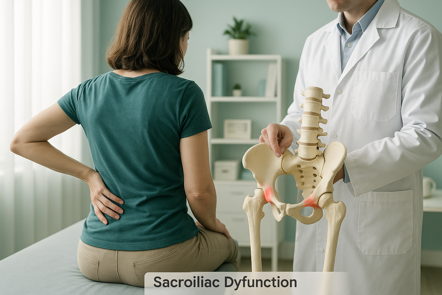
SI joint pain usually stays in the lower back, buttock and upper thigh. It rarely travels below the knee. It tends to feel like a deep ache rather than a sharp, shooting sensation.
Sciatica often involves sharp or burning pain that travels from the lower back through the buttock and all the way down the leg, sometimes to the foot. It is typically caused by a compressed nerve in the spine.
The two conditions can also exist together. When standard investigations focus only on the spine, an SI joint problem may be missed entirely. A whole-body standing image lets a clinician see the pelvis, spine and legs in one view, making it easier to identify which structure is contributing.
How is SI joint dysfunction assessed with a standing scan?
A standing scan captures your skeleton while you are upright and bearing weight, exactly as your body works in everyday life. This matters because lying down relaxes muscles and ligaments, allowing the pelvis to settle into a position that may look normal even when it is not.
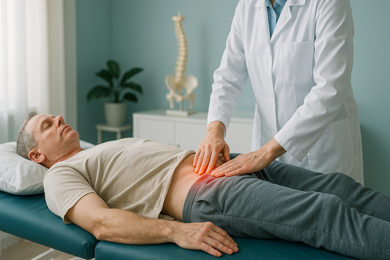
An EOS scan uses about 90% less radiation than a standard X-ray, around ten times less. It captures a full-length image from head to foot in a single pass, so the relationship between the pelvis, spine and legs is visible in one picture. This makes it much easier to spot a pelvic tilt, leg length difference or compensatory curve that could be overloading one SI joint.
Because your body is a connected chain, an alignment problem higher up in the spine or lower down in the legs can shift extra stress onto the sacroiliac joint. A single-area lying-down scan would not show this bigger picture.
When MRI is the better tool: EOS shows bones, joints and alignment under load. If your clinician suspects soft-tissue damage, disc problems or nerve compression, MRI is usually the more appropriate investigation. The two scans answer different questions and can complement each other.
What generally helps manage SI joint pain?
Accurate diagnosis is the first step. Once the SI joint has been confirmed as the source, a range of approaches may be considered by your treating clinician. ScanAlign provides diagnostic imaging only and does not offer treatment.
- Targeted physiotherapy to strengthen the muscles around the pelvis and improve stability.
- Activity modification to reduce movements that repeatedly load the joint.
- Understanding your alignment so that any contributing factors, such as a pelvic tilt or leg length difference, can be addressed alongside the joint itself.
Many people spend months treating the wrong structure because the SI joint was not identified. Having clear imaging of the whole chain, from feet to spine, can help your clinician plan the right approach from the start.
Wondering whether your lower back or buttock pain could be coming from the SI joint? A standing whole-body scan can reveal alignment issues a standard scan might miss.
Book a free video consultationFrequently asked questions about SI joint dysfunction
SI joint pain is usually felt on one side of the lower back or buttock. It tends to worsen when you sit for a long time, climb stairs, or stand on one leg. If your pain fits this pattern and has not responded to standard lower back treatment, the SI joint may be worth investigating.
Yes. Pain can spread into the buttock and down the back of the thigh. However, it rarely travels below the knee. If your leg pain extends to the calf or foot, a nerve-related cause such as sciatica may also be involved.
Not exactly. Sacroiliitis refers specifically to inflammation of the SI joint, which can be caused by conditions such as ankylosing spondylitis. SI joint dysfunction is a broader term covering any mechanical problem with the joint, including stiffness, excessive movement or wear and tear.
When you lie down, gravity no longer loads the pelvis. Muscles relax and the joint settles into a neutral position that can look normal on a scan. Standing imaging captures the pelvis under real weight, revealing tilts, asymmetries and compensations that only appear when you are upright.
An EOS scan uses about 90% less radiation than a standard X-ray, around ten times less. This makes it one of the lowest-dose imaging options available for skeletal assessment.
No. ScanAlign accepts self-referrals. You can book a free video consultation directly to discuss whether a standing scan is appropriate for your situation.
Pricing details are available on the ScanAlign FAQs page. You can also ask about costs during your free video consultation.
A standard X-ray may show bony changes at the SI joint, but because it is usually taken lying down and only covers a small area, it often misses alignment problems in the pelvis and legs that contribute to the pain. A standing whole-body image provides the bigger picture.
A disc problem involves the soft cushions between the vertebrae in the spine. SI joint dysfunction involves the joint where the spine meets the pelvis. Pain from both can be felt in the lower back and buttock, which is why they are sometimes confused. MRI is better for assessing discs, while standing imaging excels at showing joint alignment and pelvic balance.
ScanAlign is based at 19 Harley Street, London. The clinic offers private, self-pay EOS standing whole-body imaging. You can learn more about how a whole-body standing scan works on the homepage.
Still unsure what is causing your lower back or buttock pain?
A free video consultation can help you understand whether standing whole-body imaging is the right next step.
Book a free video consultationRelated pages
Author: Abbas Dhami (Specialist Diagnostic Radiographer)
Sources
- Vleeming A, Schuenke MD, Masi AT, et al. The sacroiliac joint: an overview of its anatomy, function and potential clinical implications. Journal of Anatomy. 2012;221(6):537-567.
- Cohen SP. Sacroiliac joint pain: a comprehensive review of anatomy, diagnosis, and treatment. Anesthesia & Analgesia. 2005;101(5):1440-1453.
- NHS. Back pain: causes, symptoms and treatments. nhs.uk/conditions/back-pain.
This page is general information and not a diagnosis.
ScanAlign is the trading name of CoreMed Solutions Ltd and operates under The Harley Street Hospital's CQC licence.