
Hip Impingement (FAI): What Is It and Why Does Your Hip Catch?
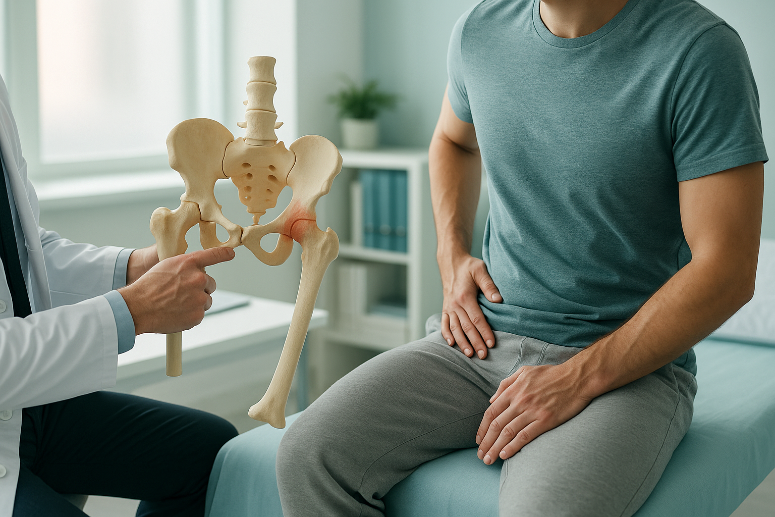
Hip impingement, also called femoroacetabular impingement (FAI), happens when extra bone on the hip joint causes the ball and socket to rub together abnormally. This pinches surrounding tissue during movement, leading to groin or hip pain, stiffness, and a catching sensation. It is one of the most common causes of hip pain in active adults.
Key takeaways
- FAI is caused by an irregular shape of the ball, the socket, or both.
- There are two main types: cam (ball-side) and pincer (socket-side). Many people have a mix of both.
- Pain usually appears in the groin or front of the hip, especially during deep bending, sitting, or twisting.
- Standard lying-down scans may not show how the joint behaves under your full body weight.
- A standing, whole-body EOS scan can reveal how hip shape affects your alignment from feet to spine.
What is the difference between cam and pincer impingement?
Cam impingement is a bump of extra bone on the ball (femoral head), while pincer impingement is extra bone on the rim of the socket (acetabulum). Cam lesions are more common in younger men and athletes. Pincer lesions are seen more often in women. Many people have a combination of both, sometimes called mixed-type FAI.
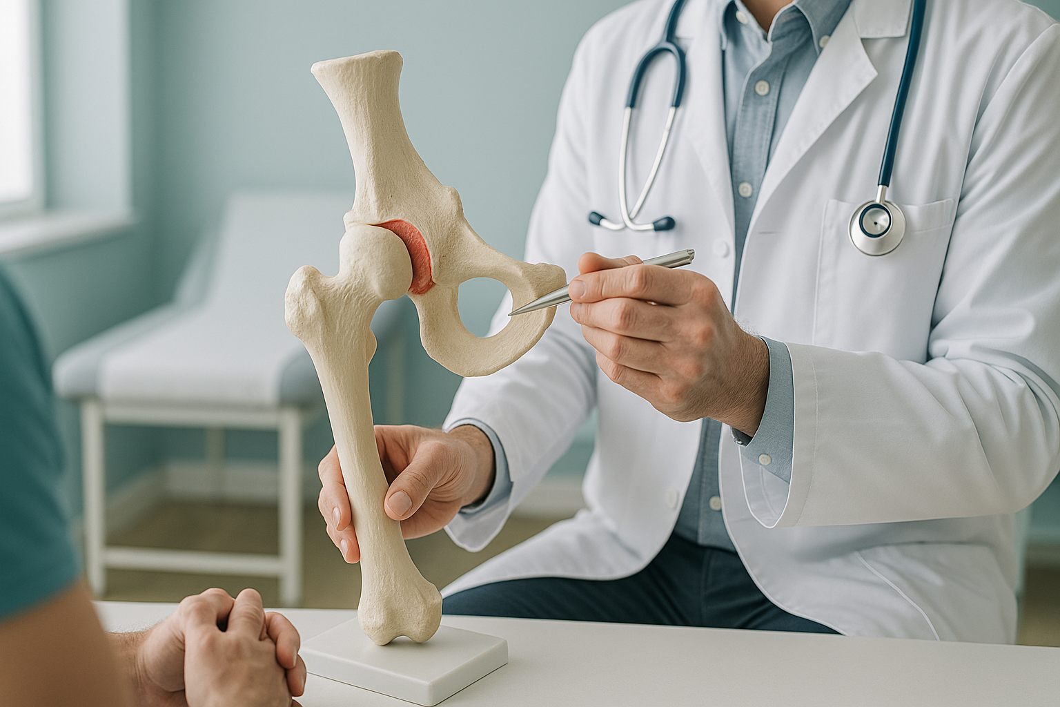
The type matters because it affects where the joint catches and which movements trigger pain. A cam bump tends to jam during deep flexion (bending the hip). A pincer overhang tends to pinch during rotation. Understanding the type helps your clinician plan the right approach.
What does hip impingement feel like?
Hip impingement usually causes a deep ache or sharp pinch in the groin or front of the hip. You might feel it most when bending, sitting for a long time, getting in and out of a car, or during sport. Some people also feel stiffness first thing in the morning or a clicking, locking sensation in the joint.
Important: If you notice sudden weakness in your leg, numbness spreading below your hip, or any loss of bladder or bowel control, please seek urgent medical attention. Contact your GP, call 111, or go to A&E. These symptoms need immediate assessment, not a routine scan.
What causes hip impingement?
The bone shape that leads to FAI usually develops during childhood and adolescence as the hip joint grows. Most people never know they have it until symptoms begin, often triggered by a change in activity level, repetitive sport, or simple wear over time.
Because the body works as a connected chain from feet to head, the position of your pelvis, spine and knees all influence how your hip joint sits under load. A problem elsewhere, such as a subtle leg length difference, can change how the ball meets the socket and push an otherwise mild impingement into painful territory.
How is hip impingement assessed when you are standing up?
Most hip scans are done while you lie flat on a table. That relaxes your muscles, unloads the joint, and lets the pelvis settle into a position it would never adopt when you are actually on your feet. A standing, weight-bearing EOS scan captures the hip under real-world conditions.
An EOS scan images your whole body from head to feet while you stand naturally. It shows true pelvic tilt, leg length, and how the hip socket is oriented under load. This gives your clinician a complete picture of the forces acting on the joint. EOS uses about 90% less radiation than a standard X-ray, around ten times less.
A conventional X-ray, CT or MRI taken lying down removes gravity and muscle tension from the equation. The pelvis may look level when it is not. The socket angle can appear normal even though it pinches when you stand. Lying-down imaging still has an important role, but it cannot show how the joint loads in real life.
This matters because hip impingement is not just about bone shape. It is about what that shape does under load. Two people with identical bone bumps can have very different symptoms if their pelvic tilt or spinal posture differ. A whole-body standing image captures these relationships in a single scan.
When MRI is the better choice: EOS excels at showing bones, joints and alignment. If your clinician suspects a labral tear, cartilage damage, or other soft-tissue injury inside the hip, MRI is usually the better tool. In many cases, both types of imaging work together to build the full picture.
Not sure which type of imaging you need? A short, free video consultation can help you decide.
Book a free video consultationHow is hip impingement usually managed?
Treatment for FAI depends on the severity and the type of impingement. ScanAlign is a diagnostic imaging clinic and does not provide treatment. However, knowing the common management paths can help you have a more informed conversation with your orthopaedic specialist or physiotherapist.
- Activity modification. Avoiding positions that trigger the pinch, such as deep squats or prolonged sitting with crossed legs, can reduce day-to-day pain.
- Physiotherapy. Strengthening the muscles around the hip (especially the gluteal muscles) and improving movement control can ease pressure on the joint.
- Injections. A guided injection into the hip joint can help confirm the diagnosis and provide temporary pain relief.
- Arthroscopic surgery. If conservative measures are not enough, keyhole surgery can reshape the bone and repair damaged tissue. The approach depends on whether the impingement is cam, pincer, or mixed.
Accurate imaging before any intervention is important. Understanding the bone shape, the pelvic position, and the alignment of the whole lower limb helps your treating clinician choose the right plan. You can read more about hip alignment and EOS imaging at ScanAlign.
Frequently asked questions about hip impingement
Hip impingement is about abnormal bone shape causing the joint to pinch during movement. Arthritis is wear of the cartilage lining the joint. However, untreated impingement can speed up cartilage wear and lead to arthritis over time. That is one reason early assessment matters.
The bone shape itself does not change without surgery. Symptoms can improve with physiotherapy and activity changes, but the underlying bump or overhang remains. Getting an accurate diagnosis helps you decide whether conservative management is enough.
Low-impact activities such as swimming, cycling with a raised seat, and targeted gluteal strengthening are often well tolerated. Avoid deep squats, lunges, and anything that forces the hip into extreme flexion or rotation until you have been assessed by a physiotherapist.
Your clinician will usually start with a physical examination, including the FADIR test (flexion, adduction, internal rotation). Imaging then confirms the bone shape. A standing EOS scan can add alignment information that conventional X-rays taken lying down may miss.
When you lie down, your pelvis tilts differently and the hip socket angle changes. A standing scan shows how the joint actually loads under gravity, which can reveal impingement that looks mild or even absent on a lying-down image.
Yes. The EOS system uses about 90% less radiation than a standard X-ray, around ten times less. You can find more details on the FAQs and safety page.
No. ScanAlign accepts self-referrals. You can book a free video consultation to discuss your symptoms and decide whether an EOS scan is appropriate for you.
Pricing details are available on the ScanAlign FAQs page. The initial video consultation is free.
Yes. A stiff or painful hip can change the way you walk and stand. Over time, your lower back, knee, or even your other hip may start to compensate. That is why a whole-body scan can be more revealing than imaging one joint in isolation.
You stand naturally inside the EOS scanner at 19 Harley Street, London. The scan takes about 20 seconds. There is no enclosed tunnel and no need to change into a hospital gown. A specialist radiographer reviews the images and prepares a detailed report. Learn more about what ScanAlign offers.
Wondering if your hip pain is caused by impingement?
A free video consultation with a specialist radiographer can help you understand whether a standing alignment scan is the right next step.
Book a free video consultationSources
- Griffin DR, Dickenson EJ, et al. "The Warwick Agreement on femoroacetabular impingement syndrome (FAI syndrome)." British Journal of Sports Medicine, 2016;50:1169-1176.
- Ganz R, Parvizi J, et al. "Femoroacetabular impingement: a cause for osteoarthritis of the hip." Clinical Orthopaedics and Related Research, 2003;(417):112-120.
- NHS. "Hip pain in adults." nhs.uk. Available at: https://www.nhs.uk/conditions/hip-pain/
Written by Abbas Dhami (Specialist Diagnostic Radiographer)
This page is general information and not a diagnosis.
ScanAlign is the trading name of CoreMed Solutions Ltd and operates under The Harley Street Hospital's CQC licence.