
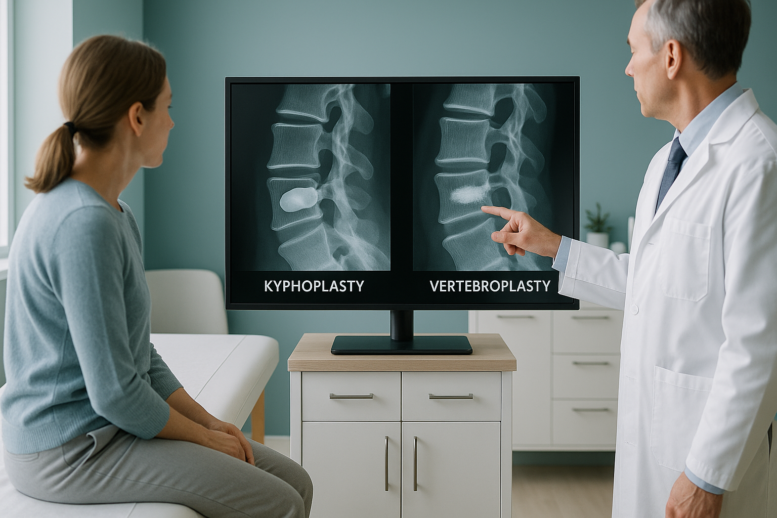
Kyphoplasty vs Vertebroplasty: What Is the Difference and Which Do You Need?
Kyphoplasty and vertebroplasty are both minimally invasive procedures that inject bone cement into a fractured vertebra to relieve pain and stabilise the bone. The key difference is that kyphoplasty uses a small balloon to restore some of the lost vertebral height before the cement is placed, while vertebroplasty injects the cement directly. Your surgeon or spinal specialist decides which is best based on imaging of your fracture.
Key takeaways
- Both procedures treat vertebral compression fractures by injecting bone cement into the broken vertebra.
- Balloon kyphoplasty inflates a small balloon first to try to restore height, then fills the space with cement.
- Vertebroplasty injects cement directly without a balloon step.
- Accurate pre-procedure imaging of spinal alignment, ideally while you are standing, helps your specialist plan the best approach.
- ScanAlign does not perform either procedure. We provide the standing whole-body imaging that helps guide your specialist's decision.
What are kyphoplasty and vertebroplasty?
Both are minimally invasive procedures used to treat vertebral compression fractures, which are small breaks in the bones of your spine. These fractures are most common in people with osteoporosis, but they can also result from trauma or, less commonly, from tumours affecting the bone.
When a vertebra collapses, it can cause severe back pain, loss of height, and a rounded upper back (sometimes called a dowager's hump). If the pain does not improve with rest, pain relief, and bracing, a specialist may recommend one of these cement-based procedures.
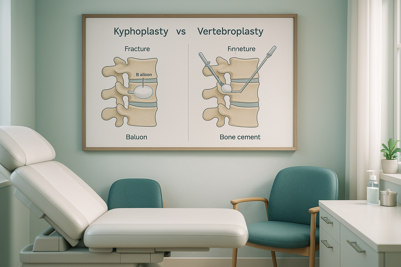
What is the difference between kyphoplasty and vertebroplasty?
The core difference is the balloon step. In vertebroplasty, bone cement is injected directly into the fracture through a needle. In balloon kyphoplasty, a small inflatable balloon is inserted first. The balloon is gently expanded to create a cavity and lift the collapsed bone back towards its original height. Then the balloon is removed and the space is filled with cement.
| Feature | Vertebroplasty | Balloon kyphoplasty |
|---|---|---|
| Balloon step | No | Yes, inflated to restore height |
| Height restoration | Limited | May partially restore vertebral height |
| Cement leakage risk | Slightly higher | Slightly lower (cavity is pre-formed) |
| Procedure time | Generally shorter | Slightly longer |
| Anaesthesia | Local or light sedation | Local or light sedation (sometimes general) |
| Pain relief | Often rapid | Often rapid |
Both procedures are typically day-case or involve a short overnight stay. Recovery is usually quick. Most people are able to walk the same day and return to light activities within a few days, though your specialist will give you specific guidance.
Which procedure is better for you depends on several factors, including the age of the fracture, how much height has been lost, and the overall alignment of your spine. That last point, alignment, is where the quality of your pre-procedure imaging really matters.
How do you know if you have a vertebral compression fracture?
A vertebral compression fracture can happen suddenly or develop gradually. Some people feel a sharp pain after a minor fall or even just bending forward. Others notice a slow, worsening ache in the mid or lower back. Here are the most common signs.
A sharp pain in the mid or lower back that comes on after bending, lifting, or a minor fall.
You seem to be getting shorter over time, or your clothes fit differently.
A visible curve or hump developing in the upper spine, sometimes called a kyphotic curve.
Discomfort that gets worse when you stand or walk, and eases when you lie down.
A note on soft-tissue imaging. EOS shows bones, joints, and alignment clearly. If your specialist needs to check for nerve compression, disc problems, or soft-tissue damage around the fracture, MRI is usually the better tool. The two types of imaging complement each other well.
Urgent symptoms. If you develop sudden numbness or weakness in your legs, or lose control of your bladder or bowels, seek urgent medical attention immediately. Contact your GP, call 111, or go to A&E. These symptoms can indicate pressure on the spinal cord and need emergency assessment.
Why does standing imaging matter before a fracture procedure?
Standing imaging gives your specialist a true picture of how your spine behaves under your own body weight. This is important because lying-down scans can mask the real extent of a compression fracture.
When you lie flat for a standard X-ray, CT, or MRI, gravity is no longer pressing down through your spine. Muscles relax, and a partially collapsed vertebra may appear more intact than it actually is. Standing up, the fracture is loaded, the collapse becomes clearer, and the effect on your overall spinal alignment can be measured properly.
Your spine is a connected chain from your head to your pelvis. A single collapsed vertebra changes the curve above and below it. Your body compensates by shifting weight, tilting the pelvis, or rounding the upper back further. These compensations can cause pain in areas far from the fracture itself. A whole-body standing image captures all of this in a single view.
EOS imaging does this with about 90% less radiation than a standard X-ray, around ten times less. You stand naturally during the scan, so the image shows your real, everyday alignment. This gives your surgeon the information needed to decide whether kyphoplasty or vertebroplasty is the right choice, and to plan the procedure accurately.
If you have been told you have a compression fracture and want a clear picture of your whole spinal alignment before seeing a specialist, a standing EOS scan can help.
Book a free video consultationFrequently asked questions
Neither is universally better. Balloon kyphoplasty may be preferred when there is potential to restore vertebral height, while vertebroplasty may be suitable for older or more stable fractures. Your spinal specialist decides based on your imaging and clinical assessment.
Most people are up and walking the same day. Light activities can usually resume within a few days. Full recovery varies, but many people feel significant pain relief within the first week. Your specialist will give you specific advice.
The cement used (polymethylmethacrylate, or PMMA) has been used in orthopaedic surgery for decades. There is a small risk of cement leakage, which is why accurate pre-procedure imaging is important to plan needle placement carefully.
Yes. EOS produces high-quality standing images of the entire spine, making compression fractures visible in the context of your overall alignment. It shows how the fracture affects the curves above and below it.
No. ScanAlign is a private, self-pay clinic. You can self-refer and book directly without a GP letter. We always recommend sharing your results with your GP or specialist afterwards.
For current pricing details, please visit our FAQs and cost page.
EOS uses about 90% less radiation than a standard X-ray, around ten times less. It is one of the lowest-dose skeletal imaging systems available.
When you lie flat, gravity is removed from the equation. A partially collapsed vertebra may look more intact because it is not bearing weight. Standing imaging shows the fracture as it actually behaves during your normal daily life.
No. ScanAlign is a diagnostic imaging clinic only. We provide the standing EOS imaging that helps your surgeon or spinal specialist plan the right procedure for you.
You stand still inside the EOS system for about 15 to 20 seconds while it scans. There is no enclosed tunnel and no loud noise. You can learn more about the process on our EOS scan overview page.
Get the full picture before your procedure
A standing whole-body EOS scan shows your specialist exactly how a compression fracture is affecting your spinal alignment, helping them plan the best approach for you.
Book a free video consultationWant to learn more about how standing imaging works for spinal conditions? Visit our spine imaging page.
Author: Abbas Dhami (Specialist Diagnostic Radiographer)
Sources
- National Institute for Health and Care Excellence (NICE). Percutaneous vertebroplasty and percutaneous balloon kyphoplasty for treating osteoporotic vertebral compression fractures. Technology Appraisal Guidance TA279, 2013.
- Defined Procedure Guidance. Interventional procedures guidance: balloon kyphoplasty for vertebral compression fractures (IPG166). NICE, 2006 (reviewed 2012).
- NHS. Vertebral compression fractures and osteoporosis. NHS Inform / NHS UK guidance on spinal fractures.
This page is general information and not a diagnosis.
ScanAlign is the trading name of CoreMed Solutions Ltd and operates under The Harley Street Hospital's CQC licence.