
Kyphosis-Lordosis Posture: What Is It and Why Does It Matter?
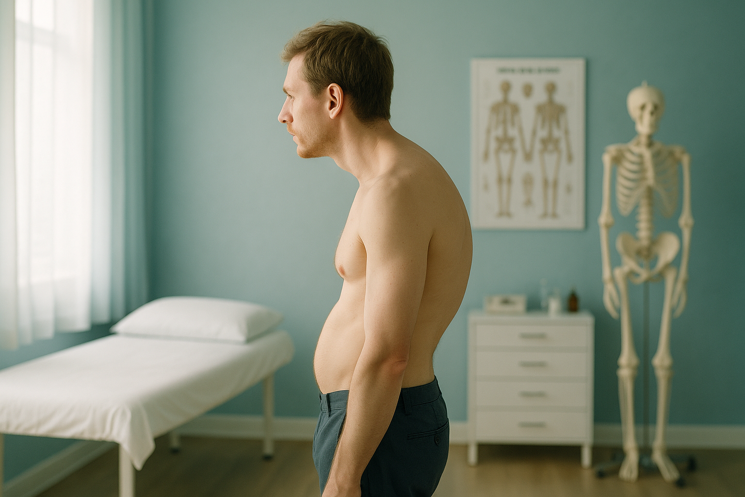
Kyphosis-lordosis posture is a combined spinal pattern where the upper back rounds forward more than normal while the lower back arches excessively. The two curves feed each other, shifting your centre of gravity and often causing pain in areas far from the original problem. A standing, whole-body image is the clearest way to see both curves together under real weight-bearing load.
Key takeaways
- Kyphosis (upper back rounding) and lordosis (lower back arching) often appear together as a single postural pattern.
- Pain may show up in the neck, mid-back, hips or knees because the body compensates along the whole chain.
- Lying-down scans relax the muscles and can hide the true extent of both curves.
- An EOS standing scan captures the full spine and lower limbs in one low-dose image, with about 90% less radiation than a standard X-ray.
- ScanAlign provides the diagnostic imaging that informs treatment decisions made by your clinician.
What is kyphosis-lordosis posture?
Kyphosis-lordosis posture, sometimes called kypholordotic posture, is a postural type where two natural spinal curves become exaggerated at the same time. The thoracic spine (upper back) rounds forward more than it should, while the lumbar spine (lower back) dips into a deeper arch.
Everyone's spine has gentle curves. They act as shock absorbers. In a kypholordotic pattern, those curves go beyond the normal range. The pelvis typically tilts forward, the head drifts ahead of the shoulders, and the abdomen may push out. Together, these shifts change how your body distributes weight from head to toe.
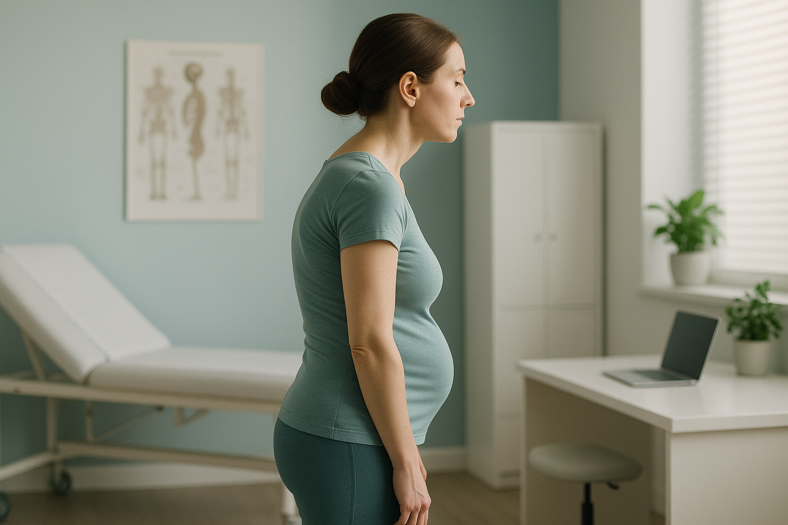
Because the spine works as a connected chain, an exaggerated curve in one region almost always creates a compensating curve elsewhere. That is why kyphosis and lordosis so often appear together rather than in isolation.
How do you know if you have a kypholordotic posture?
You may notice some of the signs below, although many people live with this pattern for years without realising the connection between their symptoms.
Shoulders roll forward and the upper back looks hunched, especially when standing relaxed.
A visible deep curve at the base of the spine, often with the abdomen pushing forward.
The head sits ahead of the shoulders rather than balanced above them, straining the neck.
Discomfort that moves between the thoracic area, the lower back and the hips.
If you experience sudden numbness, weakness, or any loss of bladder or bowel control, seek urgent medical attention through your GP, 111 or A&E rather than booking a scan.
What causes combined kyphosis and lordosis?
A kypholordotic posture usually develops gradually, driven by muscle imbalances, habitual positions, or structural changes in the vertebrae. The most common contributing factors include the following.
Hours at a desk tighten the hip flexors and weaken the upper back muscles.
Weak core and gluteal muscles allow the pelvis to tilt forward, deepening both curves.
Disc thinning and vertebral wedging can gradually increase the thoracic curve over the years.
Scheuermann's disease, spondylolisthesis, or previous spinal injury can alter vertebral shape.
Because the pelvis, spine and head work together as a single kinetic chain, a change at one level ripples up or down. A forward-tilting pelvis deepens the lumbar arch, which in turn pushes the thoracic spine into a greater curve to keep the head over the feet. Pain may appear in the neck, the mid-back or even the knees, often far from the curves themselves.
How is kyphosis-lordosis posture assessed when standing up?
The most accurate way to measure spinal curves is while you are upright and weight-bearing, because that is how your body actually carries its load. When you lie down for a conventional X-ray, CT or MRI, gravity is removed and your muscles relax. Curves that cause real problems on your feet may flatten out on the table, giving an incomplete picture.
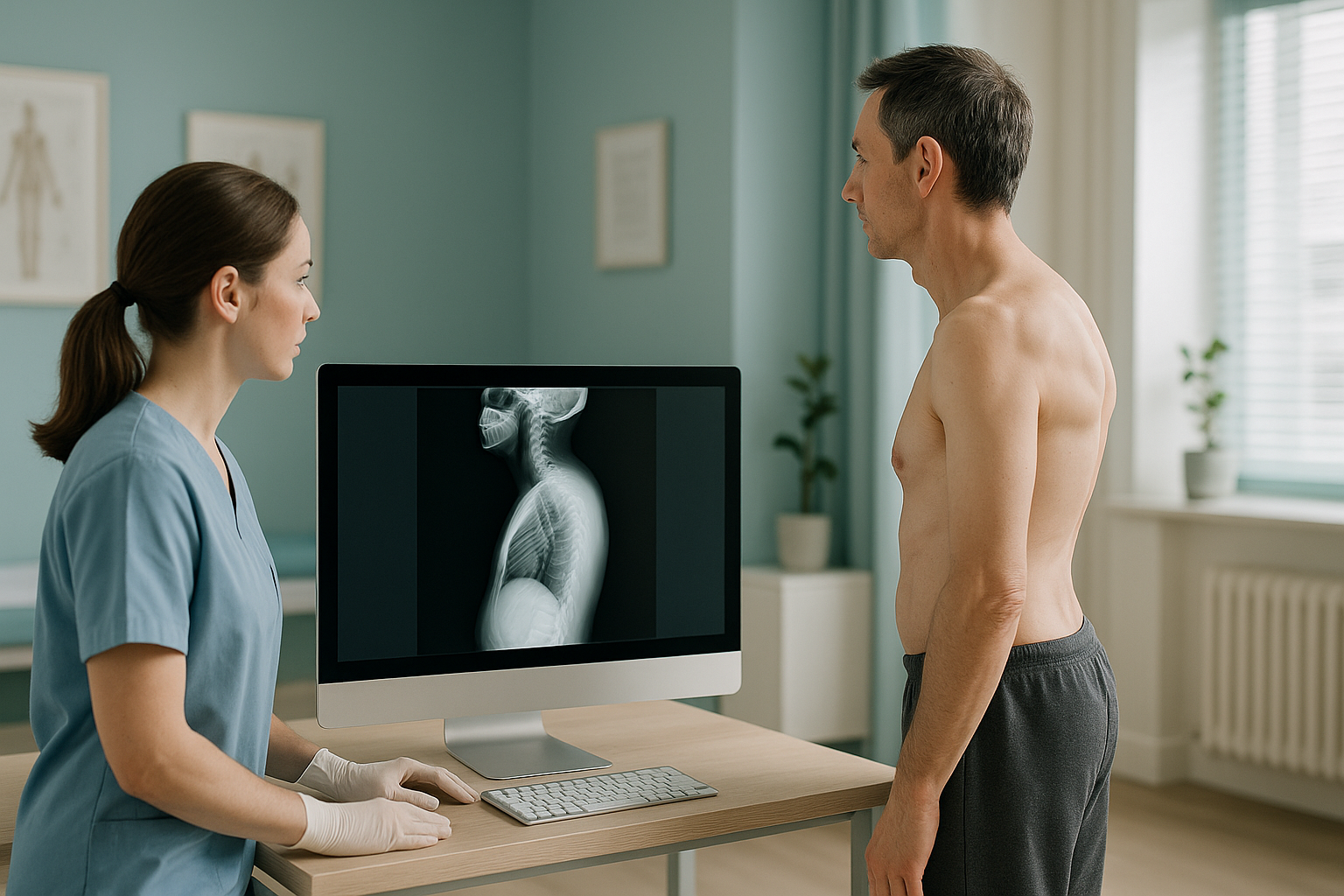
An EOS scan captures a full-length image of your skeleton from head to feet in a single standing exposure. Both the thoracic and lumbar curves are visible on one image, along with the pelvis, hips and legs. Your radiographer and reporting specialist can see exactly how the two curves relate to each other and how the rest of your body is compensating. The scan uses about 90% less radiation than a standard X-ray, around ten times less.
Wondering whether your posture could benefit from a full standing assessment?
Book a free video consultationWhen is MRI the better choice? EOS excels at showing bones, joints and overall alignment. If your clinician suspects disc problems, nerve compression or soft-tissue damage, MRI is usually the more appropriate tool. The two scans complement each other well.
What generally helps manage a kypholordotic posture?
Management usually focuses on rebalancing the muscles around the spine and pelvis. Your clinician may recommend a combination of approaches based on what the imaging shows.
- Targeted strengthening. Exercises that build core stability and upper-back strength can help support the spine in a more neutral position.
- Flexibility work. Stretching tight hip flexors, chest muscles and hamstrings may reduce the pull on the pelvis and thoracic spine.
- Posture awareness. Small adjustments to workstation setup, standing habits and movement breaks can slow further progression.
- Clinical guidance. A physiotherapist, osteopath or orthopaedic specialist can tailor a plan once they understand the full alignment picture.
ScanAlign does not provide treatment. We provide the detailed, standing diagnostic images that help your treating clinician understand exactly what is happening across your whole alignment. You can learn more about how spinal alignment is assessed with EOS.
Frequently asked questions about kyphosis-lordosis posture
Yes. In fact, the two often appear together. An increased upper-back curve shifts the body's centre of gravity forward, and the lower back compensates by arching more to keep you balanced. This combined pattern is known as kypholordotic posture.
Not exactly. Everyone's posture sits on a spectrum. Kypholordotic posture describes a specific pattern where both curves exceed the typical range. It can be partly habitual and partly structural, which is why imaging helps clarify the cause.
From the side, you might notice a rounded upper back, a forward head position, a deep arch in the lower back, and the abdomen pushed forward. The shoulders often roll inward.
It can. Because the body is a connected chain, a forward-tilting pelvis changes loading on the hips and affects how the knees track. Pain may appear well away from the spinal curves that are driving the imbalance.
No. ScanAlign is a private, self-pay clinic. You can book directly without a GP referral. If you already have a referral from a consultant or physiotherapist, you are welcome to bring it along.
The EOS system uses about 90% less radiation than a standard X-ray, around ten times less. This makes it particularly suitable when full-body imaging is needed.
When you lie down, gravity is removed and your muscles relax. Curves that cause symptoms when you are upright may flatten on the table. A standing scan captures your skeleton under real weight-bearing conditions, showing the true extent of both curves.
Targeted strengthening and flexibility work can often improve the pattern, especially if it is largely muscular. However, if structural changes in the vertebrae are involved, the approach may differ. Imaging helps your clinician understand the balance between muscular and structural factors.
You can find current pricing and what is included on our FAQs and pricing page.
Your images are reviewed by a specialist. You receive a detailed report showing how your spinal curves, pelvis and lower limbs relate to each other under load. You can then share this with your treating clinician to guide the next steps. Learn more about the EOS whole-body assessment.
See your full alignment, standing up
A free video consultation lets you discuss your symptoms, ask questions and find out whether a standing EOS scan is the right next step for you.
Book a free video consultationAuthor: Abbas Dhami (Specialist Diagnostic Radiographer)
Sources
- Kendall FP, McCreary EK, Provance PG, Rodgers MM, Romani WA. Muscles: Testing and Function with Posture and Pain. 5th ed. Lippincott Williams & Wilkins, 2005.
- Roussouly P, Pinheiro-Franco JL. Biomechanical analysis of the spino-pelvic organisation and adaptation in pathology. European Spine Journal. 2011;20(Suppl 5):609-618.
- NHS. Overview: Kyphosis. nhs.uk/conditions/kyphosis. Accessed 2024.
This page is general information and not a diagnosis.
ScanAlign is the trading name of CoreMed Solutions Ltd and operates under The Harley Street Hospital's CQC licence.