
Posterior Pelvic Tilt: What Is It and Why Does It Affect Your Whole Body?
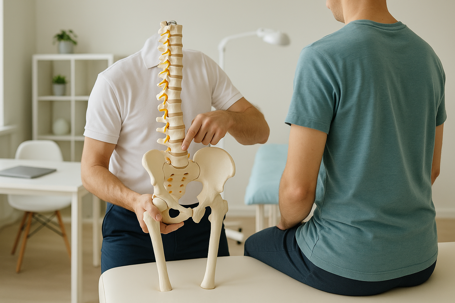
A posterior pelvic tilt is when the top of your pelvis tips backward. This flattens the natural curve of your lower back and shifts your centre of gravity. It can cause stiffness, discomfort and compensations throughout your spine, hips and knees. Standing imaging reveals the full picture because the tilt only shows reliably when you are upright and weight-bearing.
Key takeaways
- Your pelvis rocks backward, reducing or removing the normal lumbar curve.
- Pain often appears in the lower back, hips or knees rather than the pelvis itself.
- Lying-down scans can hide the tilt because gravity and relaxed muscles change your alignment.
- A whole-body standing scan captures how your skeleton actually balances under load.
- Understanding the tilt is a diagnostic step, not a treatment. It guides what comes next.
Your pelvis is the foundation of your spine. When it sits in a neutral position, it supports a gentle inward curve in your lower back (your lumbar lordosis). A posterior pelvic tilt pulls the bottom of the pelvis forward and the top backward. The result is a flatter lower back, sometimes called "flat back posture."
Many people live with a mild posterior tilt and never notice. But when the tilt is significant, it changes how weight travels through your spine, hips and legs. That is when symptoms start to appear, often far from the pelvis itself.
What are the signs of a posterior pelvic tilt?
The most common sign is a visibly flat lower back, with your bottom tucked under rather than gently curved. You may also notice stiffness when standing for long periods, or a feeling that you are leaning backward.
Some people also experience discomfort in the hips, hamstrings or even the knees. This happens because a tilted pelvis changes how your leg joints are loaded with each step.
What causes a posterior pelvic tilt?
A posterior pelvic tilt is usually caused by muscle imbalances, prolonged sitting habits or structural changes in the spine and pelvis. It is rarely a single factor. Most people develop the tilt gradually over months or years.
How does a posterior pelvic tilt affect the rest of your body?
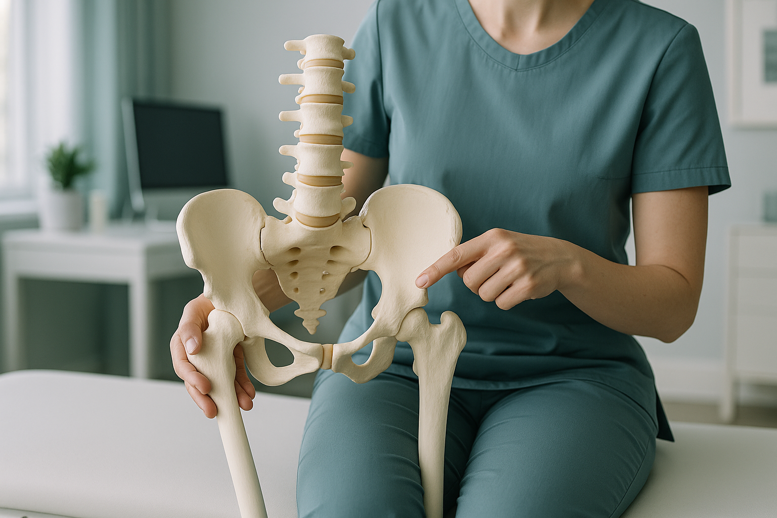
Your body is a connected chain from your feet to your head. When the pelvis tilts backward, every joint above and below it has to compensate. Your lower back loses its natural shock-absorbing curve. Your upper back may round forward to keep you balanced. Your hips sit in a slightly different position, which changes how your knees and ankles are loaded.
This is the compensation effect. Pain in your mid-back, tightness in your hamstrings, or a nagging ache in one knee could all trace back to the way your pelvis is sitting. Treating only the area that hurts, without understanding the underlying alignment, often means the problem returns.
A posterior pelvic tilt changes how forces travel through your whole skeleton. A standing whole-body scan can map those compensations from head to ankle, all in one image.
Book a free video consultationHow is a posterior pelvic tilt assessed with a standing scan?
A posterior pelvic tilt is best assessed when you are standing, because that is when your muscles, gravity and body weight all interact to produce your real alignment. Lying-down imaging, such as a standard X-ray or MRI, relaxes your muscles and lets the pelvis settle into a different position. The tilt can look less significant, or disappear entirely, on a scan taken while lying flat.
An EOS scan captures your full skeleton from head to ankle in one standing image, using about 90% less radiation than a standard X-ray, around ten times less. The image shows exactly where your pelvis sits relative to your spine, hips, knees and ankles. Your reporting clinician can then see the chain of compensations, not just the pelvis in isolation.
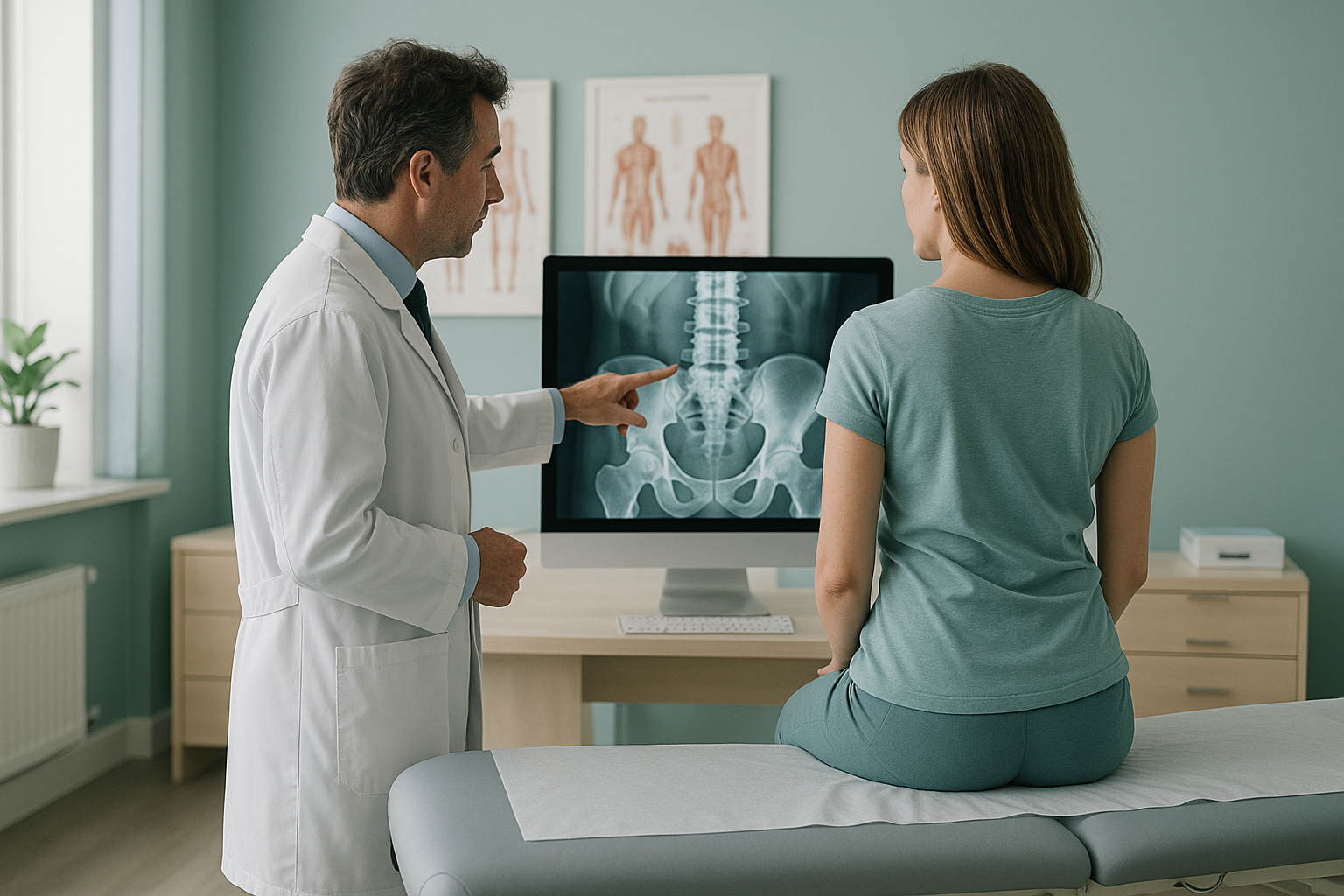
This matters because treatment decisions are better when they are based on what your body actually does while you are upright. A scan that hides the tilt can lead to incomplete answers. You can learn more about how EOS whole-body imaging works on our main page.
When MRI is the better choice: EOS shows bones, joints and alignment clearly. If your clinician suspects a disc problem, nerve compression or soft-tissue injury, MRI is usually the better tool. The two scans answer different questions, and sometimes you need both.
What helps with a posterior pelvic tilt?
Addressing a posterior pelvic tilt usually starts with understanding exactly how your pelvis and spine are positioned. Without that information, any programme of stretching, strengthening or manual therapy is based on guesswork.
Common approaches recommended by physiotherapists and musculoskeletal specialists include targeted stretching of tight hamstrings and abdominals, strengthening of the lower back and hip flexors, and ergonomic changes to your sitting and standing habits. In some cases, a structural cause such as disc degeneration or a joint problem may need further investigation.
ScanAlign does not provide treatment. We provide the diagnostic imaging that helps you and your clinician understand the full picture. With a clear standing image of your alignment, your treatment team can build a plan that addresses the root cause rather than just the symptoms. If you are already working with a physiotherapist or surgeon, your scan report can be shared directly with them.
For related hip and pelvic alignment questions, our hip alignment page explains more about how pelvic position affects the hip joint.
Frequently asked questions about posterior pelvic tilt
It is a posture pattern where the top of the pelvis tips backward. This reduces the natural inward curve of your lower back and can shift your weight distribution, sometimes causing discomfort in the back, hips or knees.
Common causes include prolonged sitting, tight hamstrings, weak lower back muscles, age-related disc changes and previous injuries. It often develops gradually rather than from a single event.
Stand side-on in front of a mirror. If your lower back looks very flat and your bottom appears tucked under, you may have a posterior tilt. A standing X-ray or EOS scan provides a precise assessment that goes beyond what you can see in a mirror.
They are closely related. A flat back describes the visible result of the pelvis tilting backward. The posterior pelvic tilt is the mechanical cause of that flattened lower back appearance.
Yes. When you lie down, gravity and muscle relaxation change your pelvic position. A tilt that is clearly present while standing can look much less significant, or not appear at all, on a scan taken lying flat.
An EOS scan uses about 90% less radiation than a standard X-ray, around ten times less. This makes it suitable for whole-body imaging in a single session.
No. ScanAlign is a private, self-pay clinic. You can book directly without a referral. We offer a free video consultation first so we can check that a scan is appropriate for your situation.
Full pricing details are available on our FAQs and cost page. Prices are not listed on individual condition pages to keep information current.
It can contribute to pain in those areas. The pelvis connects to both your spine and your legs. When it tilts, it changes the loading pattern through the hip joints and knees, which can lead to discomfort over time.
Yes. Numbness, weakness, or any loss of bladder or bowel control are red-flag symptoms. Please seek urgent medical attention from your GP, NHS 111 or A&E rather than booking a scan first.
Ready to see your whole alignment?
A free video consultation lets us understand your situation and confirm whether a standing scan is the right next step for you.
Book a free video consultationWritten by Abbas Dhami (Specialist Diagnostic Radiographer)
Sources
- Le Huec, J.C., et al. "Sagittal balance of the spine." European Spine Journal, 2019. Springer.
- NHS. "Back pain: overview, causes and treatment." nhs.uk.
- Illés, T., Somoskeöy, S. "The EOS imaging system and its uses in daily orthopaedic practice." International Orthopaedics (SICOT), 2012.
This page is general information and not a diagnosis.
ScanAlign is the trading name of CoreMed Solutions Ltd and operates under The Harley Street Hospital's CQC licence.