
Spinal Stenosis: What Is It and Why Does It Cause Pain When You Walk?
Spinal stenosis is a narrowing of the spaces inside your spine. This puts pressure on the nerves that travel through it. It most often affects the lower back or neck, and its hallmark symptom is pain, heaviness or cramping in the legs that gets worse when you walk and eases when you sit down or lean forwards.
Key takeaways
- Spinal stenosis means the canal or openings in the spine have become too narrow for the nerves passing through them.
- The most common cause in adults is normal wear and tear of the joints and discs over time.
- Symptoms often depend on posture. Standing and walking tend to make them worse, while sitting or bending forwards eases them.
- Standing, weight-bearing imaging can reveal how your spine behaves under load, something lying-down scans can miss.
- ScanAlign provides diagnostic imaging only, not treatment. Your results help guide the right next step with your specialist.
If you have been told you have spinal stenosis, or you recognise the pattern of leg pain that eases when you sit, this page explains what is happening inside your spine and how whole-body standing imaging can add to the picture.
What is spinal stenosis?
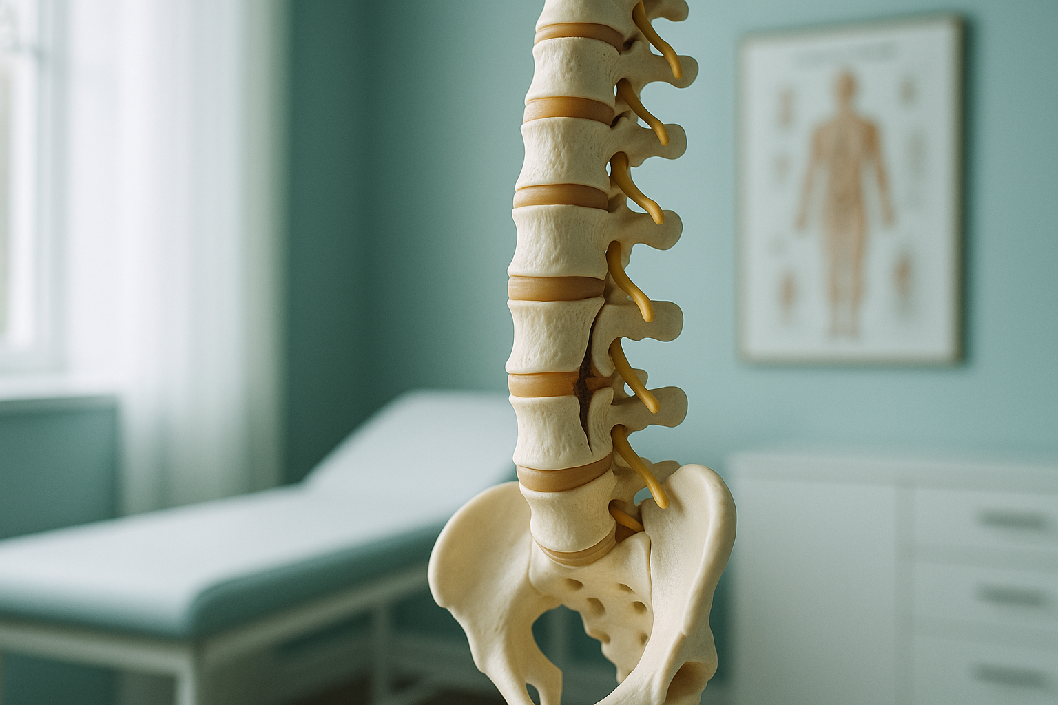
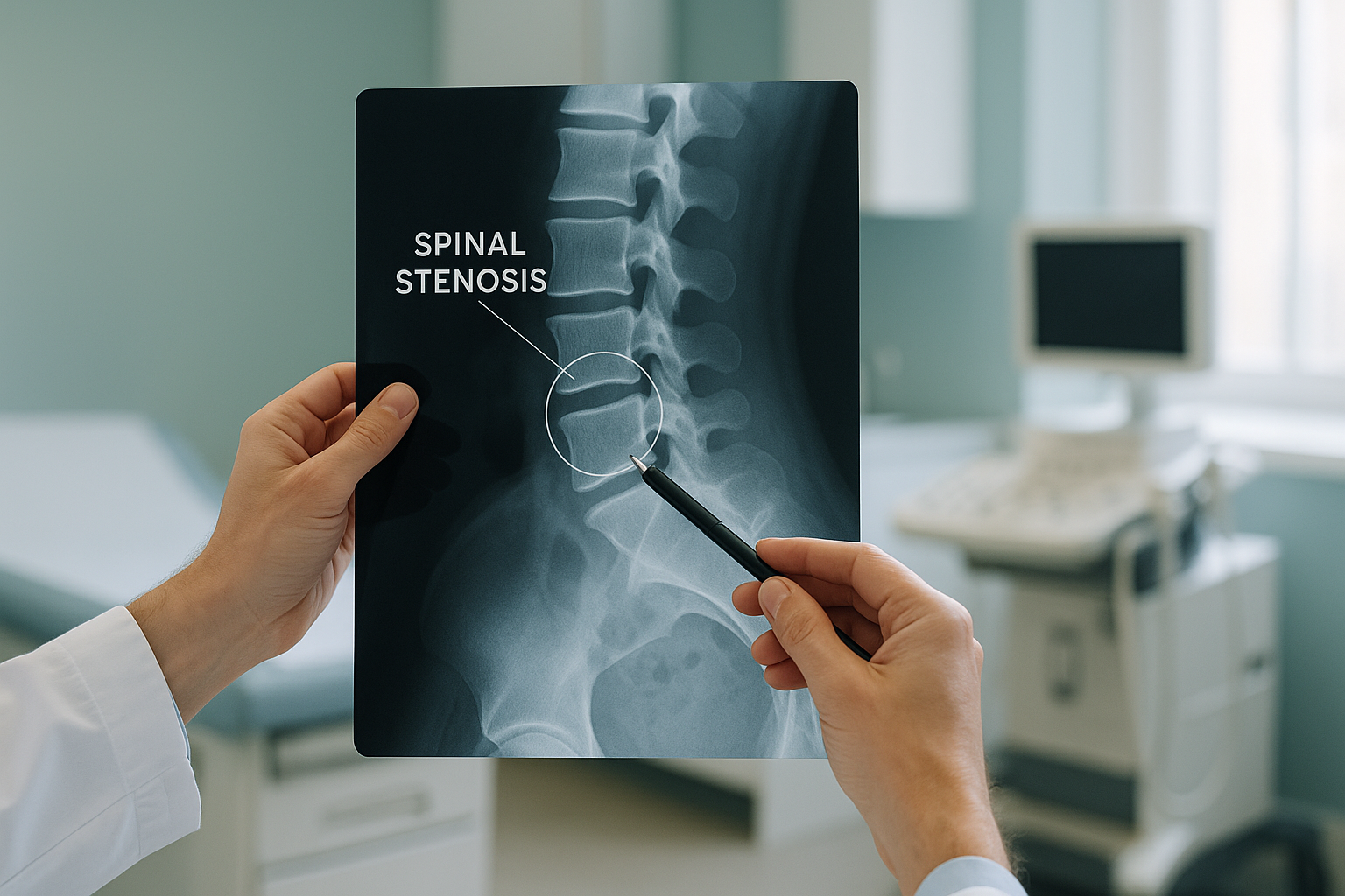
Spinal stenosis is a gradual narrowing of the spinal canal, the bony tunnel that protects your spinal cord and the nerves branching off it. When that space shrinks, the nerves get squeezed. This can happen in the central canal, in the smaller side tunnels (called foramina) where individual nerves exit, or in both.
The condition is most common in the lumbar spine (lower back), where it tends to affect the nerves that run down into your legs. It can also occur in the cervical spine (neck), where it may affect the arms or hands. Some people have narrowing in more than one area at the same time.
What are the symptoms of spinal stenosis?
Symptoms depend on which part of the spine is affected and how much the nerves are compressed. Many people with mild narrowing on a scan have no symptoms at all. When symptoms do appear, they tend to follow a recognisable pattern.
An aching, heavy or cramping feeling in one or both legs when you walk or stand, often called neurogenic claudication.
Symptoms ease quickly when you sit down, lean on a shopping trolley, or bend forwards. This opens up the spinal canal slightly.
Pins and needles, numbness, or a feeling of weakness in the legs (lumbar) or hands and arms (cervical).
Over months or years, the distance you can walk comfortably may gradually shorten.
When to seek urgent help
If you develop sudden numbness in the groin area, difficulty controlling your bladder or bowels, or rapidly worsening leg weakness, contact your GP, call 111, or go to A&E straight away. These symptoms may indicate a rare but serious condition called cauda equina syndrome, which needs immediate medical attention.
What causes spinal stenosis?
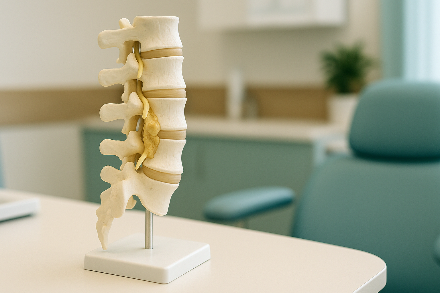
The most common cause is age-related wear and tear, sometimes called degenerative change. As the spine ages, several structures can thicken or bulge inward, reducing the space available for the nerves.
Discs lose water content and may bulge into the canal.
New bone growth at the joints can project into the canal or foramina.
The ligamentum flavum, which lines the canal, can thicken and fold inward.
The facet joints at the back of each vertebra can enlarge with arthritis.
Less commonly, stenosis can result from a spondylolisthesis (where one vertebra slips forward on the one below) or from a naturally narrow canal that a person is born with. When the spine loses its normal alignment, the narrowing can become worse under load. That is why posture and overall spinal balance matter.
How does standing imaging help assess spinal stenosis?
Standing, weight-bearing imaging shows your spine the way it actually works, under the pull of gravity and the load of your body weight. This matters for stenosis because the narrowing often changes depending on your position.
When you lie down for a standard MRI or CT scan, your muscles relax and the spine unloads. Disc bulges may reduce slightly and the canal can appear a little wider than it really is during daily life. Standing up reverses this. Gravity compresses the discs, the ligaments fold differently, and any slippage between vertebrae may increase. In other words, the very position that triggers your symptoms is the one that lying-down imaging does not capture.
An EOS scan captures your full skeleton from head to feet in a single standing image, using about 90% less radiation than a standard X-ray, around ten times less. This whole-body view also reveals how other areas of the spine and pelvis may be compensating for the stenosis. Your body is a connected chain. A narrowed segment in the lower back can change the way you stand, shifting load to your hips, knees or the opposite side of the spine.
EOS imaging does not replace MRI. It adds a different layer of information, showing bony alignment and balance under real-world conditions. Together, the two give your specialist a much clearer picture than either one alone.
Wondering whether a standing alignment scan could add to your existing imaging? We offer a free video consultation to talk it through.
Book a free video consultationA note about MRI. EOS imaging excels at showing bones, joints and overall spinal alignment. For soft tissue detail, such as discs, nerves and the ligaments inside the canal, MRI is usually the better tool. Many people with spinal stenosis benefit from both types of imaging, each answering a different question.
What helps manage spinal stenosis?
Management depends on how severe the narrowing is and how much it affects your daily life. ScanAlign is a diagnostic imaging clinic, not a treatment provider, so the detail below is general background rather than a recommendation.
- Staying active. Gentle exercise, particularly activities where the spine is slightly flexed (like cycling or swimming), often helps maintain mobility and manage symptoms.
- Physiotherapy. A physiotherapist can guide exercises that strengthen the core and improve flexibility without aggravating the nerves.
- Pain management. Your GP or a pain specialist may suggest medication or injections to help control symptoms while you remain active.
- Surgical options. When symptoms are severe or worsening despite other approaches, a spinal surgeon may discuss a decompression procedure. Detailed alignment data from standing imaging can help with surgical planning.
Whatever path is right for you, accurate diagnostic imaging is the starting point. Understanding exactly where the narrowing is, how your spine is balanced, and whether other areas are compensating helps your clinical team make better decisions.
Frequently asked questions about spinal stenosis
It can range from mild (no symptoms at all) to severe (significant difficulty walking). Most cases progress slowly over years. In rare cases involving cauda equina syndrome, it becomes a medical emergency requiring urgent attention.
The structural narrowing itself does not reverse, but symptoms can improve with appropriate exercise, physiotherapy and pain management. Many people manage well for years without surgery.
Lumbar stenosis affects the lower back and typically causes leg symptoms. Cervical stenosis affects the neck and can cause symptoms in the arms, hands, or in severe cases, affect balance and coordination.
Standing and walking extend the spine slightly, which can narrow the canal further and press on the nerves. Sitting or bending forwards opens the canal a little, relieving the pressure. This pattern is very characteristic of lumbar stenosis.
They show different things. MRI reveals soft tissues like discs and ligaments inside the canal. A standing EOS scan shows how the whole spine is aligned under load, whether a vertebra has slipped, and how the rest of the body is compensating. The two are complementary, not competing.
Yes. EOS uses about 90% less radiation than a standard X-ray, around ten times less. You can read more about safety, cost and what to expect on our FAQs page.
No. ScanAlign is a private, self-pay clinic. You can refer yourself directly. We recommend starting with a free video consultation so we can check that the scan is appropriate for your situation.
We do not list prices on individual pages. You can find current pricing on our FAQs page, or ask during your free video consultation.
Indirectly, yes. When your lower back movement is limited by stenosis, your hips and knees may take on extra load or move differently to compensate. This is one reason a whole-body alignment view is so valuable.
You stand comfortably inside the EOS scanner at our clinic at 19 Harley Street. The scan takes about 20 seconds and captures your full skeleton in one image. There is no enclosed tunnel and no need to lie down. A specialist radiographer supervises the entire process.
See your spine the way it really stands
A free video consultation with our team will help you understand whether standing alignment imaging is the right next step for your spinal stenosis.
Book a free video consultationWant to learn more about how whole-body standing imaging works? Visit our EOS scan overview page, or explore our spine imaging guide.
Sources
- NHS. Spinal stenosis. nhs.uk. Available at: https://www.nhs.uk/conditions/spinal-stenosis/
- Lurie J, Tomkins-Lane C. Management of lumbar spinal stenosis. BMJ. 2016;352:h6234.
- Illés T, Somoskeöy S. The EOS imaging system and its uses in daily orthopaedic practice. International Orthopaedics. 2012;36(7):1405-1411.
Written by Abbas Dhami (Specialist Diagnostic Radiographer)
This page is general information and not a diagnosis.
ScanAlign is the trading name of CoreMed Solutions Ltd and operates under The Harley Street Hospital's CQC licence.