

Cervical Myelopathy Treatment: What Are Your Options and When Is Surgery Needed?
Cervical myelopathy treatment depends on how severely the spinal cord in your neck is compressed. Mild cases may be managed with careful monitoring, physiotherapy and lifestyle changes. When symptoms progress, surgery to relieve cord pressure is usually the recommended approach. Standing whole-body imaging can reveal how your neck alignment changes under load.
Key takeaways
- Cervical myelopathy is compression of the spinal cord in the neck, not just a pinched nerve.
- It tends to worsen over time, so early assessment matters.
- Non-surgical management can help mild or stable cases, but surgery is often needed if symptoms are progressing.
- Standing imaging shows how gravity and posture affect neck alignment, which lying-down scans can miss.
- ScanAlign provides diagnostic imaging only. Treatment decisions are made with your surgeon or specialist.
Important. If you notice sudden weakness in your arms or legs, difficulty walking, or loss of bladder or bowel control, seek urgent medical attention. Contact your GP, call 111, or attend A&E. These symptoms need immediate assessment, not a routine scan.
What is cervical myelopathy?
Cervical myelopathy is a condition where the spinal cord inside your neck becomes compressed, or squeezed. This is different from a pinched nerve (radiculopathy), which affects a single nerve root. Because the spinal cord carries signals to your entire body below the neck, compression here can cause widespread symptoms in your hands, arms, legs and balance.
The compression usually develops gradually over months or years. It is most common in people over 50, often linked to age-related wear in the cervical spine. Because it tends to worsen rather than improve on its own, getting a clear picture of your neck alignment early is important.

What are the signs of cervical myelopathy?
The hallmark of myelopathy is problems with coordination and fine motor skills, not just pain. Many people notice changes so gradually that they dismiss them as "getting older."
Not everyone with myelopathy has neck pain. Some people only notice problems in their legs or hands. That is why a thorough assessment of the whole spine, not just the sore spot, can be valuable.
What causes cervical myelopathy?
The most common cause is age-related narrowing of the spinal canal in the neck. Several changes contribute to this.
Some people are born with a naturally narrower spinal canal, which means even modest wear-and-tear changes can cause compression sooner. Poor posture and forward head position may also play a role by changing how forces travel through the cervical spine.
How is cervical myelopathy treated?
Cervical myelopathy treatment falls into two broad categories: non-surgical management for mild or stable cases, and surgery when symptoms are progressing or already significant. Your specialist will decide based on the severity of your symptoms and what imaging shows.
Non-surgical management
For people with very mild symptoms that are not getting worse, careful monitoring and conservative measures may be appropriate. These can include:
- Regular specialist reviews to check for any change.
- Gentle physiotherapy focused on maintaining neck mobility and strength, guided by a therapist experienced in cervical spine conditions.
- Activity modification, such as avoiding high-impact sports or extreme neck positions.
- Postural awareness and ergonomic adjustments at work.
Non-surgical management does not reverse spinal cord compression. It aims to maintain function and catch any worsening early. If symptoms progress, surgery is usually recommended.
Surgical options
When myelopathy is progressing, surgery to decompress the spinal cord is typically the recommended path. The goal is to stop further damage and, where possible, allow some recovery. The main surgical approaches include:
- ACDF (anterior cervical discectomy and fusion). Approached from the front of the neck. The damaged disc is removed and the vertebrae are fused. Commonly used when one or two levels are affected.
- Laminectomy. Approached from the back of the neck. Part of the bone (lamina) is removed to create more space for the cord. Often combined with fusion for stability.
- Laminoplasty. Also from the back. Instead of removing the bone entirely, the lamina is hinged open like a door. This widens the canal without fusion, preserving more neck movement.
The choice between these depends on where the compression is, how many levels are involved, your neck curve, and your overall health. Your surgeon will explain which approach suits your specific anatomy.
A note on MRI and EOS. MRI is usually the key scan for cervical myelopathy because it shows the spinal cord, discs and soft tissues in detail. EOS shows bones, joints and overall alignment. For soft-tissue assessment, discs or nerve detail, MRI is usually the better tool. However, a standing EOS scan of your whole spine can add valuable information about how your overall alignment and posture may be contributing to the load on your neck, especially under gravity.
How does standing imaging help assess cervical myelopathy?
Standing imaging shows your spine as it actually functions, upright and bearing your body weight. When you lie down for a conventional scan, gravity is removed. Your muscles relax, your curves change, and mild instability or alignment problems can be hidden.
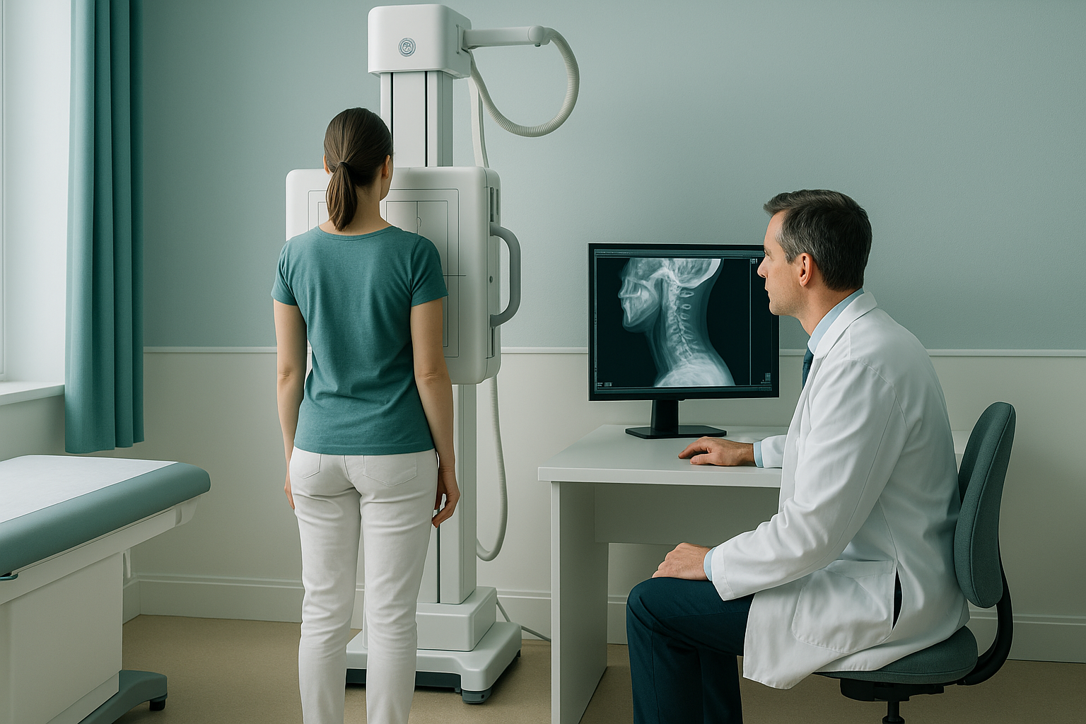
Your body is a connected chain from feet to head. If your thoracic spine (mid-back) has increased rounding, your cervical spine may compensate by extending further forward to keep your head upright. This forward head posture places extra load on the neck, which can worsen compression. A whole-body standing scan captures this full picture in a single image.
An EOS scan uses about 90% less radiation than a standard X-ray, around ten times less. That makes it practical for full-length imaging without the radiation concern of conventional radiography.
Want to understand how your whole-body alignment may be affecting your neck? A free video consultation can help you decide if standing imaging is the right next step.
Book a free video consultationFrequently asked questions about cervical myelopathy treatment
Myelopathy caused by structural compression cannot typically be reversed without surgery. Non-surgical management aims to maintain function and monitor for worsening. If the condition is mild and stable, some people manage well with conservative care, but surgery is usually needed when symptoms progress.
Surgery is typically recommended when symptoms are getting worse, when there is significant cord compression on imaging, or when daily activities like walking and using your hands are noticeably affected. Your specialist will weigh these factors together.
Both are performed from the back of the neck. Laminectomy removes part of the bone to open the canal. Laminoplasty hinges the bone open, widening the canal while keeping the bone in place. Laminoplasty may preserve more neck movement because it does not always require fusion.
Recovery varies widely between individuals. Most people can return to light activities within a few weeks, but full bone fusion typically takes several months. Your surgeon will give you a personalised timeline and advise when you can safely return to work and exercise.
Physiotherapy can help maintain neck mobility, strengthen supporting muscles and improve balance. It does not treat the underlying cord compression, so it is usually part of a broader management plan rather than a standalone solution. Always work with a physiotherapist experienced in spinal conditions.
An EOS scan shows the bony alignment of your entire spine from head to pelvis while you are standing. It reveals how your cervical curve relates to the rest of your posture. It does not show soft tissues like the spinal cord itself, which is where MRI plays its role. Learn more about how EOS whole-body imaging works.
Yes. EOS uses about 90% less radiation than a standard X-ray, around ten times less. You can find more details about safety, preparation and what to expect on our FAQs page.
No. ScanAlign is a private self-pay service. You can refer yourself directly. If you are unsure whether imaging is right for you, you can start with a free video consultation.
Pricing details are available on our FAQs and cost page.
Poor posture, especially a forward head position, increases the mechanical load on the cervical spine. Over time this may contribute to greater compression. Understanding your full spinal alignment with a standing scan can highlight postural factors your specialist may want to address. You can read more about spinal alignment assessment.
Understand your neck alignment
A free video consultation with our specialist radiographer can help you decide whether standing whole-body imaging is a useful next step for you.
Book a free video consultationWritten by Abbas Dhami (Specialist Diagnostic Radiographer)
Sources
- NHS. Cervical spondylosis. nhs.uk. Available at: https://www.nhs.uk/conditions/cervical-spondylosis/
- Fehlings MG, Tetreault LA, Riew KD, et al. A Clinical Practice Guideline for the Management of Degenerative Cervical Myelopathy. Global Spine Journal. 2017;7(3 Suppl):S21–S33.
- American Academy of Orthopaedic Surgeons (AAOS). Cervical Spondylotic Myelopathy. OrthoInfo. Available at: https://orthoinfo.aaos.org/
This page is general information and not a diagnosis.
ScanAlign is the trading name of CoreMed Solutions Ltd and operates under The Harley Street Hospital's CQC licence.