
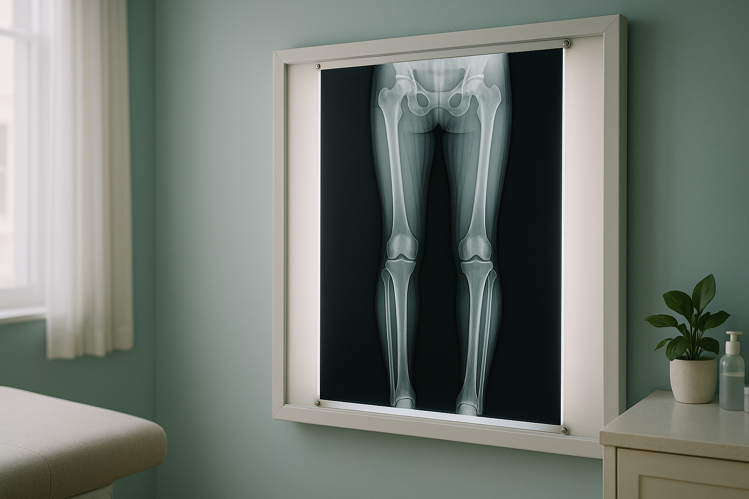
Full-Leg Standing X-ray: What Does It Show and Why Does It Matter?
A full leg x-ray captures both legs in a single image from hip to ankle while you stand. It reveals your true weight-bearing alignment, leg length difference, and joint angles. Because it is taken upright, it shows how your skeleton actually loads under gravity, something a lying-down scan cannot reproduce.
Key takeaways
- A full leg x-ray maps your alignment from the hip joint through the knee to the ankle, all in one image.
- Standing imaging shows how gravity and body weight change your joint angles. Lying down can hide these differences.
- It is commonly used before knee or hip replacement surgery, and to investigate unexplained leg, knee or hip pain.
- EOS technology captures the same view with about 90% less radiation than a standard X-ray, around ten times less.
- No referral is needed at ScanAlign. You can self-refer for a free video consultation.
What does a full leg x-ray show?
A full leg x-ray shows the complete bony anatomy of both legs in one continuous image, from the hip sockets down through the thigh bones, across the knee joints and shin bones, to the ankles. It reveals how these structures line up relative to each other when you are standing on your own two feet.
Your specialist can use this single image to assess several things at once.
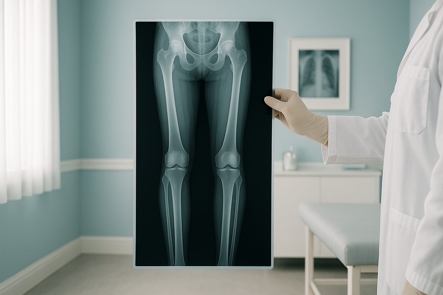
The mechanical axis is an imaginary line drawn from the centre of the hip to the centre of the ankle. In a well-aligned leg, that line passes through the middle of the knee. When the line falls to one side, it tells the clinician how much the alignment has shifted and which compartment of the knee carries extra load.
A full leg x-ray also shows leg length differences between the two sides, the condition of each joint surface, and early or progressing arthritis. Surgeons use it routinely before knee or hip replacement to plan the correct implant size and placement angle.
How do you know if you need a full leg x-ray?
You may benefit from a long leg alignment x-ray if you have symptoms or a history that suggests your leg alignment could be part of the problem. Common reasons include the following.
Inner or outer knee pain that does not respond to normal treatment may point to malalignment rather than a simple soft-tissue problem.
Visible inward or outward bowing of the legs that has worsened over time, especially with discomfort.
When one shoe sole wears down much faster than the other, it often signals a difference in leg length or alignment.
Your surgeon has requested a hip-to-ankle view before knee replacement, hip replacement, or corrective osteotomy.
Why does standing up for the x-ray make a difference?
Standing up puts your full body weight through your joints. This is the position you actually live in. When you lie down for a standard X-ray or CT scan, gravity is removed, your muscles relax, and your joints decompress slightly. That can mask the true picture of how your bones line up under load.
Think of your body as a connected chain running from your feet through your ankles, knees, hips and spine. A shift at one level forces another area to compensate. A standing full leg x-ray captures these real-world forces in action, so your clinician can see exactly where the chain is breaking down.
Lying-down imaging has real value for many things. But for questions about alignment, leg length, and joint loading, a standing leg length x-ray gives a more accurate answer. That is why orthopaedic surgeons increasingly prefer it for pre-operative planning.
Want to understand how your whole-body alignment looks under weight? ScanAlign captures standing images from head to toe using low-dose EOS technology.
Learn about EOS imagingHow does an EOS scan capture a full leg x-ray?
An EOS scanner takes your image while you stand naturally inside a small open cabin. Two low-dose X-ray beams sweep vertically from head to toe at the same time, capturing front and side views in a single pass. The result is a full-length image of both legs, with precise measurements, all taken under real weight-bearing conditions.
Because EOS uses a slot-scanning technique rather than a wide cone of radiation, the dose is about 90% less than a standard X-ray, around ten times less. This is especially relevant if you need repeated imaging to monitor alignment over time.
At ScanAlign, the scan itself takes only a few minutes. You stand still, wearing comfortable clothing with no metal. Afterwards, a specialist diagnostic radiographer analyses the images and produces a detailed report covering your mechanical axis, any leg length difference, and the alignment of each joint.
Who benefits most from a standing leg alignment scan?
A full leg x-ray taken standing up is useful for a wide range of adults. Here is a quick guide.
- Adults with ongoing knee, hip or ankle pain that has not been explained
- Anyone planning a knee or hip replacement
- People with visible bow legs or knock knees
- Runners or athletes with recurring one-sided injuries
- Adults who feel one leg is shorter than the other
- Your main concern is a torn ligament, meniscus, or other soft tissue
- You need detailed disc or nerve imaging in your spine
- You have a suspected stress fracture needing very detailed bone imaging
When MRI is the better choice: EOS is excellent for bones, joints and alignment. If your clinician suspects a soft-tissue injury, a disc problem, or nerve compression, MRI is usually the better tool. The two tests complement each other. Your ScanAlign consultation will help you work out which is right for you.
For more information on what to expect, costs and safety, visit the FAQs page.
What other areas does alignment imaging cover?
Your legs do not work in isolation. A standing alignment assessment can extend to include the full picture.
Frequently asked questions about full leg x-rays
It is used to assess the alignment of your legs from hip to ankle, measure any leg length difference, and evaluate joint condition. Surgeons commonly request it before knee or hip replacement to plan the correct implant positioning.
Standing puts your full weight through your joints, which is how you spend most of your day. Lying down removes gravity and can relax your muscles, hiding alignment problems that only appear when you bear weight.
An EOS scan uses about 90% less radiation than a standard X-ray, around ten times less. This makes it a particularly good option if you need follow-up imaging over time.
No. You can self-refer. Your journey starts with a free video consultation where we discuss your symptoms and confirm whether this scan is the right next step for you.
The scan itself takes only a few minutes. You stand still inside an open cabin while the EOS system captures your images. Most people find it quick and comfortable.
Yes. The image shows joint space narrowing, bone spurs and changes in bone density that indicate arthritis. Because it is taken standing, it also shows how arthritis is changing the way your leg bears weight.
A normal knee x-ray only shows the knee joint in isolation. A full leg x-ray captures the hip, thigh bone, knee, shin bone and ankle in one image, so the relationship between all these structures becomes clear.
Pricing details are on our FAQs and cost page. Your free video consultation will confirm exactly what imaging you need before any charge applies.
Yes. One of its primary uses is to measure the exact difference in length between your two legs. Even a small difference can alter your gait and create compensatory strain in your knee, hip or lower back.
No. X-rays, including EOS, show bones and joints. For soft-tissue injuries such as ligament tears, cartilage damage, or disc problems, MRI is usually the better tool. Your consultation will help determine which scan suits your needs.
See how your legs really line up
A free video consultation with our specialist radiographer will help you decide whether a standing full leg x-ray is the right next step.
Book a free video consultationWritten by Abbas Dhami (Specialist Diagnostic Radiographer)
Sources
- Paley, D. Principles of Deformity Correction. Springer, 2002. A widely referenced textbook on limb alignment and mechanical axis planning.
- Deschênes, S. et al. "EOS stereoradiography system: a critical review." European Journal of Orthopaedic Surgery & Traumatology, 2010. Overview of EOS technology, dose reduction and clinical applications.
- NHS. "X-ray." NHS.uk, 2023. General patient guidance on X-ray imaging, safety and what to expect.
This page is general information and not a diagnosis.
ScanAlign is the trading name of CoreMed Solutions Ltd and operates under The Harley Street Hospital's CQC licence.