
Flatback Syndrome: What Happens When You Lose Your Natural Lumbar Curve?
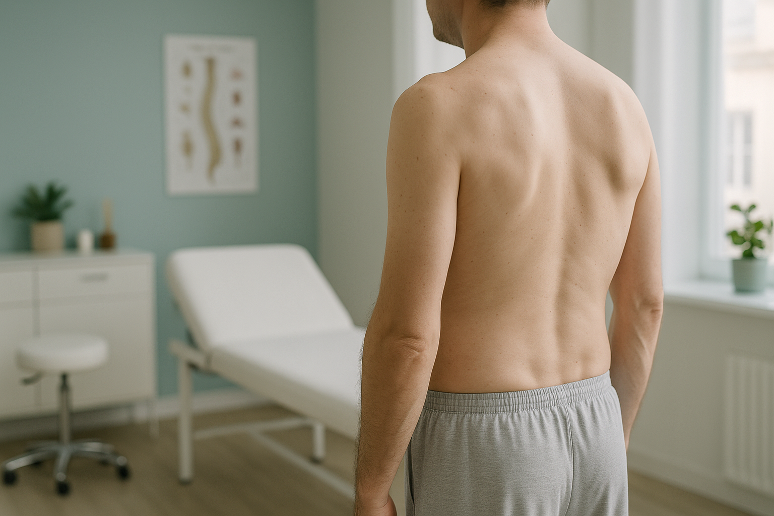
Flatback syndrome is a loss of the normal inward curve (lordosis) in the lower back. Without this curve, the spine cannot balance the head over the pelvis, so you lean forward. Standing and walking become tiring. Because this imbalance only shows under gravity, standing whole-body imaging is the clearest way to measure it.
Key takeaways
- A healthy lumbar spine curves inward. When that curve flattens, the body tilts forward.
- Fatigue, back pain and difficulty standing upright are the hallmark symptoms.
- Causes include previous spinal surgery, disc degeneration and long-term posture habits.
- Lying-down scans can miss the problem because gravity is removed.
- A standing whole-body EOS scan shows how the entire spine balances under your own weight.
What is flatback syndrome?
Flatback syndrome is a postural condition where the normal inward curve of the lumbar spine is significantly reduced or lost. A healthy lower back has a gentle lordotic curve that acts like a spring, helping you stand upright with minimal effort. When that curve flattens, the upper body shifts forward of the pelvis, and you have to work harder just to stand straight.
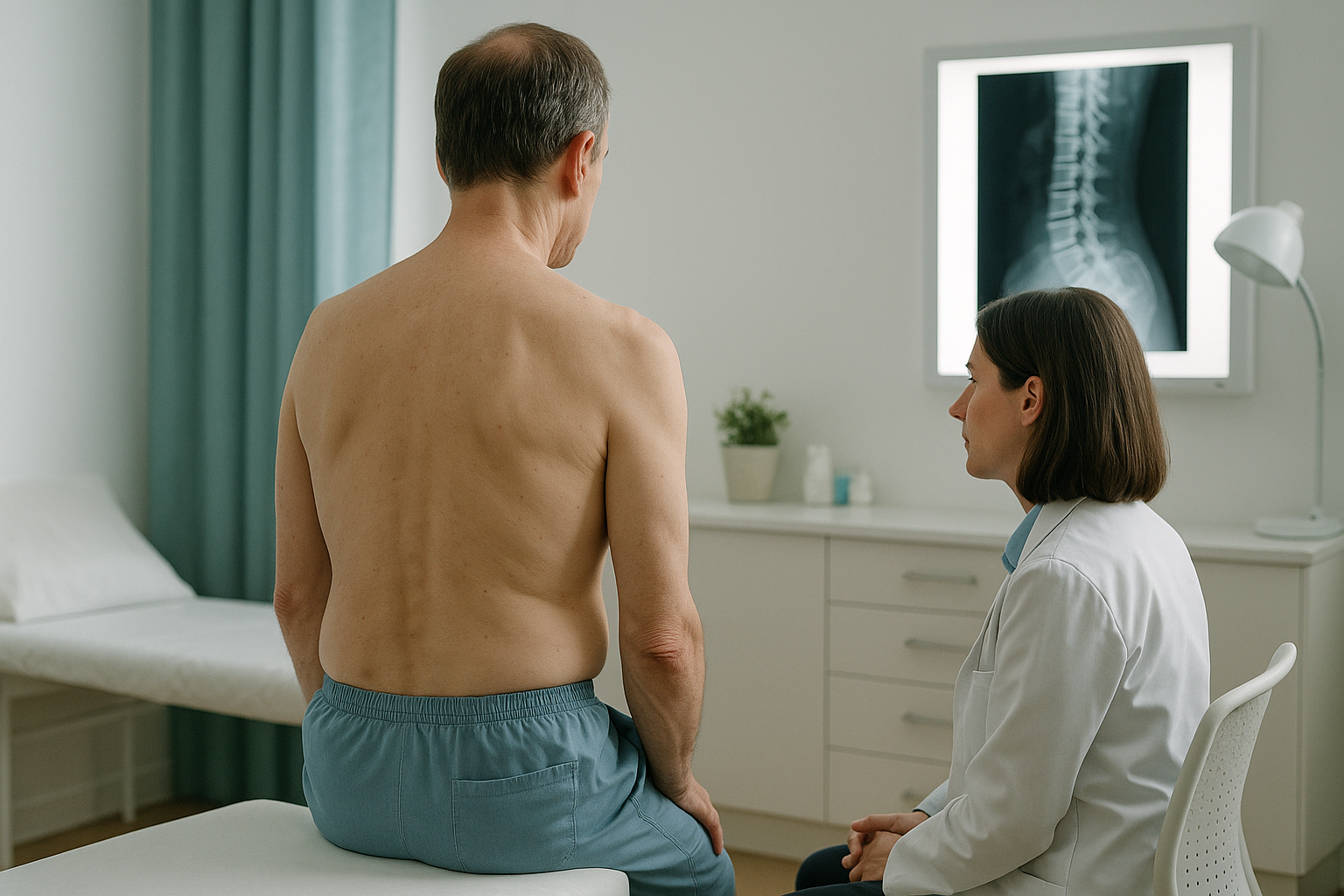
The body is a connected chain from your feet to your head. When the lower back flattens, other areas compensate. Your thoracic spine may round more, your hips and knees may flex slightly, and your muscles work overtime to keep you upright. This is why the pain you feel might be in your mid-back, hips or even your legs, not just in the lower back itself.
What are the symptoms of flatback syndrome?
The most common symptom is a feeling that you cannot stand upright without effort. Pain and fatigue tend to worsen as the day goes on because your muscles tire from constantly fighting to keep you balanced.
You feel as though you are constantly falling forward, especially when standing still or walking.
Muscles in the back, buttocks and thighs ache more as the day progresses.
You may need to stop and rest, lean on a trolley, or bend your knees to feel relief.
Symptoms often improve when you sit down, lean against something, or bend forward.
A note on nerve symptoms. If you experience numbness, tingling, weakness in your legs, or any loss of bladder or bowel control, please seek urgent medical attention from your GP, call 111, or attend A&E. These symptoms need immediate assessment.
What causes flatback syndrome?
Flatback syndrome can develop for several reasons. Sometimes it follows spinal surgery, particularly older fusion procedures. In other cases it develops gradually through disc degeneration or long-standing posture patterns.
Older fusion hardware sometimes fixed the spine in a straighter position than ideal, reducing the natural curve over time.
As discs lose height, especially at the front, the cumulative loss flattens the curve over years.
Vertebral fractures from osteoporosis can cause the front of a vertebra to collapse, gradually flattening the spine.
Spending years in a slouched or flexed position can gradually weaken the structures that maintain the curve.
In many cases, there is more than one contributing factor. A scan that captures the full spine and pelvis in a standing position helps to identify the combination of causes at play.
Why does standing imaging matter for flatback syndrome?
Standing imaging matters because flatback syndrome is fundamentally a problem of balance under gravity. When you lie down for a standard X-ray, CT or MRI, the muscles relax, the spine settles into the surface, and the degree of curve loss can appear much less severe than it really is.
An EOS scan takes a full-length image of your skeleton while you stand in your natural posture. It captures the spine, pelvis, hips, knees and ankles in one shot, so the radiologist can see exactly how each region is compensating for the lost lumbar curve. Your hips might be pushing forward, your knees might be slightly bent, or your thoracic spine might be rounding more than it should. All of these compensations become visible only when standing.
EOS also uses about 90% less radiation than a standard X-ray, around ten times less. This makes it practical for follow-up imaging if your specialist needs to track changes over time.
Want to find out whether a standing alignment scan could help explain your symptoms?
Book a free video consultationHow is flatback syndrome usually managed?
Management depends on the severity and cause. Your clinician will use the imaging findings alongside your symptoms to decide the best path. Common approaches include:
- Physiotherapy and targeted exercises. Strengthening the core and hip extensors can help support the spine and reduce muscle fatigue.
- Postural awareness. Understanding your alignment pattern can help you make adjustments in daily life.
- Pain management. Your GP or pain specialist may recommend medication, injections or other approaches for symptom relief.
- Surgical review. In more significant cases, a spinal surgeon may consider corrective surgery to restore the lumbar curve. Detailed standing imaging is typically essential for planning this.
ScanAlign is a diagnostic imaging clinic. We do not provide treatment. Our role is to give you and your clinician a clear, weight-bearing picture of your alignment so that any treatment decisions are based on the most accurate information possible.
When MRI is the better tool. EOS shows bones, joints and overall alignment. If your clinician suspects nerve compression, disc problems or soft-tissue injury, MRI is usually the more appropriate scan. The two types of imaging complement each other well.
Frequently asked questions about flatback syndrome
Yes. This is one of the most common causes. Older fusion techniques sometimes straightened the lumbar spine more than intended. The curve loss may not become noticeable for years as adjacent discs degenerate and compensations build up.
Not quite. Poor posture can contribute, but flatback syndrome usually involves structural changes in the spine itself, such as disc height loss, vertebral fractures or surgical changes. It is a medical condition, not simply a habit.
Your muscles are working constantly to keep you upright against the forward pull. By the end of the day, they fatigue, and the imbalance becomes harder to compensate for. This is why many people with flatback syndrome feel better in the morning and worse by evening.
Exercises can help manage symptoms and improve support, especially strengthening the hip extensors and core. However, if the cause is structural, exercises alone may not fully restore the curve. A proper assessment helps clarify what is realistic for your situation.
No. ScanAlign accepts self-referrals. You can book a free video consultation directly, and if a scan is appropriate, it can be arranged without waiting for a GP letter.
An EOS scan captures the entire skeleton from head to toe in one standing image, using about 90% less radiation than a standard X-ray, around ten times less. A conventional X-ray typically covers only one small area and may be taken lying down.
Yes. The radiation dose is about 90% lower than a standard X-ray, around ten times less. You can find more detail on the FAQs and safety page.
Current pricing is listed on the FAQs page. The initial video consultation is free and there is no obligation to proceed.
It can. When the lumbar curve is lost, the hips and knees often flex to help rebalance the body. This places extra load on those joints and can produce pain away from the spine itself. A whole-body scan helps identify these compensations.
ScanAlign is based at 19 Harley Street, London. It is a private self-pay clinic operating under The Harley Street Hospital's CQC licence.
Understand your alignment, standing up
A free video consultation with our specialist radiographer can help you decide whether a standing whole-body scan is the right next step for your symptoms.
Book a free video consultationLearn more about how EOS imaging works on our EOS scan overview page, or read about spine alignment assessment.
Written by Abbas Dhami (Specialist Diagnostic Radiographer)
Sources
- Glassman SD, Bridwell K, Dimar JR, Horton W, Berven S, Schwab F. The impact of positive sagittal balance in adult spinal deformity. Spine. 2005;30(18):2024-2029.
- Schwab F, Lafage V, Boyce R, Skalli W, Farcy JP. Gravity line analysis in adult volunteers: age-related correlation with spinal parameters, pelvic parameters, and foot position. Spine. 2006;31(25):E959-E967.
- NHS. Back pain: causes, treatment and when to see a GP. nhs.uk/conditions/back-pain.
This page is general information and not a diagnosis.
ScanAlign is the trading name of CoreMed Solutions Ltd and operates under The Harley Street Hospital's CQC licence.