

Hip Dysplasia in Adults: What Is It and How Do You Know You Have It?
Dysplasia in the hip means the socket is too shallow to cover the ball of the thigh bone properly. In adults, this creates extra stress on the joint, leading to groin pain, stiffness and early wear. Many people are not diagnosed until their twenties or later because symptoms build gradually and standard lying-down imaging can underestimate the problem.
Key takeaways
- Hip dysplasia is a structural problem where the hip socket does not fully cover the ball of the femur.
- It is a leading cause of hip arthritis in younger adults, often missed for years.
- Symptoms include groin pain, a feeling of instability and stiffness after sitting.
- Standing, weight-bearing imaging shows how the joint behaves under load, which lying-down scans can miss.
- Early diagnosis helps you and your clinician plan the right next steps before further joint damage occurs.
Hip dysplasia is not just a condition found in babies. Many adults live with an undiagnosed shallow hip socket for decades. The joint may cope well during your teens and twenties, but over time the uneven loading wears down cartilage faster than normal. By the time pain becomes constant, significant wear may already be present.
Understanding what dysplasia in the hip actually looks like, and how it is properly assessed, puts you in a stronger position to get the right help.
What are the signs of hip dysplasia in adults?
The most common sign is a deep ache in the groin or front of the hip, especially after walking, standing for a long time, or exercise. Many people also notice a catching or clicking sensation in the joint.
A deep ache that worsens with activity and eases with rest. Often mistaken for a muscle strain.
Your hip feels tight or locked when you get up from a chair, especially after long periods.
A mechanical click or snapping feeling during movement, often when turning or twisting the leg.
A sense that the hip might give way, especially on uneven ground or when changing direction quickly.
Some people also develop pain in the side of the hip, lower back or knee. This happens because the body compensates for the unstable joint. Pain in one place can have its true cause somewhere else in the chain.
What causes hip dysplasia in adults?
In most cases, hip dysplasia in adults is a developmental condition that has been present since birth but was never picked up. The socket did not form deeply enough during infancy. Other contributing factors include genetics, ligament laxity and, less commonly, injury or repetitive loading during growth.
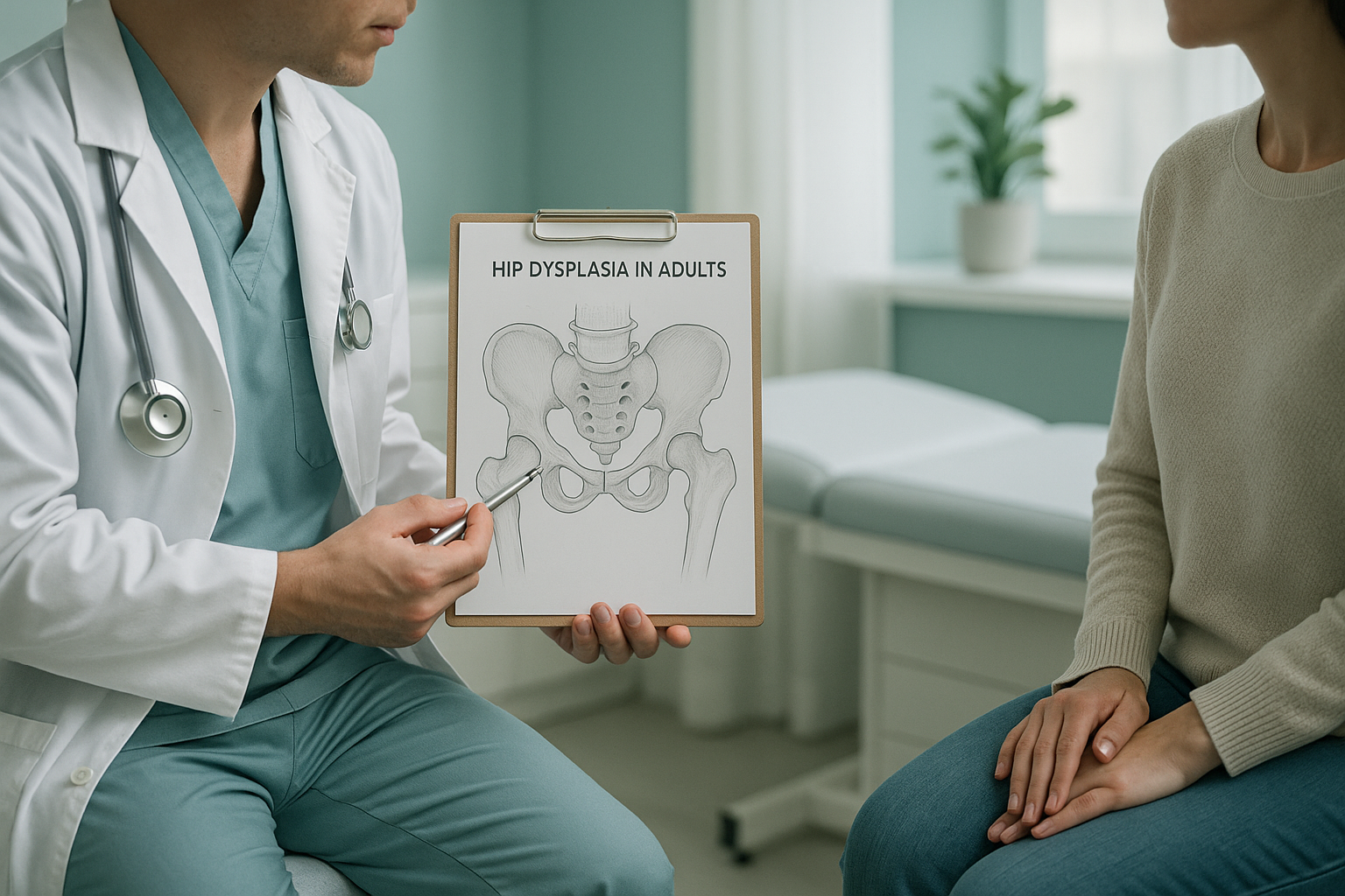
Because the socket is shallow, the ball of the thigh bone is not held as securely. This uneven contact concentrates force on a smaller area of cartilage, which accelerates wear. Hip dysplasia is one of the most common structural causes of hip arthritis in people under fifty.
It affects women more often than men, and sometimes runs in families. If a close relative has needed a hip replacement at a relatively young age, dysplasia may be worth investigating.
How is hip dysplasia diagnosed?
Diagnosis starts with a clinical examination. Your clinician will check your range of movement, test for impingement signs and ask about the pattern of your pain. Imaging is then used to confirm the shape of the socket and how well it covers the femoral head.
Standard X-rays taken lying down are useful, but they have an important limitation. When you lie flat, gravity is removed. The muscles around the hip relax, and the joint may look better aligned than it really is. The amount of socket coverage can appear adequate on a lying-down image, even when the joint is under-covered during everyday standing and walking.
This is where weight-bearing imaging adds real value. A standing X-ray captures the hip under the same load it experiences throughout your day, giving a more accurate picture of coverage and joint spacing.
When is MRI more useful? EOS shows bones, joints and alignment clearly. For soft-tissue problems such as labral tears, cartilage damage or nerve issues, MRI is usually the better tool. Many people with hip dysplasia benefit from both types of imaging.
Why does a standing EOS scan matter for hip dysplasia?
A standing EOS scan captures your entire skeleton, from head to feet, in one image while you are upright and weight-bearing. This matters for hip dysplasia because the shallow socket is under the most stress when you are standing, not when you are lying on a table.
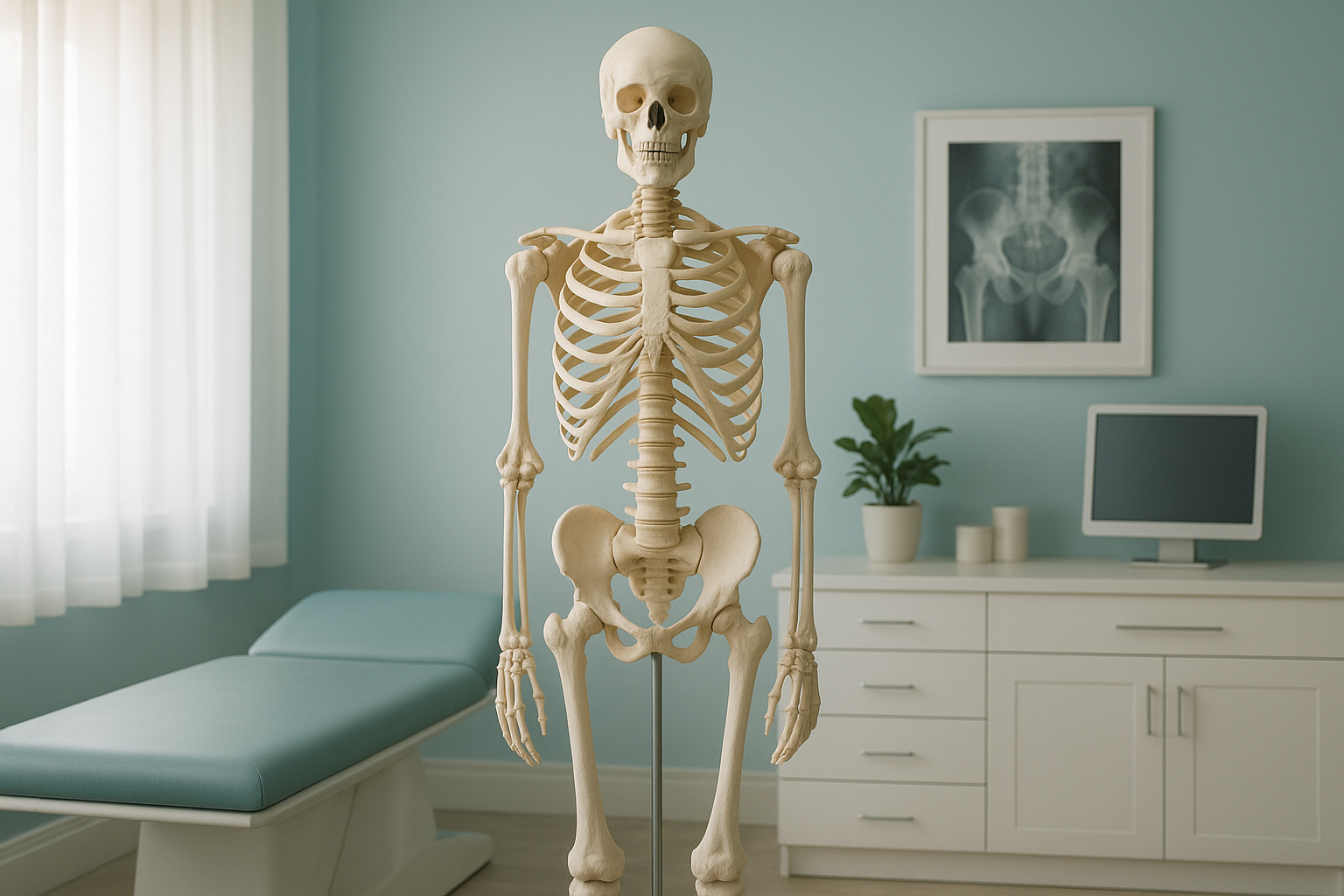
The EOS system also uses about 90% less radiation than a standard X-ray, around ten times less. That makes it a practical option if repeat imaging is needed to monitor changes over time.
Because the scan captures your full skeleton, your clinician can see whether a shallow hip is also affecting your pelvis angle, spine curvature or leg-length balance. Your body is a connected chain. A problem at the hip often forces the spine or knee to compensate, and pain may show up in those areas instead. A whole-body standing image reveals the full picture in one scan.
Want to understand how your hip alignment looks under weight? A standing EOS scan can give you and your clinician the full picture.
Book a free video consultationWhat are the next steps after a hip dysplasia diagnosis?
The right approach depends on the severity of the dysplasia, the amount of cartilage wear already present and your symptoms. Your treating clinician will use your imaging alongside clinical findings to advise you. Common pathways include:
- Physiotherapy and strengthening. Targeted exercises to improve hip stability and reduce loading on the damaged cartilage.
- Activity modification. Adjusting sports or daily habits to protect the joint while maintaining fitness.
- Surgical assessment. For more significant dysplasia, procedures such as a periacetabular osteotomy (PAO), which repositions the socket, may be discussed. In later stages where arthritis is advanced, hip replacement may be considered.
ScanAlign is a diagnostic imaging clinic. We do not provide treatment. What we do provide is the detailed, weight-bearing imaging that helps your surgeon or physiotherapist plan the most appropriate course of action. Accurate imaging early on can make a real difference to the options available to you.
Frequently asked questions about hip dysplasia
Most adult hip dysplasia has been present from birth but was not picked up during childhood screening. It is rare for the socket shape to change in adulthood, though symptoms often only appear once cartilage starts to wear.
Most people describe a deep ache at the front of the hip or groin. It tends to worsen with prolonged walking, standing or exercise and eases with rest. Some also feel a catching sensation during movement.
No. Dysplasia is the structural shape of the socket. Arthritis is the cartilage wear that can develop over time because of that shape. Dysplasia is a cause, arthritis is a potential consequence.
Yes. When the hip is unstable, other joints compensate. The pelvis may tilt, the spine may curve differently, or the knee may take extra load. Pain can appear in these areas even though the root cause is at the hip.
No referral is needed. ScanAlign is a private, self-pay service. You can book a free video consultation directly to discuss whether a scan is appropriate for you.
The EOS system uses about 90% less radiation than a standard X-ray, around ten times less. This makes it a practical choice for imaging that may need to be repeated over time.
When you lie down, gravity is removed and the muscles relax. The hip joint may appear better covered than it truly is during daily life. A standing scan shows the socket under the same load it experiences when you walk, stand and move.
Pricing information is available on our FAQs and cost page.
In milder cases, physiotherapy, strengthening and activity modification can manage symptoms effectively. Your treating clinician will advise based on the severity of the dysplasia and any cartilage changes visible on imaging.
You stand inside the EOS scanner at our Harley Street clinic. The scan takes about 20 seconds and captures your full skeleton in one image. A specialist radiographer oversees the process. Your results and report are shared with you and, if you wish, with your treating clinician. More details are on our what to expect page.
See your hip alignment under real-world load
A free video consultation lets you discuss your symptoms with our team and find out whether a standing EOS scan is the right next step for you.
Book a free video consultationWritten by Abbas Dhami (Specialist Diagnostic Radiographer)
Sources
- NHS, "Hip dysplasia," nhs.uk, 2023.
- International Hip Dysplasia Institute, "Adult hip dysplasia overview," hipdysplasia.org.
- Clohisy JC, et al., "A systematic approach to the plain radiographic evaluation of the young adult hip," The Journal of Bone & Joint Surgery, 2008;90(Suppl 4):47-66.
This page is general information and not a diagnosis.
ScanAlign is the trading name of CoreMed Solutions Ltd and operates under The Harley Street Hospital's CQC licence.