
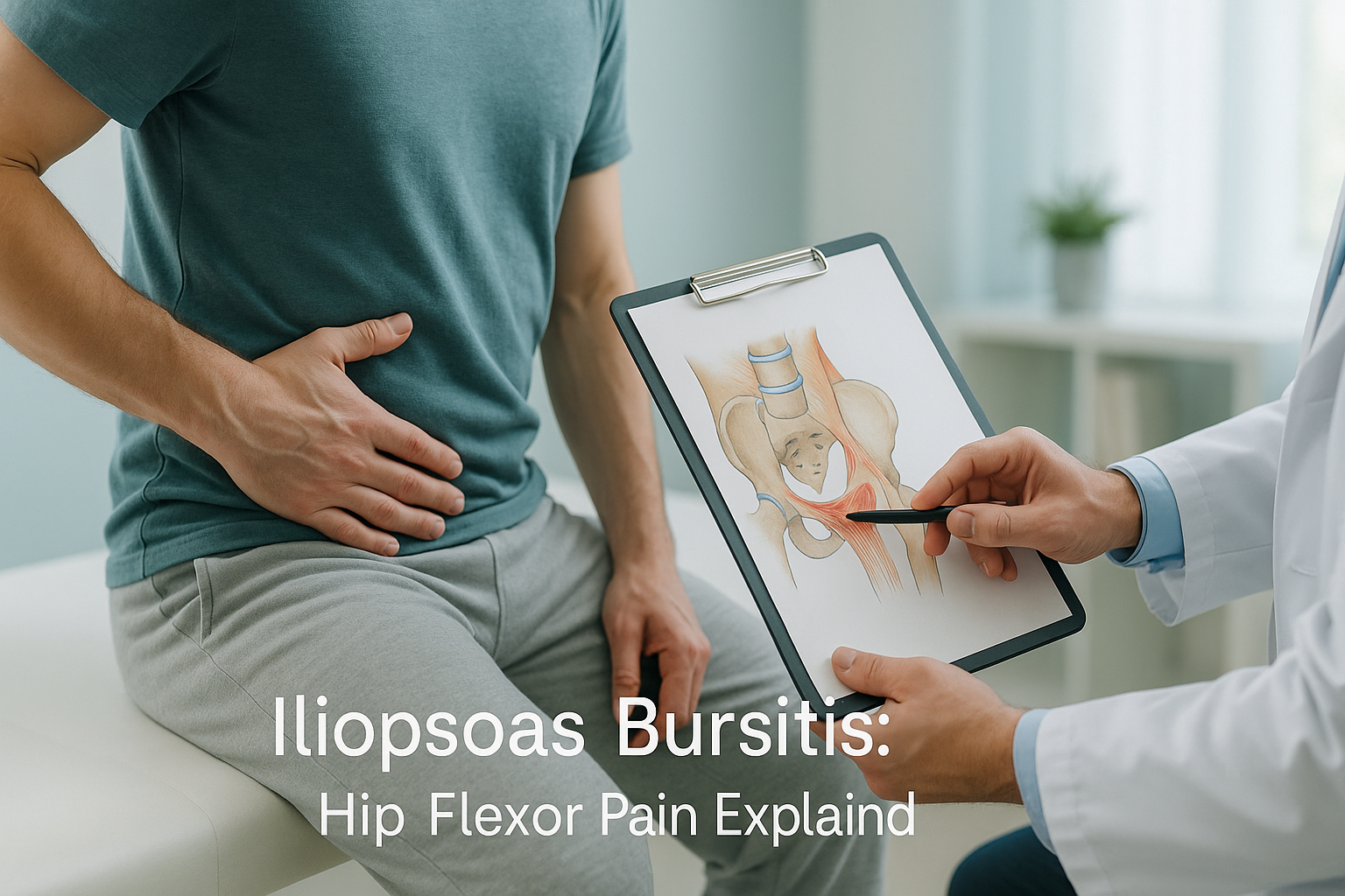
Iliopsoas Bursitis: What Is It and Why Does Your Hip Flexor Hurt?
Iliopsoas bursitis is inflammation of a small, fluid-filled sac that sits between the iliopsoas muscle and the front of the hip joint. It causes a deep ache or sharp pain in the groin or front of the hip, often made worse by walking, climbing stairs, or sitting for a long time. It is sometimes linked to snapping hip syndrome or iliopsoas tendinitis.
Key takeaways
- Iliopsoas bursitis causes pain at the front of the hip, not the side or back.
- It often develops alongside iliopsoas tendinitis or snapping hip syndrome.
- Lying-down scans can miss alignment problems that contribute to the strain.
- Standing whole-body imaging shows how your pelvis, spine, and legs work together under load.
- ScanAlign provides diagnostic imaging only, not treatment.
The iliopsoas is a powerful hip flexor muscle. It runs from the lower spine, through the pelvis, and attaches to the top of the thigh bone. A bursa (a thin, slippery cushion) sits between the muscle's tendon and the bone to reduce friction. When that bursa becomes irritated and swollen, the result is iliopsoas bursitis.
This condition is sometimes called hip flexor bursitis. It can overlap with iliopsoas tendinitis, where the tendon itself is inflamed, or with iliopsoas syndrome, a broader term covering pain from the muscle, tendon, or bursa. In some people, the tendon flicks over a bony prominence and produces an audible click. That is known as snapping hip syndrome.
What does iliopsoas bursitis feel like?
Iliopsoas bursitis typically produces a deep, nagging ache in the groin or the front of the hip. The pain is often hard to pinpoint with one finger. It can be sharp when you first stand up after sitting, then settle into a dull throb as you keep moving.
A deep ache that feels like it comes from inside the joint, often worse after rest.
Lifting the knee against gravity loads the iliopsoas and presses on the inflamed bursa.
An audible snap as the tendon catches over the front of the hip, sometimes called snapping hip syndrome.
The hip flexor shortens while you sit. Standing up stretches the inflamed area and triggers pain.
When to seek urgent help. If you develop sudden severe hip pain with numbness, weakness in the leg, or any loss of bladder or bowel control, contact your GP, call 111, or go to A&E straight away. These symptoms need immediate medical attention.
What causes iliopsoas bursitis?
Iliopsoas bursitis is usually caused by repetitive friction between the iliopsoas tendon and the front of the hip joint. Anything that increases that friction, whether it is overuse, alignment imbalance, or a change to the joint itself, can trigger it.
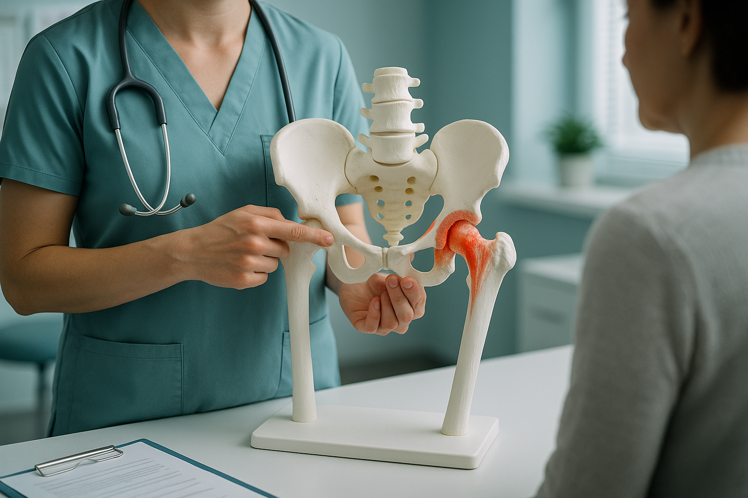
Running, cycling, dancing, or any exercise that repeatedly flexes the hip can irritate the bursa over time.
A tilted pelvis or curved spine can put extra load on one hip flexor, increasing friction on the bursa.
Hours in a chair shortens the iliopsoas. When you stand, the tight muscle tugs harder on the bursa.
Changes to joint geometry after surgery can alter how the tendon tracks, sometimes causing iliopsoas bursitis after hip replacement.
Because the iliopsoas crosses both the spine and the hip, problems further up or down the body can contribute. A leg-length difference, a stiff lower back, or a rotated pelvis can all shift how the muscle loads during movement. That is why looking at one area in isolation sometimes misses the bigger picture.
How does a standing scan help with anterior hip pain?
A standing, weight-bearing scan shows your bones and joints in the position that actually causes symptoms. When you lie down for a conventional X-ray or CT, gravity is removed, muscles relax, and small alignment shifts can disappear. That means the scan may look normal even when something is clearly wrong when you are on your feet.
EOS imaging captures a full-length view from your ankles to your skull in a single standing exposure, using about 90% less radiation than a standard X-ray, around ten times less. The image reveals how your spine, pelvis, hips, and legs relate to each other under real load.
For someone with iliopsoas bursitis, that whole-body view matters. If a pelvic tilt is putting extra strain on one hip flexor, or if a subtle leg-length difference is forcing one side to overwork, those patterns show up on a standing scan. A scan of the hip alone, taken lying down, may miss them entirely.
ScanAlign uses EOS technology at 19 Harley Street to provide this kind of full-body standing assessment. You do not need a referral.
Book a free video consultationEOS and MRI do different jobs. EOS shows bones, joints, and alignment beautifully. For soft tissue detail, such as the bursa itself, tendons, or nerves, MRI is usually the better tool. Your clinician may recommend both, depending on your situation.
What can be done about iliopsoas bursitis?
Treatment for iliopsoas bursitis is managed by your GP, physiotherapist, or orthopaedic specialist. ScanAlign does not provide treatment. What it does provide is the detailed alignment information that helps those clinicians make better decisions.
Common approaches include physiotherapy to stretch and strengthen the hip flexor, activity modification to reduce irritation, and sometimes guided injections. If the bursitis has developed because of an underlying alignment problem, addressing that root cause is often key to lasting improvement.
For people experiencing iliopsoas bursitis after hip replacement, standing alignment imaging can help the surgical team assess whether the new joint's position is contributing to the tendon irritation. This is information that a lying-down X-ray may not capture accurately.
Understanding how your whole body lines up, from your feet through your knees, hips, pelvis, and spine, gives your treatment team a clearer starting point. Pain in the front of the hip does not always start at the hip. Learn more about how hip alignment assessment works.
Frequently asked questions about iliopsoas bursitis
Bursitis is inflammation of the bursa, the fluid-filled cushion near the tendon. Tendinitis is inflammation of the tendon itself. They often occur together because irritation of one structure tends to affect the other. The term iliopsoas syndrome is sometimes used to cover both.
They are closely related. Snapping hip syndrome happens when the iliopsoas tendon catches over a bony prominence at the front of the hip. That repeated snapping can inflame the bursa underneath, leading to bursitis. Some people have the snap without pain, while others develop significant discomfort.
Sitting shortens the iliopsoas muscle. When you stand, the muscle stretches and the tendon pulls across the inflamed bursa, which can cause a sharp jolt of pain. This pattern of pain after prolonged sitting is one of the hallmarks of iliopsoas bursitis.
Yes. After hip replacement surgery, the geometry of the joint changes. This can alter how the iliopsoas tendon tracks over the front of the hip, sometimes increasing friction on the bursa. Standing alignment imaging can help assess whether the implant position is contributing.
No. ScanAlign is a private, self-pay clinic. You can book directly without a GP or consultant referral. You start with a free video consultation to discuss whether the scan is right for you.
EOS uses about 90% less radiation than a standard X-ray, around ten times less. It is one of the lowest-dose skeletal imaging options available. You can find more detail on the FAQs and safety page.
ScanAlign is a private, self-pay service. Current pricing information is available on the cost and FAQs page.
A lying-down image removes gravity. Muscles relax and the skeleton settles into a position it does not hold in real life. A standing scan captures your alignment under full body weight, showing tilts, shifts, and compensations that only appear when you are upright. Learn more about how EOS whole-body imaging works.
Yes. The iliopsoas attaches to the lower spine and crosses the hip. If the lower back is excessively curved or the pelvis is tilted, the muscle may be held in a shortened or overloaded position. That extra strain increases friction on the bursa. A full standing image of the spine and pelvis together can reveal this connection.
EOS is excellent at showing bones, joints, and whole-body alignment. The bursa itself is a soft-tissue structure that is best seen on MRI or ultrasound. However, EOS can reveal the alignment factors that may be causing the bursa to become irritated in the first place.
Find out what is behind your hip flexor pain
A free video consultation lets you discuss your symptoms with our team and find out whether a standing alignment scan is the right next step.
Book a free video consultationSources
- NHS, "Hip pain in adults," nhs.uk (accessed 2024).
- Domb BG, Shindle MK, McArthur B, et al. "Iliopsoas impingement: a newly identified cause of labral pathology in the hip." HSS Journal, 7(2), 2011.
- Illés T, Somoskeöy S. "The EOS imaging system and its uses in daily orthopaedic practice." International Orthopaedics, 36(7), 2012.
Written by Abbas Dhami (Specialist Diagnostic Radiographer)
This page is general information and not a diagnosis.
ScanAlign is the trading name of CoreMed Solutions Ltd and operates under The Harley Street Hospital's CQC licence.