
What Is Bursitis? Inflammation of a Bursa Explained
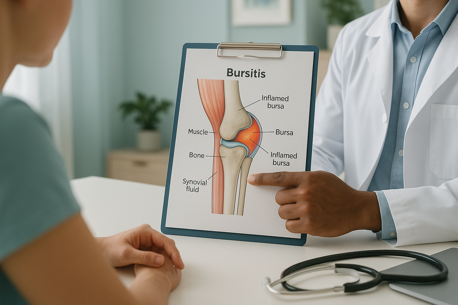
Bursitis is the inflammation of a bursa, a small fluid-filled sac that sits between bones, tendons and muscles around your joints. Its job is to reduce friction. When a bursa becomes irritated, it swells and causes localised pain, stiffness and tenderness. Bursitis most commonly affects the hip, knee, shoulder and elbow.
Key takeaways
- A bursa is a cushioning sac. Inflammation of a bursa is called bursitis.
- Common sites are the hip, knee, shoulder and elbow.
- Repetitive movement, prolonged pressure or poor alignment can all trigger it.
- Pain from bursitis sometimes has a hidden cause elsewhere in the body's alignment chain.
- Standing whole-body imaging can reveal alignment issues that lying-down scans may miss.
You have over 150 bursae in your body. Most of the time you never notice them. They sit quietly at your joints, providing a slippery cushion so tendons and muscles glide smoothly over bone. But when one becomes inflamed, the pain can be surprisingly sharp and persistent.
Bursitis is often treated as a simple overuse injury. Rest, ice, anti-inflammatories. That works for many people. But when bursitis keeps coming back, or when standard treatment does not settle it, there is usually a deeper question worth asking: why is that particular bursa under so much stress in the first place?
What does bursitis feel like?
Bursitis typically causes a dull ache or tenderness around the affected joint, which often worsens with movement or pressure. You may also notice swelling, warmth or stiffness in the area.
A deep ache at or near the joint, often worse when you press on it.
Visible puffiness over the bursa, particularly at the knee or elbow.
Reduced range of movement, especially first thing in the morning.
The skin over the bursa may feel warm, sometimes with mild redness.
A note on red flags. If your joint is very hot, severely swollen, you have a fever, or you notice sudden weakness or loss of function, seek urgent medical attention. Contact your GP, call 111, or go to A&E. These may indicate infection (septic bursitis) or another serious condition.
What causes bursitis?
Bursitis is most often caused by repetitive movement or sustained pressure on a joint. But poor overall alignment is a commonly overlooked trigger. When your body is out of balance, certain joints absorb more load than they should, and the bursae at those joints pay the price.
Running, kneeling, throwing or any activity that loads the same joint repeatedly.
Leaning on elbows, kneeling on hard surfaces, or sitting for long periods.
A pelvic tilt or uneven leg length shifts load onto one side, stressing specific bursae.
Arthritis, gout or previous injury can irritate nearby bursae over time.
Your body works as a connected chain, from your feet up through your knees, hips, spine and head. When one part is out of position, others compensate. A subtle imbalance in the pelvis, for example, may overload the bursa at your hip or knee. That is why the pain often shows up away from its true cause.
Where does bursitis occur most often?
Bursitis can develop at almost any joint, but some sites are far more common than others. Below are the areas most frequently affected.
Pain on the outside of the hip, often worse when lying on that side or climbing stairs.
Swelling and tenderness at the front of the kneecap. Common in people who kneel frequently.
A visible lump at the tip of the elbow, sometimes called "student's elbow".
Tenderness below and to the inside of the knee, often linked to altered leg alignment.
How does standing imaging help with bursitis?
Standing whole-body imaging shows your skeleton under the same gravitational load you experience every day. This is important because bursitis is often a symptom of a broader alignment problem that only appears when you are upright and weight-bearing.
When you lie down for a conventional X-ray, CT or MRI, your muscles relax and your joints decompress. A leg-length difference, a pelvic tilt, or a subtle shift in the way your knee tracks over your foot can all flatten out on the table, and the scan looks normal. But standing up, those same imbalances come back, and one bursa takes the brunt.

An EOS scan captures your full skeleton from head to feet in a single standing image, using about 90% less radiation than a standard X-ray, around ten times less. Your consultant can then see the complete alignment picture and understand whether a structural imbalance is contributing to the bursa irritation.
If bursitis keeps returning, a standing whole-body assessment may help identify the underlying cause.
Book a free video consultationWhen MRI is the better choice. EOS shows bones, joints and overall alignment. For soft tissue, discs, nerves or confirming whether a bursa itself is inflamed, MRI is usually the better tool. In many cases both types of imaging work together to give the full picture.
What helps manage bursitis?
Most bursitis settles with conservative steps. Rest, avoiding the aggravating activity, ice and over-the-counter anti-inflammatory medication are the standard first-line approach. Physiotherapy can also help strengthen the muscles around the joint and reduce future flare-ups.
When bursitis recurs despite these measures, the question shifts from "how do I calm this down?" to "why does it keep happening?" That is where understanding your overall alignment becomes valuable. If a structural imbalance is quietly overloading one side of your body, addressing the alignment issue may reduce the repetitive stress on the affected bursa.
ScanAlign is a diagnostic imaging clinic. It does not provide treatment. What it does provide is the detailed alignment information your treating clinician needs to plan the next step, whether that involves physiotherapy, orthotics, injection therapy or surgical referral.
For more about what the imaging involves, visit the EOS scan overview.
Frequently asked questions about bursitis
Bursitis is inflammation of a bursa (a fluid-filled cushioning sac), while tendonitis is inflammation of a tendon (the cord connecting muscle to bone). They often occur at the same joint and can feel similar, but they affect different structures.
Acute bursitis may settle within a few weeks with rest and appropriate care. Chronic bursitis, where the bursa becomes repeatedly irritated, can persist for months. The duration often depends on whether the underlying cause is identified and addressed.
Yes. When your body is out of alignment, certain joints take on extra load. A pelvic tilt, uneven leg length or altered gait can all place repetitive stress on a bursa, even without an obvious injury.
Recurrent bursitis often means the root cause has not been resolved. If a structural imbalance is quietly overloading one joint, the bursa will keep flaring up no matter how many times you rest it. Whole-body standing imaging can help identify these hidden causes.
No. ScanAlign is a private self-pay clinic, so you can self-refer. You start with a free video consultation to discuss your symptoms and confirm whether a standing scan is appropriate for you.
Yes. The EOS system uses about 90% less radiation than a standard X-ray, around ten times less. You can find more detail on the FAQs and safety page.
Pricing information is available on the FAQs page. A free video consultation is always the first step, so you can confirm the scan is right for you before committing.
The EOS scan shows bones, joints and whole-body alignment rather than soft tissue inflammation. It will not image the bursa itself, but it can reveal the structural misalignment that may be causing the bursa to become irritated. For soft-tissue detail, MRI is the usual choice.
When you lie down, gravity is removed and your muscles relax. Alignment problems that appear when you stand and bear weight can flatten out, making the scan look normal. Standing imaging captures your skeleton under real-world load, showing issues that lying-down scans can miss.
ScanAlign is at 19 Harley Street, London, within The Harley Street Hospital. You can begin with a free video consultation from anywhere before attending in person.
Bursitis that keeps returning deserves a closer look
A free video consultation lets you discuss your symptoms and find out whether a standing whole-body assessment could help identify the cause.
Book a free video consultationWritten by Abbas Dhami (Specialist Diagnostic Radiographer)
Sources
- NHS. "Bursitis." nhs.uk. Available at: https://www.nhs.uk/conditions/bursitis/
- American Academy of Orthopaedic Surgeons (AAOS). "Hip Bursitis." orthoinfo.aaos.org.
- Dubousset J, Charpak G, Dorion I, et al. "A new 2D and 3D imaging approach to musculoskeletal physiology and pathology with low-dose radiation in standing position: the EOS system." Bulletin de l'Académie Nationale de Médecine, 2005;189(2):287-297.
This page is general information and not a diagnosis.
ScanAlign is the trading name of CoreMed Solutions Ltd and operates under The Harley Street Hospital's CQC licence.