
Infrapatellar Bursitis: What Is It and How Is It Treated?
Infrapatellar bursitis is swelling of a small fluid-filled sac (bursa) just below the kneecap. Treatment usually begins with rest, ice and avoiding kneeling. If symptoms persist, imaging can check for deeper causes, including alignment problems in the leg that put extra load on the knee.
Key takeaways
- A bursa is a tiny cushion between bone and soft tissue. When the one below your kneecap gets irritated, it swells and hurts.
- Repeated kneeling is the most common trigger, which is why this condition is sometimes called clergyman's knee or pastor's knee.
- There are two types: superficial (just under the skin) and deep (behind the patellar tendon).
- Most cases settle with simple measures, but persistent pain may point to a leg alignment issue that keeps overloading the knee.
- Standing, weight-bearing imaging can reveal alignment problems that lying-down scans miss.
If you have swelling or tenderness just below the kneecap that does not go away, understanding what is happening inside the joint is the first step. This page explains what infrapatellar bursitis is, what causes it and how it is managed. It also covers when alignment imaging may be worth considering.
What are the symptoms of infrapatellar bursitis?
The main symptoms are pain, swelling and tenderness in the area just below the kneecap. You may notice a soft, puffy lump at the front of the knee. Pain usually gets worse when you kneel, squat or press on the area.
If your knee is red, hot and very painful, or you feel unwell with a fever, seek medical attention promptly. This could indicate an infection in the bursa (septic bursitis), which needs urgent care from your GP or A&E.
What causes infrapatellar bursitis?
Repeated pressure or friction on the front of the knee is the most common cause. The bursa becomes irritated, inflames and fills with extra fluid. Some people are more at risk because of their occupation, sport or the way their legs are aligned.
What is the difference between superficial and deep infrapatellar bursitis?
There are two infrapatellar bursae at the front of each knee. They sit in slightly different places, and the distinction matters because it affects how the problem feels and how it is investigated.
- Sits just under the skin, in front of the patellar tendon
- Swelling is usually visible and easy to feel
- Caused mainly by direct pressure from kneeling
- This is the classic "clergyman's knee" presentation
- Sits behind the patellar tendon, between the tendon and the shinbone
- Swelling is deeper and harder to see from outside
- Often linked to overuse, running or jumping
- Can be confused with patellar tendonitis on examination
How is infrapatellar bursitis different from patellar tendonitis?
Both conditions cause pain just below the kneecap, but they involve different structures. Patellar tendonitis (sometimes called jumper's knee) is irritation of the tendon itself. Infrapatellar bursitis is inflammation of the fluid-filled cushion next to or behind that tendon. The treatment approach can differ, so getting the right diagnosis matters.
With bursitis, you tend to feel pain mainly when pressing on the area or kneeling. With tendonitis, pain usually flares during activities that load the tendon, such as jumping, running or climbing stairs. A clinician can often tell the difference through examination, but imaging helps confirm which structure is involved.
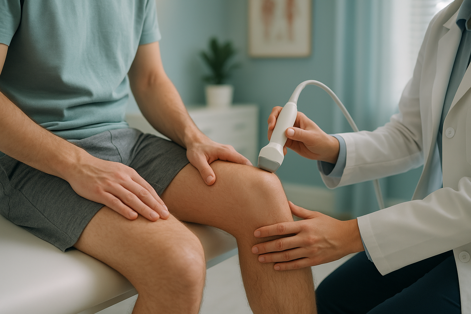
When is MRI more helpful? EOS and weight-bearing imaging are excellent for showing bone position and joint alignment. However, for soft tissue detail, such as confirming whether a bursa or a tendon is the problem, MRI is usually the better tool. Your clinician can advise which type of imaging you need.
What is the treatment for infrapatellar bursitis?
Treatment for infrapatellar bursitis usually starts with simple, conservative measures. Most cases improve within a few weeks once the source of irritation is removed.
Common approaches
- Rest and activity modification. Avoid kneeling or putting direct pressure on the front of the knee.
- Ice. Applying a cold pack for short periods can help reduce swelling and pain.
- Knee pads or cushioning. If your work or hobby requires kneeling, padded knee protection can prevent further irritation.
- Anti-inflammatory medication. Over-the-counter options (taken as directed) may ease pain and swelling. Speak to your pharmacist or GP.
- Physiotherapy. Targeted exercises can strengthen the muscles around the knee and improve how load is distributed.
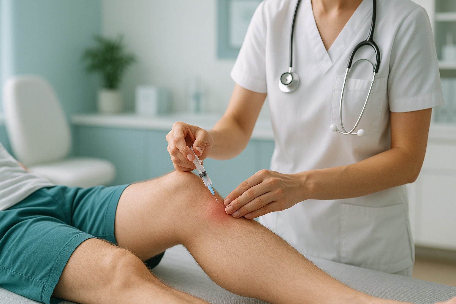
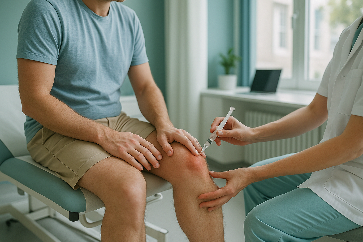
In persistent or severe cases, a clinician may consider draining the fluid (aspiration) or a corticosteroid injection. Surgery to remove the bursa is uncommon and usually reserved for cases that do not respond to other measures. Your GP or specialist will guide you on next steps.
ScanAlign does not provide treatment. It is a diagnostic imaging service. The imaging and alignment assessment it provides can help your treating clinician understand why the problem keeps coming back.
How does leg alignment affect infrapatellar bursitis?
Your leg works as a connected chain, from hip to ankle. If one part of that chain is out of alignment, the knee can end up absorbing more force than it should. A subtle tilt in the pelvis, a rotated thighbone or a difference in leg length can all change the angle at which your kneecap tracks, putting extra pressure on the bursa beneath it.
This is why some people find their bursitis keeps returning even after treatment. The irritation stops temporarily, but the underlying mechanical cause remains.
Why standing imaging matters
Most scans are taken while you lie down. In that position, gravity is removed, muscles relax and your joints settle into a different position compared with standing. A misalignment that causes problems when you walk or kneel may not be visible when you are lying flat.
An EOS scan captures your entire leg, from hip to ankle, while you stand. It uses about 90% less radiation than a standard X-ray, around ten times less. Because it images you upright and weight-bearing, it shows the real-world alignment your knee experiences every day. This gives your treating clinician a much clearer picture of whether alignment is contributing to your bursitis.
If your infrapatellar bursitis keeps coming back, a standing alignment assessment could reveal a mechanical cause that other imaging has missed.
Book a free video consultationFrequently asked questions about infrapatellar bursitis
Clergyman's knee is another name for infrapatellar bursitis. It refers to swelling of the bursa just below the kneecap, typically caused by prolonged kneeling on hard surfaces. It is also sometimes called pastor's knee.
Most cases improve within a few weeks once you stop the activity that caused it. Chronic or recurring cases can take longer and may benefit from further investigation to find an underlying cause.
Low-impact activities like swimming or cycling are usually manageable if they do not cause pain. Avoid exercises that involve kneeling, deep squats or direct pressure on the front of the knee until the swelling settles. Your physiotherapist can help you find safe alternatives.
No. Prepatellar bursitis (housemaid's knee) involves the bursa on top of the kneecap, while infrapatellar bursitis affects the bursa below the kneecap. They are separate conditions, though both can result from kneeling.
No. ScanAlign is a private, self-pay clinic. You can refer yourself without a GP or consultant referral. Your first step is a free video consultation to discuss whether an alignment assessment is appropriate for you.
Pricing details are available on the ScanAlign FAQs and cost page. The free video consultation will also cover what is included.
Yes. EOS uses about 90% less radiation than a standard X-ray, around ten times less. It is a very low-dose imaging system. You can read more on the FAQs page.
Yes. If your leg alignment is off, the knee joint may absorb uneven forces. Over time, this can irritate the bursae, tendons and other structures around the knee. Correcting the underlying alignment issue may help prevent the bursitis from returning.
You stand inside the EOS scanner in a natural, upright position. The machine captures a full-length image of your skeleton in under a minute. The image is then analysed for alignment from your spine down to your ankles.
See your GP or visit A&E if the knee is very red, hot or swollen, if you have a fever, or if you cannot bear weight on it. These may be signs of infection or a more serious injury that needs prompt attention.
Wondering why your knee bursitis keeps coming back?
A standing alignment assessment can show whether your leg mechanics are putting extra stress on your knee. Start with a free, no-obligation video consultation.
Book a free video consultationRelated reading
Sources
- NHS. "Bursitis." nhs.uk, National Health Service, 2023.
- Rishor-Olney, C.R. and Pozun, A. "Prepatellar Bursitis." StatPearls, National Library of Medicine, 2023.
- Illés, T. and Somoskeöy, S. "The EOS imaging system and its uses in daily orthopaedic practice." International Orthopaedics, 36(7), 2012, pp. 1437–1443.
Written by Abbas Dhami (Specialist Diagnostic Radiographer)
This page is general information and not a diagnosis.
ScanAlign is the trading name of CoreMed Solutions Ltd and operates under The Harley Street Hospital's CQC licence.