
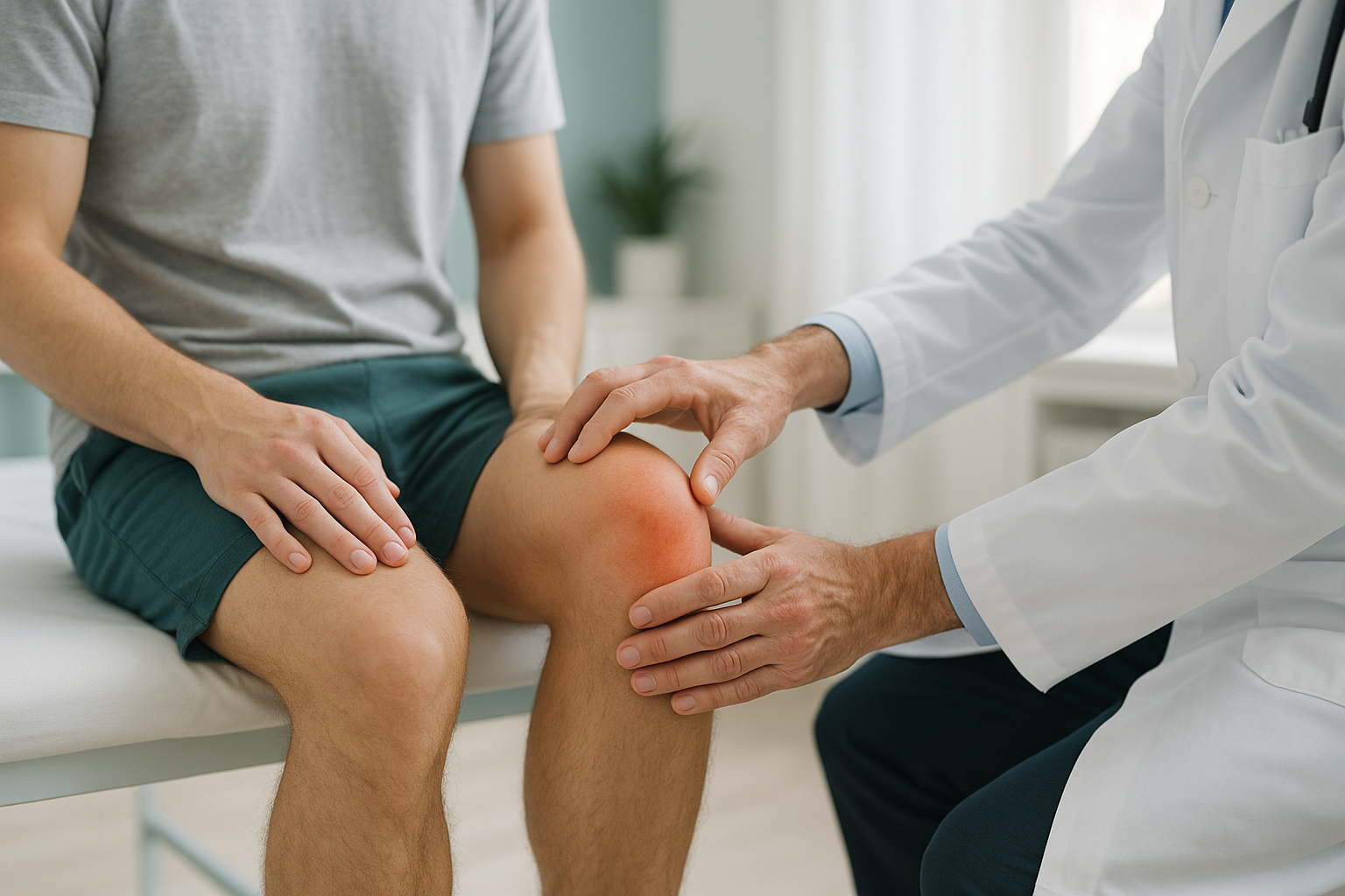
Patellar Tendonitis: What Causes It and How Do You Recover?
Patellar tendonitis is pain and inflammation in the tendon that connects your kneecap to your shinbone. It usually comes from repetitive stress on the knee, especially jumping or running. Recovery focuses on load management, strengthening exercises and identifying any alignment factors that may be adding extra strain to the tendon.
Key takeaways
- Patellar tendonitis is an overuse injury of the tendon just below the kneecap.
- Jumping, running and sudden increases in training load are common triggers.
- Leg alignment, hip rotation and foot position can all increase tendon strain.
- Standing, weight-bearing imaging can reveal alignment problems that lying-down scans miss.
- EOS imaging shows bones, joints and alignment. For soft-tissue detail, MRI is usually better.
The patellar tendon is a thick band of tissue that runs from the bottom of your kneecap (patella) to the top of your shinbone (tibia). Every time you bend or straighten your knee, this tendon is working. When it is repeatedly overloaded, tiny areas of damage build up faster than the body can repair them. The result is pain, stiffness and tenderness at the front of the knee, just below the kneecap.
It is often called "jumper's knee" because it is especially common in sports that involve a lot of jumping, such as basketball, volleyball and athletics. But you do not have to be an athlete to develop it. Anyone who rapidly increases their activity level, or who has alignment issues that place extra load on the tendon, can be affected.
What does patellar tendonitis feel like?
Patellar tendonitis typically starts as a dull ache at the front of the knee, right at the point where the kneecap meets the tendon below it. In the early stages the pain may only appear during or just after activity. If it progresses, it can become constant.
A sharp or aching pain at the very bottom of the kneecap, worse during activity.
The knee feels stiff and sore after sitting for a long time or first thing in the morning.
Going downstairs, squatting or lunging is particularly uncomfortable.
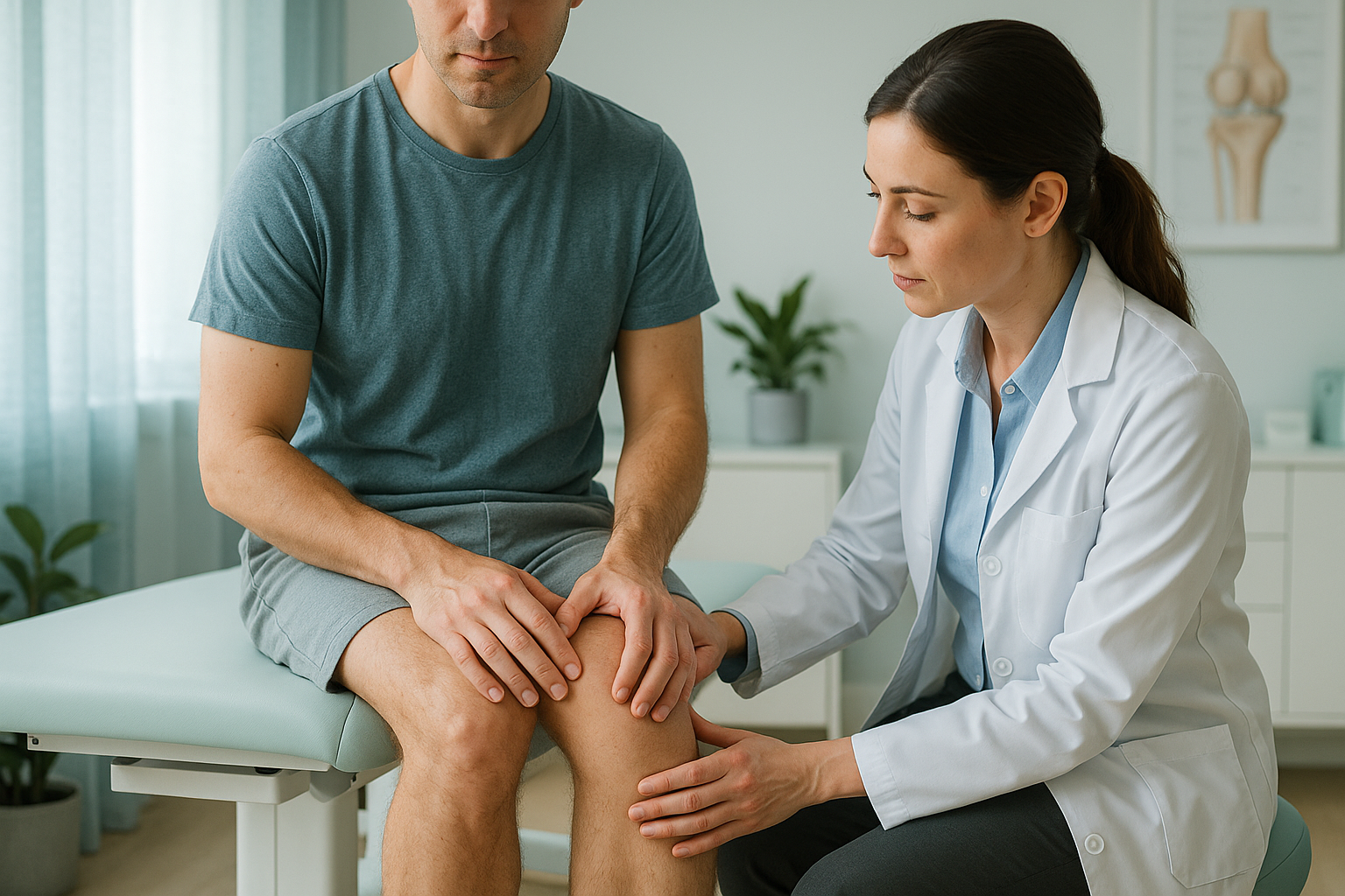
Pressing on the tendon just below the kneecap feels sore or tender.
What causes patellar tendonitis?
Patellar tendonitis is caused by repeated overloading of the patellar tendon, usually from activities that involve jumping, sprinting or sudden changes of direction. But the load on your tendon is not just about what you do. It is also shaped by how your body is aligned.
A sudden increase in volume, intensity, or jumping frequency.
Knock knees, bowed legs or rotational differences can shift load onto the tendon.
Tight quadriceps, weak glutes or hamstring imbalance increase patellar tendon stress.
Training on concrete, hard courts or poorly cushioned flooring.
Your body works as a connected chain from your feet to your spine. If your hips are slightly rotated, or one leg is a fraction longer than the other, the kneecap may track differently on one side. That places more strain on the patellar tendon even during everyday walking, not just sport.
What are the stages of patellar tendonitis?
Patellar tendonitis tends to progress through recognised stages, from mild discomfort that settles quickly to persistent pain that limits daily life. Clinicians often describe four stages.
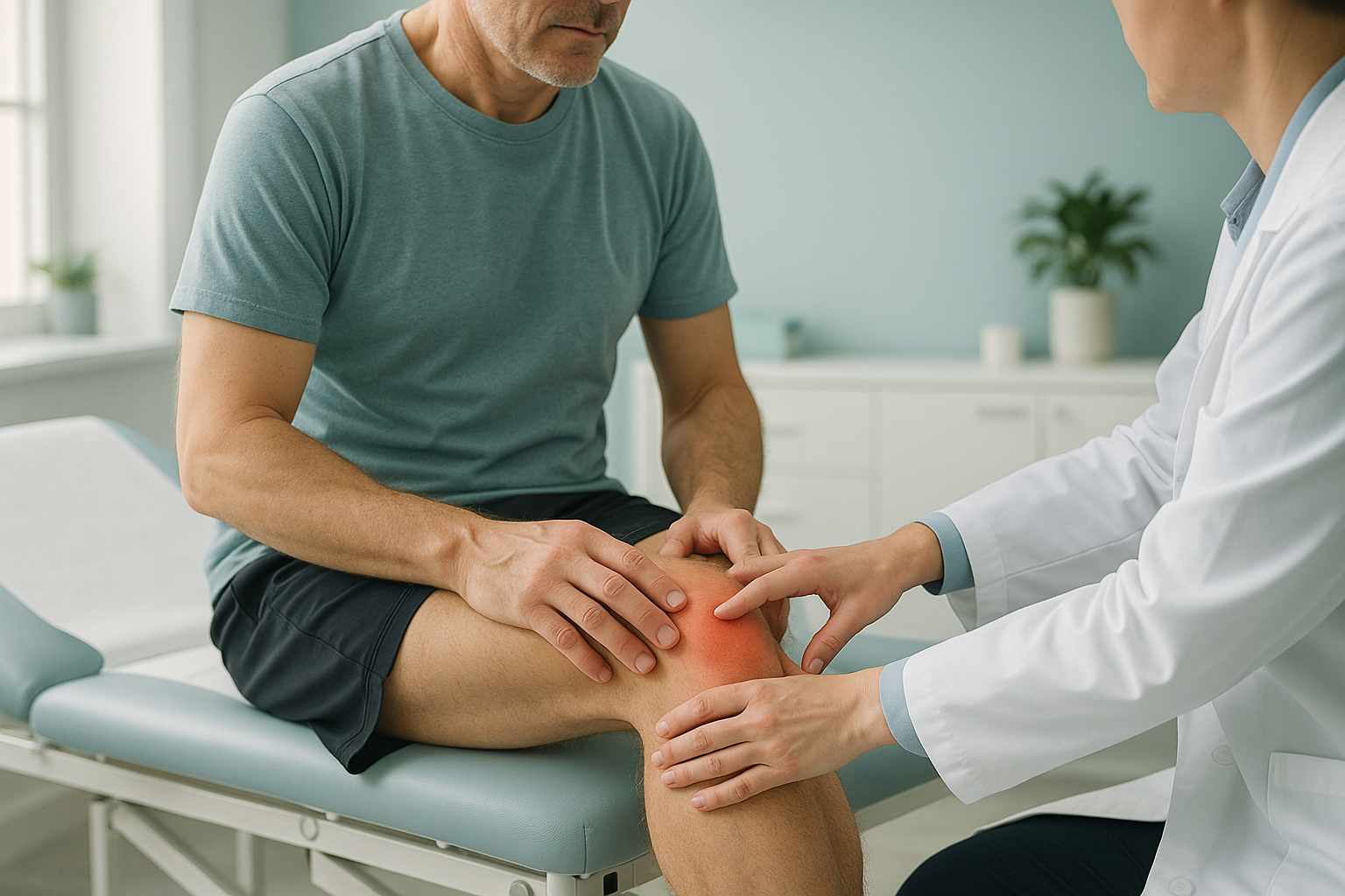
Stage 1: Pain only after exercise, settling quickly with rest. Performance is unaffected.
Stage 2: Pain during and after exercise, but you can still push through. It may warm up after a few minutes.
Stage 3: Pain during activity that limits what you can do. Performance starts to drop.
Stage 4: Constant pain even with daily activities like walking or climbing stairs. The tendon may be significantly weakened.
The earlier patellar tendonitis is identified and managed, the more straightforward recovery tends to be. At later stages, the tendon structure can change (this is sometimes called patellar tendinopathy rather than tendonitis), and the problem can become more stubborn.
How does alignment affect patellar tendonitis?
Alignment plays a bigger role than many people realise. The angle at which your kneecap sits, the rotation of your thighbone and the position of your foot all influence how force travels through the patellar tendon. Even a small difference in leg alignment can mean one tendon works harder than the other, every single step.
Standard imaging for knee pain is usually done lying down. An X-ray or MRI taken while you are flat on a table relaxes the muscles around the knee and removes the effect of gravity. The bones settle into a neutral resting position that may look perfectly normal. But when you stand up and load the joint, the picture can change. A subtle rotation, a slight tilting of the pelvis or a difference in leg length can become visible only under your own body weight.
This is where EOS imaging is useful. An EOS scan captures your whole body while you are standing naturally, using about 90% less radiation than a standard X-ray, around ten times less. Your clinician can see the full chain from your feet through your knees, hips and spine in one image. If one part of that chain is out of line, the knock-on effect on the patellar tendon becomes much easier to understand.
Want to find out whether alignment is contributing to your knee pain?
Book a free video consultationWhen is MRI the better option? EOS is excellent for seeing bones, joints and overall alignment. But the patellar tendon itself is soft tissue. If your clinician needs to assess the tendon's internal structure, check for tears or rule out other soft-tissue causes, MRI is usually the better tool. The two scans complement each other well.
How do you recover from patellar tendonitis?
Recovery from patellar tendonitis centres on gradually building the tendon's tolerance to load. Rest alone is rarely enough. The tendon needs controlled, progressive loading to heal properly and become resilient again.
What generally helps
- Load management. Reduce the activities that aggravate the tendon (especially jumping and sprinting) while keeping the knee active with lower-impact movement.
- Eccentric and isometric exercises. Exercises like slow single-leg squats and wall sits place a controlled load on the tendon, which research shows encourages repair. These are typically guided by a physiotherapist as part of a structured patellar tendon rehab programme.
- Strengthening the whole chain. Weak glutes, tight calves and poor hip control can all increase patellar tendon stress. A broader strengthening programme helps share the load.
- Addressing alignment. If imaging shows a rotational issue, a leg-length difference or poor kneecap tracking, your treating clinician can factor this into your recovery plan.
ScanAlign does not provide treatment. It provides the detailed, weight-bearing imaging that helps your physiotherapist, surgeon or sports-medicine consultant understand exactly what is happening in your knee and across your whole lower limb. That information can make a real difference to how your recovery programme is designed.
Is patellar tendonitis the same as patellofemoral pain?
No. Patellar tendonitis and patellofemoral pain syndrome are different conditions, though both cause pain at the front of the knee. Patellar tendonitis affects the tendon below the kneecap. Patellofemoral pain involves the joint surface between the kneecap and the groove in the thighbone (femur). The location of pain, the triggers and the rehab approach differ.
Both conditions can be influenced by alignment. Standing imaging helps distinguish between the two by showing exactly where the kneecap sits relative to the groove and how the lower limb lines up under load. You can learn more about how alignment assessment works on the ScanAlign knee page.
Frequently asked questions about patellar tendonitis
It depends on the stage. Early-stage tendonitis may settle in a few weeks with load management. More established tendinopathy can take several months of structured rehabilitation. Identifying and addressing alignment factors can help avoid repeated flare-ups.
Usually, yes. The goal is to modify activity rather than stop completely. Low-impact exercises and a controlled strengthening programme are generally recommended. Your physiotherapist can advise what is safe for your stage.
Tendonitis refers to acute inflammation. Tendinopathy describes longer-term changes in the tendon's structure. Over time, a tendonitis can progress to a tendinopathy if the tendon is not given time and the right loading to recover.
Many cases are diagnosed clinically. However, if symptoms persist or keep returning, imaging can help identify underlying factors such as alignment issues or structural changes in the tendon. An EOS scan shows standing alignment; an MRI shows soft-tissue detail.
An EOS scan captures your full body while standing, showing how your feet, knees, hips and spine line up under your own weight. This reveals alignment factors that may be overloading one tendon. Lying-down imaging can miss these because gravity and muscle tension are removed. Learn more about how EOS works.
Yes. EOS uses about 90% less radiation than a standard X-ray, around ten times less. You can find more detail on the FAQs page.
No. ScanAlign is a private, self-pay clinic. You can book directly without a GP referral. The process starts with a free video consultation.
Pricing information is available on the FAQs and cost page.
Yes, it can. Recurrence is common, especially if the underlying cause (such as training errors or alignment issues) is not addressed. A thorough assessment, including weight-bearing imaging, can help reduce the risk of it returning.
Not quite. "Runner's knee" usually refers to patellofemoral pain syndrome, which affects the joint surface behind the kneecap. Patellar tendonitis affects the tendon below it. Both cause front-of-knee pain, but they are different conditions with different management approaches.
Could alignment be behind your knee pain?
A free video consultation with ScanAlign can help you understand whether standing, whole-body imaging could give you and your clinician a clearer picture.
Book a free video consultationAuthor: Abbas Dhami (Specialist Diagnostic Radiographer)
Sources
- Lian OB, Engebretsen L, Bahr R. Prevalence of jumper's knee among elite athletes from different sports. American Journal of Sports Medicine. 2005;33(4):561-567.
- Malliaras P, Cook J, Purdam C, Rio E. Patellar tendinopathy: clinical diagnosis, load management, and advice for challenging case presentations. Journal of Orthopaedic & Sports Physical Therapy. 2015;45(11):887-898.
- NHS. Tendonitis. nhs.uk/conditions/tendonitis.
This page is general information and not a diagnosis.
ScanAlign is the trading name of CoreMed Solutions Ltd and operates under The Harley Street Hospital's CQC licence.