
Knock Knee Surgery: What Are Your Options for Genu Valgum Correction?
Knock knee surgery in adults usually involves a distal femoral osteotomy, where the thighbone is carefully reshaped to straighten the leg's alignment. Surgery is typically considered when non-surgical approaches have not helped and knee pain or joint wear is progressing. Standing whole-body imaging before surgery helps your surgeon plan the correction accurately.
Key takeaways
- Knock knees (genu valgum) cause the knees to angle inward, shifting load to the outer knee joint.
- The main surgical option for adults is a distal femoral osteotomy. Joint replacement may be considered if arthritis is advanced.
- Accurate, weight-bearing imaging of the whole leg is essential for planning any correction.
- EOS standing imaging shows the true alignment under load, something lying-down scans can miss.
- ScanAlign provides the diagnostic imaging that helps your surgeon decide if and how surgery could help.
What is genu valgum and why does it matter?
Genu valgum is the medical term for knock knees, where the knees angle inward so that when you stand with your knees together, your ankles sit apart. A small amount of inward angle is normal. It becomes a problem when the angle is large enough to shift how weight passes through your knee joint.
When your leg alignment is off, the outer (lateral) compartment of the knee takes more load than it was designed for. Over time, this uneven loading can wear down cartilage, cause pain, and lead to early arthritis. It can also affect your hip, ankle and lower back, because the whole chain from foot to pelvis has to compensate.
How do you know if your knock knees need surgery?
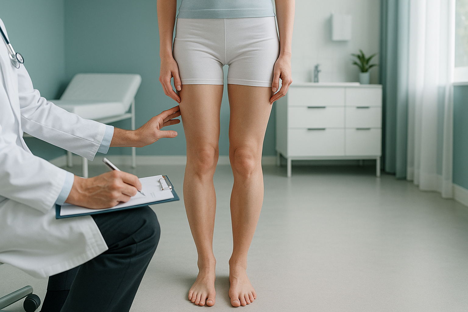
Not every person with knock knees needs an operation. Surgery is usually considered when symptoms are significant and have not responded to physiotherapy, strengthening exercises, or bracing. The following signs may suggest the alignment has become a real problem.
Pain on the outside of the knee that gets worse with walking or standing for long periods.
The inward angle is getting visibly worse over months or years.
Imaging shows cartilage thinning or early arthritis in the outer knee compartment.
The alignment is stopping you from exercising or managing daily tasks comfortably.
What causes knock knees in adults?
Knock knees in adults can develop for several reasons. Sometimes the alignment was present since childhood and never fully corrected. In other cases, it develops later due to injury or joint changes.
A childhood knock knee pattern that persisted into adulthood.
A fracture near the knee that healed with the bone at an angle.
Cartilage loss on the outer side of the knee shifts alignment inward.
Conditions like rickets or metabolic bone disease that affect bone shape.
What are the main surgical options for knock knee correction?
The right procedure depends on the cause, the severity of the misalignment, your age, and whether arthritis has already developed. Your orthopaedic surgeon will decide on the best approach based on thorough imaging and clinical assessment. Here are the most common options.
Distal femoral osteotomy
This is the most common corrective surgery for knock knees in adults who still have healthy cartilage. The surgeon makes a precise cut in the lower part of the thighbone and repositions it to straighten the leg. A plate and screws hold the bone in its new position while it heals. The goal is to shift weight-bearing back to the centre of the knee, protecting the worn compartment and slowing down joint damage.
High tibial osteotomy
Less common for knock knees than for bow legs, this involves reshaping the top of the shinbone. It may be considered when the misalignment originates in the tibia rather than the femur.
Partial or total knee replacement
If arthritis is already advanced and the cartilage is significantly worn, an osteotomy may not be enough. In these cases, a surgeon might recommend replacing part or all of the knee joint. Accurate alignment imaging is equally important here, because the replacement needs to be positioned to restore proper leg alignment.
When MRI is the better tool: EOS standing imaging shows bones, joints, and whole-leg alignment clearly. For soft tissue problems such as ligament tears, meniscal damage, or cartilage detail, MRI is usually the better option. Your surgeon may recommend both types of imaging for a complete picture.
Why does standing imaging matter before knock knee surgery?
Standing imaging matters because your leg alignment changes when you stand up and put weight through your joints. When you lie down for a standard X-ray or CT scan, your muscles relax and your joints settle into a different position. The true alignment, the one your surgeon needs to correct, only shows up when you are standing and weight-bearing.
This is where the body's compensation chain becomes important. A knock knee does not just affect the knee in isolation. The hip tilts, the ankle rolls, the pelvis shifts, and the spine adjusts. An EOS scan captures the full picture, from your ankles to your spine, in a single standing image. This gives your surgeon measurements of the whole chain, not just one joint.
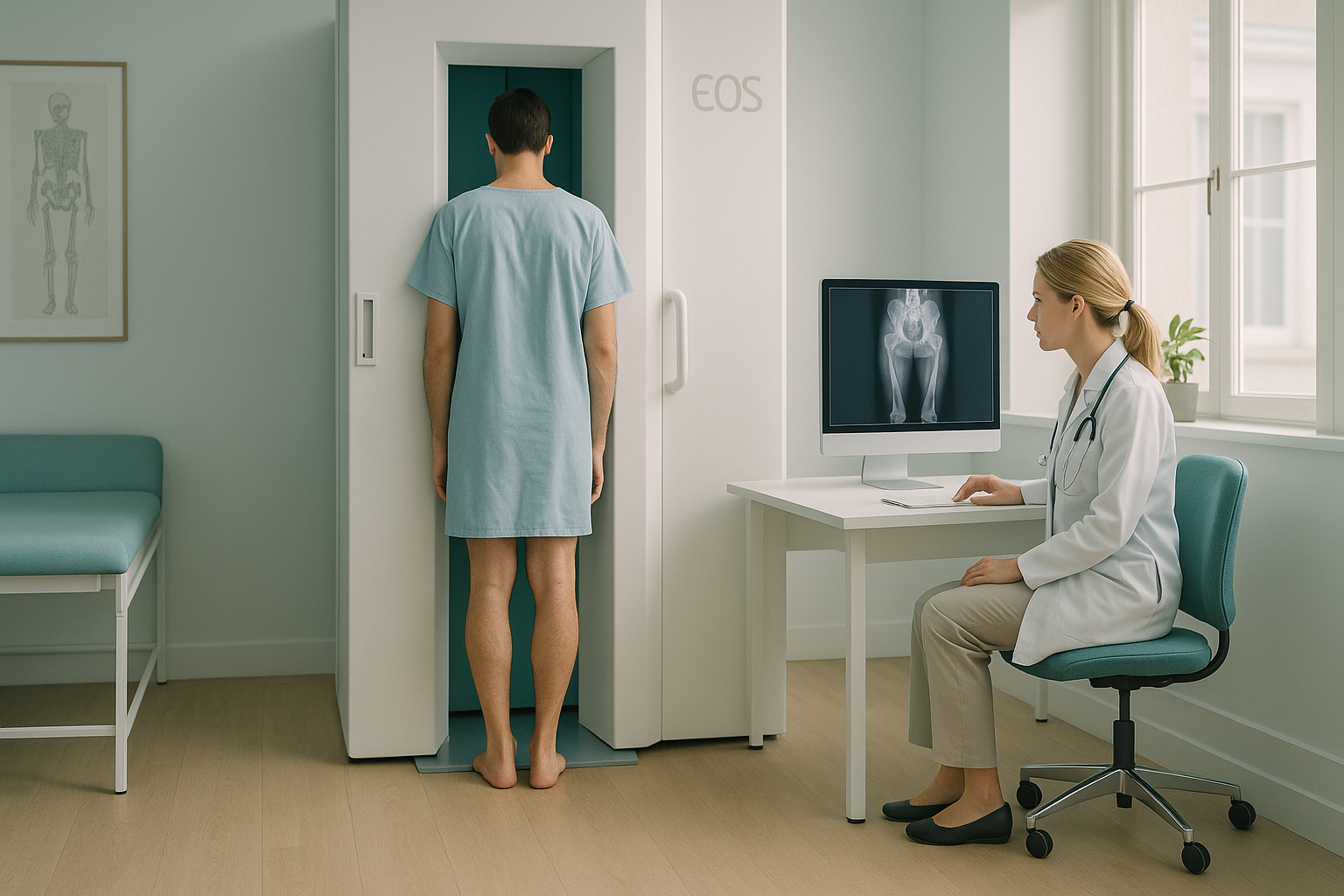
An EOS scan also uses about 90% less radiation than a standard X-ray, around ten times less. That makes it particularly suitable when multiple images or follow-up scans are needed.
Considering surgery for knock knees? A standing alignment scan can give your surgeon the measurements they need to plan the correction accurately.
Book a free video consultationWhat non-surgical approaches are tried before surgery?
Most surgeons will want to see that conservative measures have been given a fair try before recommending an operation. These may include targeted strengthening of the muscles around the hip and knee, physiotherapy programmes, custom orthotics or insoles, and sometimes bracing. Activity modification and weight management can also reduce the load through the affected compartment.
If these approaches are not enough and symptoms continue or worsen, that is typically when surgical correction becomes a conversation. A detailed alignment assessment at that stage helps determine which procedure is most appropriate and gives your surgeon the baseline measurements for planning.
Frequently asked questions about knock knee surgery
For adults with significant pain, progressive misalignment, or early arthritis caused by the valgus angle, surgery can be very effective. The decision depends on your symptoms, the degree of misalignment, and whether conservative treatment has helped. Detailed imaging and a thorough assessment by an orthopaedic surgeon are the best way to find out if it is the right choice for you.
The most common procedure is a distal femoral osteotomy, where the lower thighbone is precisely cut and repositioned to straighten the leg. It is typically recommended when cartilage is still reasonably healthy and the goal is to prevent further joint damage.
Recovery varies depending on the procedure and the individual. Most people use crutches for several weeks and begin physiotherapy soon after surgery. Full recovery, including return to sport, can take several months. Your surgeon will give you a personalised timeline based on your situation.
The cost of surgery varies widely depending on the procedure, the surgeon, and the hospital. ScanAlign provides the diagnostic imaging that supports surgical planning, not the surgery itself. For details on scan pricing, visit our FAQs and cost information page.
No. ScanAlign is a self-referral clinic. You can book directly without a GP referral. We offer a free video consultation to discuss whether a scan is appropriate for your situation before you commit.
An EOS scan takes images while you stand, capturing your whole body from head to foot in a single pass. It uses about 90% less radiation than a standard X-ray, around ten times less. Because you are standing, it shows your true alignment under load, which conventional lying-down X-rays cannot do. Learn more on our EOS scan overview page.
Yes. Because the body works as a connected chain, an inward knee angle can change how your hip sits, how your pelvis tilts, and how your spine curves. Pain can appear in the hip or lower back even though the root cause is the knee alignment. A whole-body standing scan helps identify these connections.
Yes. The EOS system uses about 90% less radiation than a standard X-ray, around ten times less. It is one of the lowest-dose imaging systems available. More safety details are on our FAQs page.
An EOS scan provides the precise alignment measurements your surgeon needs to decide whether surgery is appropriate and which procedure would be best. ScanAlign is a diagnostic imaging clinic. We provide the imaging information, and your orthopaedic team makes the treatment decisions.
Generally yes, because EOS uses X-ray technology rather than magnets. Metal implants are not usually a problem. If you have any concerns, mention them during your free video consultation and we can advise before your appointment.
Sources
- Brinkman, J.M. et al. "Osteotomies around the knee: patient selection, stability of fixation and bone healing in high tibial osteotomies and distal femoral osteotomies." The Journal of Bone and Joint Surgery (British Volume), 2008.
- Illés, T. and Somoskeöy, S. "The EOS imaging system and its uses in daily orthopaedic practice." International Orthopaedics, 2012.
- NHS. "Knee replacement: when it is needed." NHS.uk, 2022.
Written by Abbas Dhami (Specialist Diagnostic Radiographer)
Considering knock knee correction?
A standing whole-body alignment scan gives your surgeon the precise measurements needed to plan the right procedure. Start with a free, no-obligation video consultation.
Book a free video consultationThis page is general information and not a diagnosis.
ScanAlign is the trading name of CoreMed Solutions Ltd and operates under The Harley Street Hospital's CQC licence.