
Patellofemoral Pain Syndrome: What Causes It and How Do You Find the Real Problem?
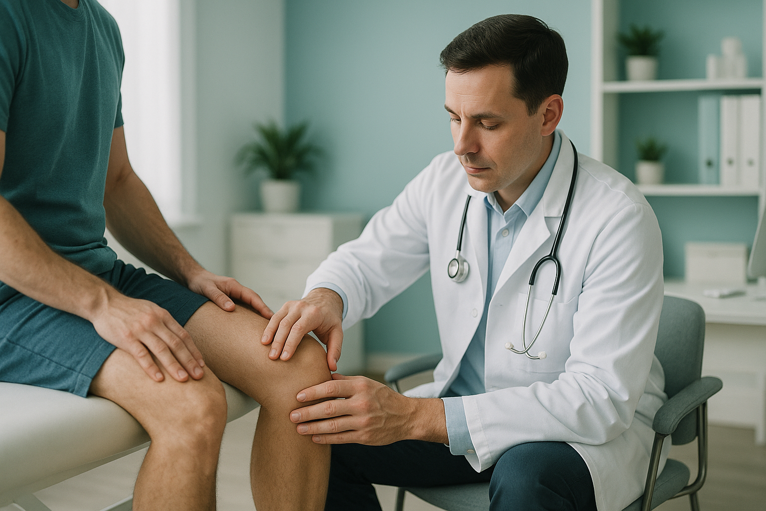
Patellofemoral pain syndrome (PFPS) is a dull, aching pain behind or around the kneecap. It happens when the kneecap does not track smoothly in its groove during movement. The cause is often not in the knee itself. Alignment problems in the hip, pelvis or leg can change how force passes through the joint, and standing imaging can reveal what lying-down scans miss.
Key takeaways
- PFPS is the most common cause of anterior knee pain in active adults.
- The kneecap relies on alignment from the hip down to the foot, so the cause often sits away from where it hurts.
- Lying-down imaging relaxes the muscles and can hide tracking problems that only appear when you stand and bear weight.
- An EOS standing scan images the whole leg in one pass, using about 90% less radiation than a standard X-ray.
- ScanAlign provides diagnostic imaging only. Treatment is carried out by your clinician or physiotherapist.
You might know PFPS by other names. "Runner's knee" and "moviegoer's knee" both describe the same condition. The pain often appears during activities that load the bent knee, such as running, squatting, climbing stairs or sitting for a long time with the knees bent.
What does patellofemoral pain syndrome feel like?
PFPS typically causes a dull ache behind or around the front of the kneecap. It tends to come on gradually rather than after a single injury. You may also notice a grinding or clicking feeling when you bend or straighten the knee.
A deep, dull pain at the front of the knee, often worse after sitting for a while or climbing stairs.
Going up or down stairs or walking on slopes increases the load on the kneecap and makes the pain worse.
A grating sensation when bending or straightening the knee, sometimes audible.
Stiffness and pain after sitting with bent knees for a long time, such as in a cinema or on a long drive.
If you experience sudden swelling, the knee giving way, or you cannot bear weight at all, see your GP, call 111 or attend A&E. These symptoms may point to a different problem that needs urgent attention.
What causes patellofemoral pain syndrome?
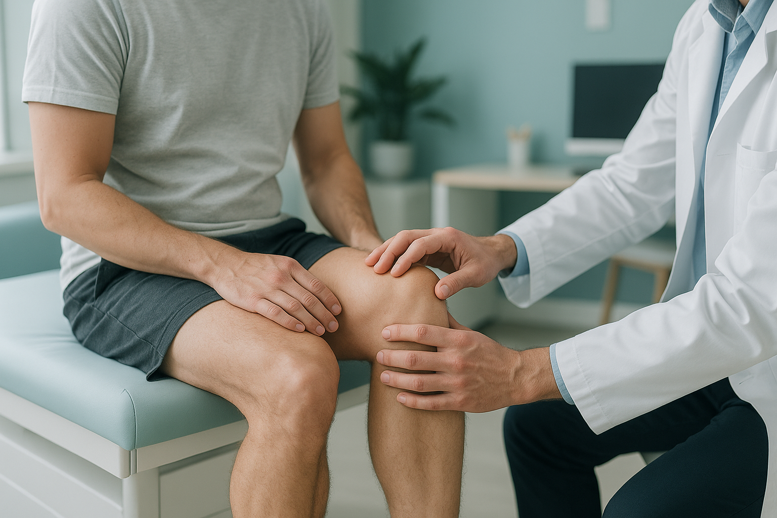
PFPS happens when the kneecap (patella) does not sit or move correctly in the groove at the front of the thighbone (femur). Several factors can contribute, and they often work together.
Weak hip or thigh muscles let the kneecap drift out of its groove during movement.
Knock knees, a rotated shin, or a flat foot can all change the angle at which the kneecap sits.
A sudden increase in running, cycling or squatting loads the joint faster than it can adapt.
A tilted pelvis shifts how weight passes down the leg, putting extra stress on one knee.
Notice how several of those causes sit above or below the knee. Your body is a connected chain from feet to head. When one link is out of place, the joints above and below it compensate, and pain often appears somewhere different from the real problem.
Why does whole-leg alignment matter for anterior knee pain?
Because the kneecap sits at the meeting point of forces from the hip, thigh and lower leg, its tracking depends on the alignment of the entire limb. A problem at the hip or ankle can redirect force through the kneecap unevenly.
Standard investigations usually look at the knee in isolation, often with the patient lying down. But lying down relaxes muscles and removes the effect of gravity. The kneecap may sit perfectly in its groove when you are relaxed on a table, then shift when you stand and your full weight presses through the joint.
This is why many people with patellofemoral pain have normal-looking knee scans but still hurt. The problem is not visible unless it is measured under real, weight-bearing conditions, with the whole leg in the picture.
How does an EOS standing scan assess patellofemoral pain?
An EOS scan takes a full-length image of both legs while you stand naturally. It captures the hip, knee and ankle in a single exposure, showing exactly how the bones and joints line up under your own body weight.
Because the image is taken standing, the kneecap is loaded the way it is in real life. Your clinician or surgeon can see whether the kneecap sits off-centre, whether the thighbone is rotated, whether the lower leg angles inward, and whether a hip or pelvic tilt higher up is contributing.
The EOS system uses about 90% less radiation than a standard X-ray, around ten times less. The scan takes only a few seconds. You do not need a GP referral to book, and the images are reported by a specialist radiologist.
When is MRI the better choice? EOS shows bones, joints and alignment clearly. If your clinician suspects cartilage damage behind the kneecap, a torn ligament, or swelling in the soft tissue, MRI is usually the better tool. EOS and MRI answer different questions, and sometimes you need both.
Not sure whether a standing alignment scan is the right next step for your knee pain?
Book a free video consultationWhat helps manage patellofemoral pain syndrome?
Most people with PFPS improve with conservative measures guided by a physiotherapist or sports medicine clinician. The focus is usually on strengthening the muscles that control the kneecap and correcting any alignment or movement patterns that are contributing.
Common approaches include targeted exercises for the quadriceps and hip stabilisers, activity modification to reduce flare-ups, and sometimes orthotic insoles if the foot position is part of the problem. In persistent cases, more detailed imaging helps the treating clinician understand exactly what is going on structurally before deciding on the next step.
ScanAlign provides the diagnostic imaging that informs these decisions. We do not prescribe exercises or carry out treatment. Your report goes to you and, if you wish, directly to your treating clinician so they can plan your care with the full picture.
You can learn more about how EOS imaging fits into the assessment of knee problems.
Frequently asked questions about patellofemoral pain syndrome
Yes, runner's knee is a common name for PFPS. "Moviegoer's knee" and "anterior knee pain syndrome" are other names for the same condition. They all describe pain behind or around the kneecap caused by poor tracking or overload.
Mild cases sometimes settle with rest and activity modification. However, if there is an underlying alignment issue, the pain often returns once you increase your activity again. Identifying and addressing the cause gives the best chance of long-term improvement.
Going downstairs places a higher braking force through the kneecap. If the kneecap is not tracking smoothly, this extra load increases friction and pain. Some people feel it more going up. It depends on where the kneecap sits and which muscles are weakest.
Standard X-rays and MRI can be useful, but they are taken lying down and may not show how the kneecap tracks under load. A standing EOS scan captures the full leg alignment under your own body weight. If soft-tissue damage is suspected, MRI is usually the better choice for that specific question.
You stand still inside the EOS cabinet for a few seconds while two low-dose beams capture a full-length image. There is no tunnel, no noise, and no injection. You can read more about the process on the FAQs page.
Yes. The EOS system uses about 90% less radiation than a standard X-ray, around ten times less. It is one of the lowest-dose skeletal imaging options available.
No. ScanAlign is a private, self-pay clinic. You can book directly without a referral. If you are unsure whether the scan is right for you, start with a free video consultation.
Pricing details are on the ScanAlign FAQs page.
Yes. Weak hip muscles or a tilted pelvis can change the angle of the thighbone, which in turn pulls the kneecap off-track. This is one reason why imaging the knee alone can miss the real cause. A full-leg standing scan captures the hip, knee and ankle together.
ScanAlign is at 19 Harley Street, London, inside The Harley Street Hospital. You can find directions and practical details on the ScanAlign homepage.
Wondering if your knee pain is really a knee problem?
A free video consultation with our specialist radiographer can help you decide whether a standing alignment scan is the right next step.
Book a free video consultationWritten by Abbas Dhami (Specialist Diagnostic Radiographer)
Sources
- Crossley KM, Stefanik JJ, Selfe J, et al. 2016 Patellofemoral pain consensus statement from the 4th International Patellofemoral Pain Research Retreat. British Journal of Sports Medicine. 2016;50(14):839-843.
- NHS. Knee pain overview. nhs.uk/conditions/knee-pain.
- Deschamps F, Courvoisier A, Amzallag J, et al. The EOS imaging system and its uses in daily orthopaedic practice. International Orthopaedics. 2012;36(7):1405-1412.
This page is general information and not a diagnosis.
ScanAlign is the trading name of CoreMed Solutions Ltd and operates under The Harley Street Hospital's CQC licence.