
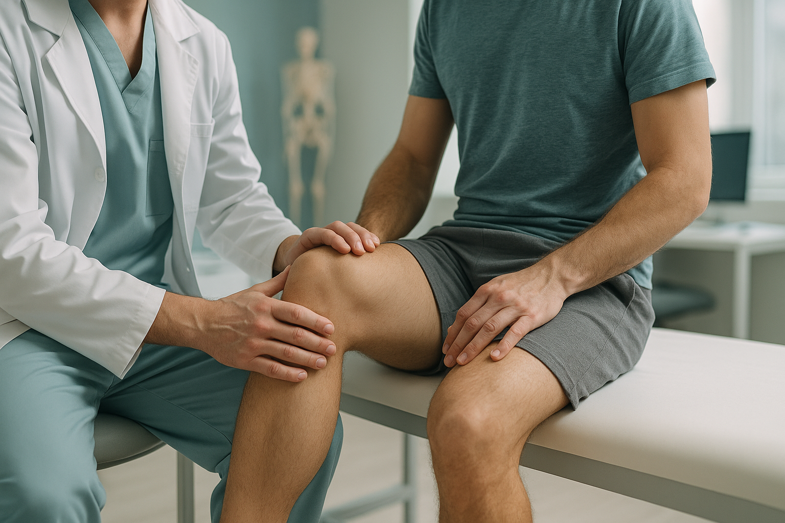
PCL Strain: What Is a Posterior Cruciate Ligament Injury and How Do You Know You Have One?
A posterior cruciate ligament strain is a stretch or tear of the strong ligament at the back of your knee. It usually happens when a force pushes the shin bone backwards, such as hitting the dashboard in a car accident or landing hard on a bent knee. Symptoms include deep knee pain, swelling and a feeling that the knee gives way.
Key takeaways
- The PCL is the strongest ligament in the knee, but it can still be stretched or torn by a direct blow or fall.
- Many PCL strains are missed because the knee can feel stable enough to walk on, even when the ligament is damaged.
- Lying-down scans can miss the way a PCL injury changes your standing alignment and how your whole leg absorbs weight.
- An EOS standing scan captures your full leg alignment under load, giving your specialist a clearer picture for planning next steps.
The posterior cruciate ligament (PCL) sits behind the anterior cruciate ligament (ACL), deep inside the knee joint. Its job is to stop the shin bone from sliding too far backwards relative to the thigh bone. Because it is thicker and stronger than the ACL, PCL injuries are less common. But they are also more often overlooked.
If you have persistent knee pain, instability during activities, or a knee that just does not feel right after a knock, a PCL strain may be worth investigating.
What does a PCL tear feel like?
A PCL tear often feels less dramatic than an ACL tear. You may not hear a "pop." Instead, you notice a deep ache at the back of the knee, swelling that develops over hours, and a vague sense of looseness when walking downhill or on stairs.
Urgent warning. If your knee is severely swollen, you cannot bear weight at all, or you have numbness or tingling in your lower leg, seek medical attention straight away. Contact your GP, call 111, or go to A&E.
What causes a posterior cruciate ligament strain?
A PCL strain is usually caused by a direct blow to the front of the shin while the knee is bent. This forces the shin bone backwards and stretches or tears the ligament.
What are the grades of a PCL sprain?
PCL injuries are graded by how much the ligament is damaged and how far the shin bone can shift backwards. Understanding the grade helps guide the best next steps.
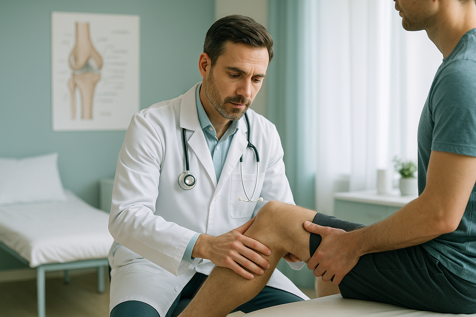
- Grade I (mild). The ligament is stretched but not torn. The knee still feels stable. Recovery is typically managed with rest and physiotherapy.
- Grade II (partial tear). Some of the ligament fibres are torn. The knee feels looser than normal and you may notice it giving way under load.
- Grade III (complete tear). The ligament is fully ruptured. The knee is noticeably unstable. Surgical reconstruction may be discussed if other ligaments are also damaged or the knee remains unreliable.
How does a standing scan help after a PCL injury?
A standing, weight-bearing scan shows how your leg alignment actually behaves under your body weight. This is something a lying-down scan cannot do.
When you lie down for a standard X-ray or MRI, gravity is removed. Your muscles relax. The knee joint settles into a position that may look more normal than it really is. When you stand up and walk, the forces change completely. A damaged PCL allows the shin bone to sag backwards, and that shift can alter the alignment of your entire leg, from the hip down to the ankle.
Your body is a connected chain. If your PCL is lax, the thigh bone may rotate or your hip may tilt to compensate, which can cause discomfort far from the knee itself. A whole-body standing image, like the one produced by an EOS scanner, captures these compensations in a single low-dose exposure, using about 90% less radiation than a standard X-ray, around ten times less.
This full picture helps your surgeon or physiotherapist understand not just the ligament damage, but how the injury has changed your overall mechanics. It is especially useful if you have already had an MRI and your knee still does not feel right.
When MRI is the better tool. EOS shows bones, joints and alignment under load. For soft-tissue detail of the ligament itself, cartilage, meniscus or nerve involvement, MRI is usually the better choice. The two are complementary.
Wondering whether your knee alignment is part of the problem? A short video call can help you decide if an EOS scan is the right next step.
Book a free video consultationHow is a PCL injury managed without surgery?
Many PCL injuries, especially grade I and grade II strains, are managed without surgery. Treatment is guided by the grade of injury, overall knee stability and your activity goals. Common approaches include rest, bracing, and a structured physiotherapy programme to strengthen the muscles around the knee, particularly the quadriceps.
Grade III tears, or injuries where other ligaments are also damaged, may require surgical reconstruction. Your orthopaedic specialist will make that decision based on clinical examination and imaging.
Understanding your leg alignment before and during recovery can be valuable. If the knee is compensating in a way that puts extra stress on the hip or ankle, targeted rehabilitation can address those patterns early.
Learn more about how standing imaging supports knee assessment and alignment.
Frequently asked questions about PCL strains
Grade I and some grade II tears can heal with rest, bracing and physiotherapy. The ligament may stretch slightly but regain enough stability for daily activity. Complete tears rarely heal fully without intervention, though not everyone needs surgery.
Recovery varies by grade. A mild strain may settle in a few weeks with guided rehabilitation. A partial or complete tear can take several months. Your specialist will tailor a timeline based on your specific injury and progress.
A dashboard knee injury happens when the front of the shin hits the car dashboard during a collision. The knee is bent, and the impact drives the shin bone backwards, stretching or tearing the PCL. It is one of the most common mechanisms for this injury.
Not necessarily. PCL tears are often less immediately disabling because the knee may still feel stable enough to walk on. However, untreated PCL laxity can lead to long-term problems, including cartilage wear and pain in the kneecap area. Each injury needs proper assessment.
No. ScanAlign is a private, self-pay clinic. You can refer yourself directly. You are welcome to bring results from any clinician already involved in your care.
An EOS scan uses about 90% less radiation than a standard X-ray, around ten times less. It captures full-length, weight-bearing images in a single low-dose exposure.
EOS shows bones and joint alignment, not the soft-tissue ligament itself. What it does show is how a PCL injury changes the position of the shin bone relative to the thigh bone when you are standing, along with any compensatory changes up and down the leg. For imaging the ligament directly, MRI is usually the better tool.
For current pricing, visit the ScanAlign FAQs and cost page.
When the PCL is lax, the shin bone can sag backwards, changing how your leg bears weight. Your hip may compensate by tilting or rotating to keep you balanced. Over time, this can cause discomfort in the hip, even though the original injury is in the knee. A full-leg standing scan can reveal these compensatory patterns.
You stand in the EOS scanner for a short, comfortable image. No preparation is needed. The scan itself takes only seconds. You receive detailed alignment images and a report that you can share with your treating clinician. Full details are on the FAQs page.
See how your knee really bears weight
A short, free video consultation can help you decide if a standing EOS scan is the right next step for your PCL injury.
Book a free video consultationSources
- Schulz MS, Russe K, Weiler A, Eichhorn HJ, Strobel MJ. Epidemiology of posterior cruciate ligament injuries. Archives of Orthopaedic and Trauma Surgery. 2003;123(4):186-191.
- NHS. Knee ligament injuries. nhs.uk. Available at: https://www.nhs.uk/conditions/knee-ligament-surgery/
- Illés T, Somoskeöy S. The EOS imaging system and its uses in daily orthopaedic practice. International Orthopaedics. 2012;36(7):1405-1411.
Written by Abbas Dhami (Specialist Diagnostic Radiographer)
This page is general information and not a diagnosis.
ScanAlign is the trading name of CoreMed Solutions Ltd and operates under The Harley Street Hospital's CQC licence.