

MCL Injury: What Is MCL Damage and How Do You Know You Have It?
MCL damage is a sprain or tear of the medial collateral ligament, the strong band of tissue on the inner side of your knee. It usually happens when the knee is pushed inward by a blow, a twist or a sudden change of direction. Severity ranges from a mild stretch to a complete tear, and most cases heal without surgery.
Key takeaways
- The MCL stabilises the inner knee and is one of the most commonly injured knee ligaments.
- MCL sprains are graded 1 to 3, from a mild stretch to a complete tear.
- Most MCL injuries recover well with rest, bracing and rehabilitation.
- Leg alignment problems such as knock knees can increase stress on the MCL over time.
- A standing whole-body scan can reveal alignment factors that lying-down imaging may miss.
Your MCL runs along the inner edge of your knee, connecting the thigh bone to the shin bone. Its job is to stop the knee from bending inward too far. When the ligament is stretched beyond its limit, the fibres are damaged. This is what clinicians mean by MCL damage, MCL sprain or MCL tear.
MCL injuries are common in sports like football, rugby and skiing, but they also happen during everyday activities. A stumble, an awkward landing or a sudden twist can be enough. Understanding the grade of your injury, and whether your overall leg alignment is putting extra strain on the ligament, helps guide the right next steps.
What are the symptoms of an MCL sprain?
The main symptom is pain on the inner side of the knee. It may come on suddenly after an injury or build up gradually if the ligament is stressed over time.
Tenderness or sharp pain along the inner edge of the knee, often worse when pressing on the area.
The inner knee may swell within hours of the injury, making the joint feel stiff and warm.
A feeling that the knee might give way, especially when changing direction or stepping sideways.
Difficulty fully bending or straightening the knee, particularly in the first few days after injury.
Seek urgent medical attention if you experience sudden severe knee pain with an inability to bear any weight, numbness or tingling below the knee, or noticeable deformity of the joint. Contact your GP, call 111 or go to A&E.
What are the grades of an MCL tear?
MCL tears are classified into three grades based on the extent of fibre damage. The grade determines how the knee feels, how stable it is and how long recovery typically takes.
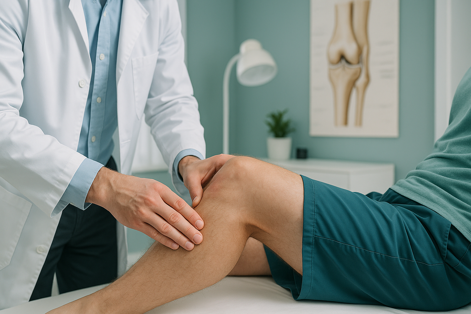
Grade 1 means the MCL fibres are stretched but not torn. The knee usually feels sore on the inside but remains stable. Most people can still walk.
Grade 2 means some of the fibres are torn. There is more swelling and pain, and the knee may feel slightly loose when tested.
Grade 3 is a complete tear. The ligament can no longer hold the inner knee steady, and the joint may feel noticeably unstable. A Grade 3 tear sometimes occurs alongside other ligament damage, such as an ACL injury.
What causes MCL damage?
MCL damage happens when a force pushes the knee inward beyond its normal range. This can be a single traumatic event or the result of repeated low-level stress on the ligament.
A direct blow to the outer knee, common in football and rugby tackles.
Sudden direction changes while the foot is planted, such as in skiing or tennis.
Landing off-balance from a jump or step with the knee buckling inward.
Knock-knee alignment (valgus) places ongoing extra load on the inner ligament.
The last point is important but often overlooked. Your body is a connected chain from your feet to your head. If your hips, pelvis or ankles sit out of alignment, the knee compensates. Over time, that compensation increases strain on the MCL, even without a dramatic injury event.
How does standing imaging reveal hidden causes of MCL stress?
Standing whole-body imaging shows your skeleton under its own weight, exactly as it works in real life. This matters because alignment problems that load the inner knee often disappear when you lie down for a standard scan.
When you lie on a table for a conventional X-ray, CT or MRI, gravity is removed and your muscles relax. A subtle knock-knee angle, a pelvic tilt or a rotated hip may straighten out. The image looks normal, but the stress on your MCL when you stand and walk is still there.
EOS imaging captures your entire body in one standing view, from head to feet, using about 90% less radiation than a standard X-ray, around ten times less. This lets a specialist see whether your leg alignment, hip position or pelvic balance is adding strain to the inner knee. For many people with recurring or slow-healing MCL problems, this whole-body perspective explains what single-area imaging misses.
Wondering whether your alignment could be contributing to your MCL problem? A free video consultation can help you decide whether a standing scan is the right next step.
Book a free video consultationWhen MRI is the better choice: EOS imaging excels at showing bones, joints and standing alignment. If your clinician suspects soft-tissue damage to the MCL itself, cartilage or meniscus tears, MRI is usually the better tool for that. The two scans complement each other well.
How long does MCL injury recovery take?
Recovery time depends on the grade of the tear. Most MCL injuries heal without surgery because the ligament has a good blood supply, which supports natural repair.
- Grade 1: Typically a few weeks. Rest, ice and a supportive knee brace are usually enough.
- Grade 2: Often several weeks to a couple of months. A hinged knee brace and guided rehabilitation exercises help protect the ligament while it heals.
- Grade 3: Recovery can take several months. Some complete tears heal with a structured brace and physiotherapy programme. Surgery is mainly considered when other ligaments are also damaged.
These are general patterns, not guarantees. Your own recovery depends on your overall health, the exact nature of the injury and whether there are other contributing factors such as alignment. A knee brace for a torn MCL is a common part of treatment, helping to limit sideways movement while the fibres repair.
What helps during recovery?
ScanAlign is a diagnostic imaging clinic, not a treatment provider. However, treatments your clinician may recommend typically include ice and elevation in the early stages, a hinged brace to protect the healing ligament, and a progressive physiotherapy programme to rebuild strength and stability. Addressing any underlying alignment factors may also help reduce the risk of re-injury.
Frequently asked questions about MCL damage
With a Grade 1 or mild Grade 2 tear, most people can still walk, though it may be uncomfortable. A Grade 3 tear often makes the knee feel unstable, and walking without support can be difficult. A knee brace and crutches are commonly used in the early stages.
The MCL is on the inner side of the knee and resists inward forces. The ACL is inside the knee joint and prevents the shin bone from sliding forward. ACL tears more commonly need surgery, while MCL tears often heal with bracing and rehabilitation. The two injuries can happen together in more severe cases.
No. Most MCL tears, including many Grade 3 tears, heal without surgery. The MCL has a strong blood supply that supports natural healing. Surgery is usually only considered when other knee ligaments are also damaged or when the knee remains unstable despite proper rehabilitation.
Yes. If your legs angle inward (sometimes called knock knees or valgus alignment), the inner side of the knee is under more stress with every step. This can make the MCL more vulnerable to sprains and slow down healing after an injury.
A standing scan shows your whole body under load. It can reveal alignment factors, such as a pelvic tilt, hip rotation or knock-knee angle, that place extra strain on the inner knee. These factors often disappear when you lie down for a conventional scan.
EOS uses about 90% less radiation than a standard X-ray, around ten times less. It captures your entire body in one low-dose scan. You can find more details on the FAQs and safety page.
No. ScanAlign is a private, self-pay clinic, and you can refer yourself. You start with a free video consultation to discuss whether a standing whole-body scan is appropriate for your situation.
Pricing details are available on the FAQs and cost page. The initial video consultation is free.
A mild Grade 1 sprain may heal with rest alone, but a brace is generally recommended for Grade 2 and Grade 3 injuries. It limits the sideways movement that could re-injure the healing ligament. Your physiotherapist or doctor can advise on how long to wear it.
Repeated MCL injuries may suggest an underlying alignment issue that is placing ongoing stress on the ligament. A standing whole-body assessment can identify whether your hip, pelvis or leg alignment is a contributing factor, giving your treatment team the full picture.
Find out what is really loading your knee
A free video consultation with our specialist radiographer can help you understand whether a standing whole-body scan is the right next step for your MCL problem.
Book a free video consultationSources
- NHS. "Sprains and strains." nhs.uk, 2023.
- Phisitkul P, James SL, Wolf BR, Amendola A. "MCL injuries of the knee: current concepts review." Iowa Orthopedic Journal, 2006; 26: 77-90.
- Illés T, Somoskeöy S. "The EOS imaging system and its uses in daily orthopaedic practice." International Orthopaedics, 2012; 36(7): 1405-1411.
Written by Abbas Dhami (Specialist Diagnostic Radiographer)
This page is general information and not a diagnosis.
ScanAlign is the trading name of CoreMed Solutions Ltd and operates under The Harley Street Hospital's CQC licence.