
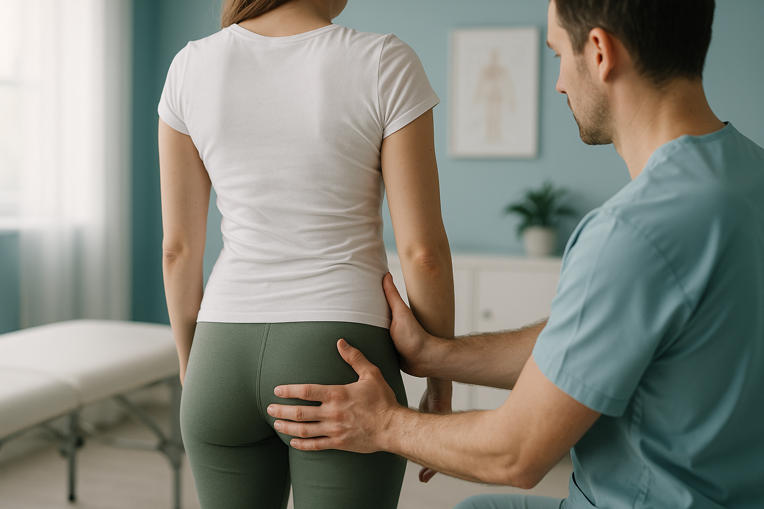
Hip Tilt: Why Do Your Hips Look Uneven and How Can You Fix It?
A hip tilt means your pelvis sits at an angle rather than level, either tipping forward (anterior hip tilt), backward, or with one side higher than the other. It changes the way forces travel through your spine, knees and feet. Standing whole-body imaging can show exactly how far the pelvis has shifted and which areas are compensating.
Key takeaways
- An anterior hip tilt tips the top of the pelvis forward, increasing the curve in your lower back.
- A lateral hip tilt means one hip sits higher than the other, which can affect your whole posture chain.
- Pain often appears in the back, knee or foot rather than the hip itself, because nearby joints compensate.
- Lying-down scans relax the muscles and can hide a tilt that only shows under your full body weight.
- An EOS standing scan captures your entire alignment in one image, using about 90% less radiation than a standard X-ray.
What is a hip tilt?
A hip tilt is a shift in the position of your pelvis away from its neutral, balanced alignment. Your pelvis is the foundation between your spine above and your legs below. When it tilts, every structure connected to it has to adjust, often without you noticing.
Think of the pelvis as a bowl of water. If the bowl tips forward, the water spills out the front. If it tips to one side, the water flows sideways. In either case the body above and below must compensate to keep you upright.
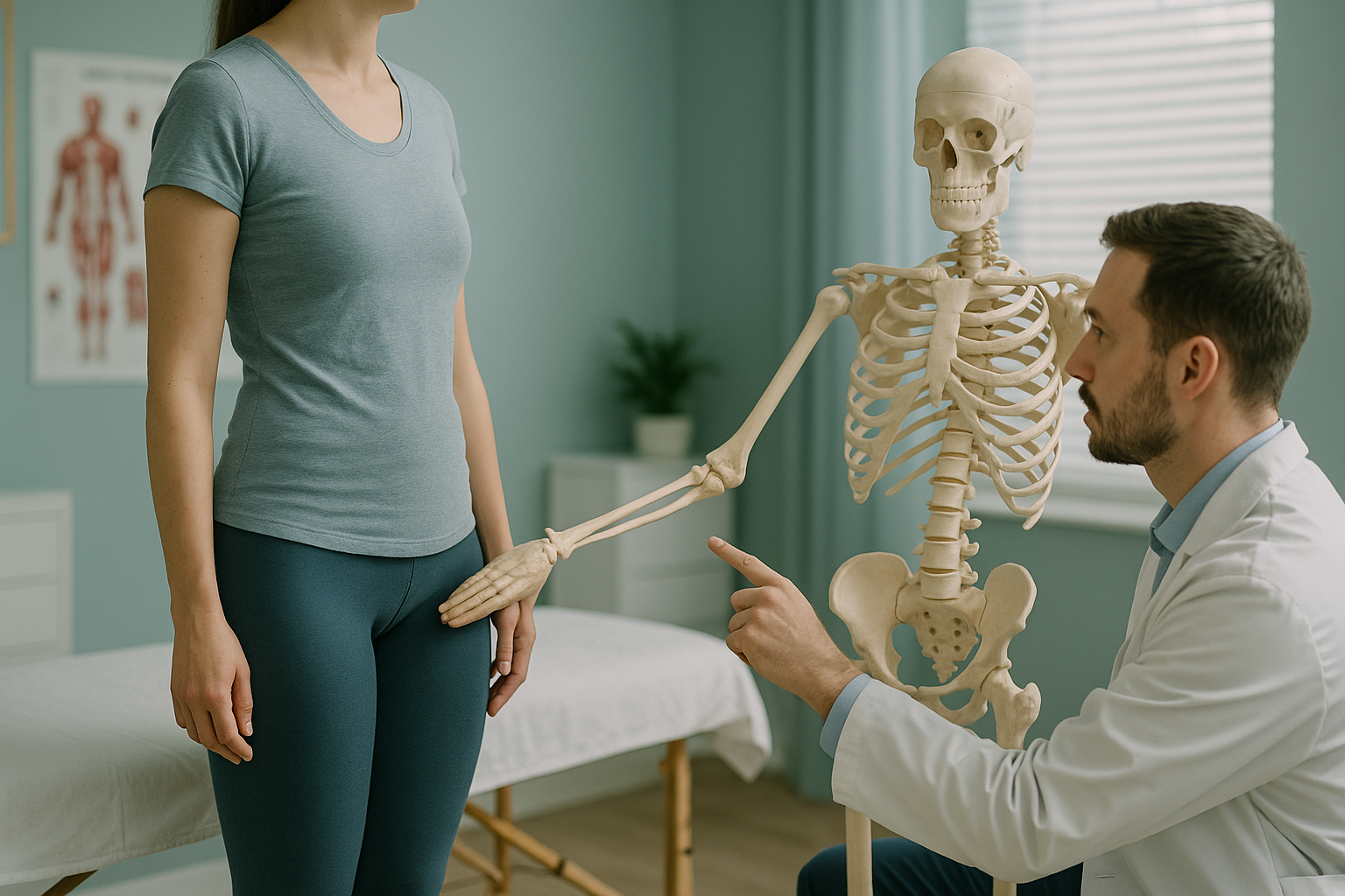
What is the difference between lateral and anterior hip tilt?
The two most common patterns are anterior hip tilt and lateral hip tilt. They look different, feel different, and create different compensation patterns through the body.
The top of the pelvis tips forward while the sit-bones rise. This deepens the lower back curve and pushes the belly forward. It is very common in people who sit for long hours.
One hip sits higher than the other. The spine often curves to compensate, and the shoulder on the lower side may drop. This can make one leg feel shorter even when both legs are the same length.
The pelvis tucks under, flattening the lower back. Less common on its own, it often appears alongside a rounded upper back and can cause stiffness through the hamstrings.
Many people have a combination. For example, a pelvis that is tipped forward and also higher on one side. Without a full-length standing image, it is difficult to tell which pattern is present.
How do you know if you have a hip tilt?
Some signs are visible when you look in a mirror. Others show up as pain or tightness in areas you might not connect to the pelvis at all.
Your belt or waistband sits higher on one side, or trousers seem to pull to one direction.
A persistent, dull ache across the lower back, especially after standing or walking for a while.
The front of the hip feels stiff or pinched when you stand up after sitting, a classic sign of anterior hip tilt.
One knee takes more load than the other, or one foot rolls inward more, because the pelvis shifts your weight unevenly.
A simple self-test is to stand with your back flat against a wall. Slide your hand behind the curve of your lower back. If a whole fist fits easily, your pelvis may be tipping forward more than it should. This is a rough guide, not a diagnosis. A proper assessment needs imaging.
What causes uneven hips?
Uneven hips develop for several reasons. Sometimes it is one main factor, but often it is a combination building up over months or years.
Sitting for hours shortens the hip flexors at the front and weakens the glutes at the back, pulling the pelvis forward.
Stronger muscles on one side of the body can pull the pelvis out of its neutral position over time.
A true or functional leg length difference tilts the pelvis to one side. Even a small difference can cause noticeable compensation.
An old ankle, knee or hip injury can change how you stand and walk, gradually shifting the pelvis off centre.
Because the body is a connected chain from your feet to your head, a problem at one level often causes symptoms at another. That is why treating the painful spot alone does not always work. You need to see the whole picture.
How is hip alignment assessed when standing up?
The most accurate way to assess hip alignment is to image the body in the position it actually uses, standing upright under its own weight. When you lie down for a standard X-ray, CT or MRI, gravity is removed. The muscles relax, the pelvis settles into the table, and a tilt that is obvious when you stand can disappear completely.
An EOS scan captures your full skeleton from head to feet in a single standing image. It uses about 90% less radiation than a standard X-ray, around ten times less. This means your clinician can see exactly how far the pelvis has shifted, whether one hip is higher, and how the spine and legs have adjusted in response.
This full-chain view is what makes standing imaging so useful for hip tilt. Rather than guessing whether the pelvis is involved, you can see it directly, and so can whoever plans your treatment.
Want to find out whether a hip tilt is behind your symptoms? A free video consultation can help you decide if an EOS standing scan is the right next step.
Book a free video consultationWhat helps correct a hip tilt?
Correction starts with understanding exactly what type of tilt you have and how the rest of your body has adapted. Without that information, exercises and treatments are often aimed at the wrong area.
General approaches a clinician might recommend include targeted stretching for shortened muscles, strengthening work for weak areas, and sometimes orthotics or heel raises if a leg length difference is contributing. The specific plan depends entirely on what imaging and assessment reveal.
ScanAlign provides the diagnostic imaging that informs these decisions. We do not provide treatment ourselves. Your scan results and report are shared with you and your chosen clinician so that any correction plan is based on clear evidence of what is happening in your body.
When MRI is the better tool. EOS excels at showing bone position, joint alignment and how the whole skeleton lines up under load. If your clinician suspects a soft-tissue problem, a disc issue, or nerve involvement, MRI is usually the more appropriate scan. If you are unsure which you need, the free video consultation can help clarify.
Frequently asked questions about hip tilt
Yes. When the pelvis tips forward, the lower back curve deepens, placing extra load on the lumbar joints and muscles. Many people with persistent lower back pain find a forward-tilted pelvis is a contributing factor once it is properly assessed.
Stand in front of a mirror and place your hands on the bony points at the front of each hip. If one hand sits noticeably higher, you may have a lateral hip tilt. This is a rough check. Accurate measurement requires imaging that shows the whole pelvis and legs together.
They are essentially the same thing. The pelvis and the hips are part of the same bony structure. "Hip tilt" and "pelvic tilt" are used interchangeably in everyday language.
Targeted exercises can improve muscle balance and reduce the severity of a tilt in many cases. However, the right exercises depend on what is causing the tilt. If a structural issue like a leg length difference is involved, exercises alone may not be enough. That is why accurate imaging matters before starting a programme.
When you lie flat, gravity pulls your body evenly against the table, and your muscles relax. A tilt that appears under your standing body weight can flatten out completely. Standing imaging captures the pelvis in its natural, loaded position, showing the tilt as it actually exists day to day.
No referral is needed. ScanAlign is a private, self-pay clinic. You can book directly. A free video consultation helps confirm whether an EOS scan is suitable for your situation before you commit.
An EOS scan uses about 90% less radiation than a standard X-ray, around ten times less. This makes it one of the lowest-dose skeletal imaging options available.
It can. When the pelvis tilts or shifts to one side, the leg underneath takes more load. This changes the angle at the knee and can contribute to pain on the inner or outer side of the joint. A whole-body standing scan helps show whether the knee pain originates from the hip alignment above.
Pricing details are available on the ScanAlign FAQs page.
You stand still inside the EOS scanner for around 15 to 20 seconds while it captures a full-length image from head to feet. There is no enclosed tunnel and no loud noise. You stay fully clothed in most cases, though you may be asked to remove metal items like belts or jewellery.
See what your hips are really doing
A free video consultation can help you understand whether a standing EOS scan is the right next step for your hip alignment.
Book a free video consultationSources
- Dubousset, J. et al. "EOS: A new imaging system with low dose radiation in standing position for spine and bone & joint disorders." European Journal of Radiology, 2010.
- NHS. "Back pain: causes, treatment and prevention." nhs.uk, 2023.
- Roussouly, P. & Pinheiro-Franco, J.L. "Biomechanical analysis of the spino-pelvic organisation and adaptation in pathology." European Spine Journal, 2011.
Written by Abbas Dhami (Specialist Diagnostic Radiographer)
Related reading: Learn more about hip alignment and EOS imaging, or explore the full ScanAlign EOS scan overview.
This page is general information and not a diagnosis.
ScanAlign is the trading name of CoreMed Solutions Ltd and operates under The Harley Street Hospital's CQC licence.