
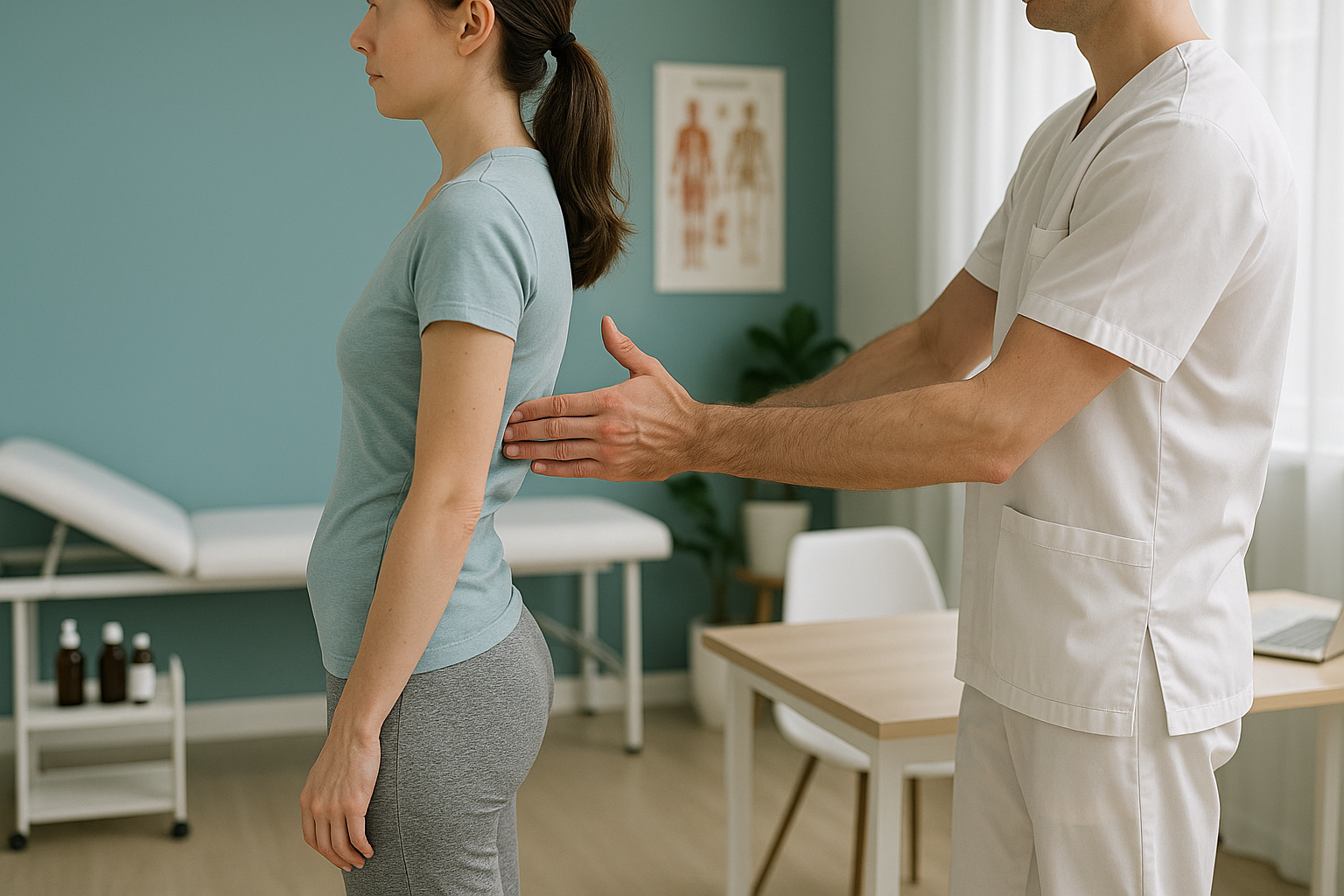
Sway Back Posture: What Is It and How Do You Know You Have One?
Sway back posture happens when the pelvis pushes forward and the upper back rounds backward to compensate. It shifts your centre of gravity, placing extra load on the lower spine, hips and knees. Because it develops slowly, many people live with it for years without realising it is the root cause of their pain.
Key takeaways
- Sway back shifts the hips forward of the ankles, flattening the lower spine and rounding the upper back.
- It often causes pain far from its origin, in the knees, hips or mid-back, because the body compensates along the whole chain.
- Lying-down scans relax the muscles and can hide the misalignment. Standing, weight-bearing imaging shows the full picture.
- EOS whole-body imaging captures your skeleton from head to toe in a single standing scan, with about 90% less radiation than a standard X-ray.
- ScanAlign offers a free video consultation so you can find out whether an alignment assessment is right for you.
If you have been told your scans look "normal" but you still ache after standing or walking, sway back posture could be the missing piece. It is one of the most common postural patterns, yet it rarely gets picked up on standard imaging because those scans are usually taken lying down.
What does sway back posture actually look like?
Sway back posture is a postural pattern where the pelvis drifts forward of the ankles while the shoulders lean backward. The lower back loses its natural inward curve and becomes flatter, while the upper back rounds more than usual. From the side, you look like you are leaning back even though your feet are underneath you.
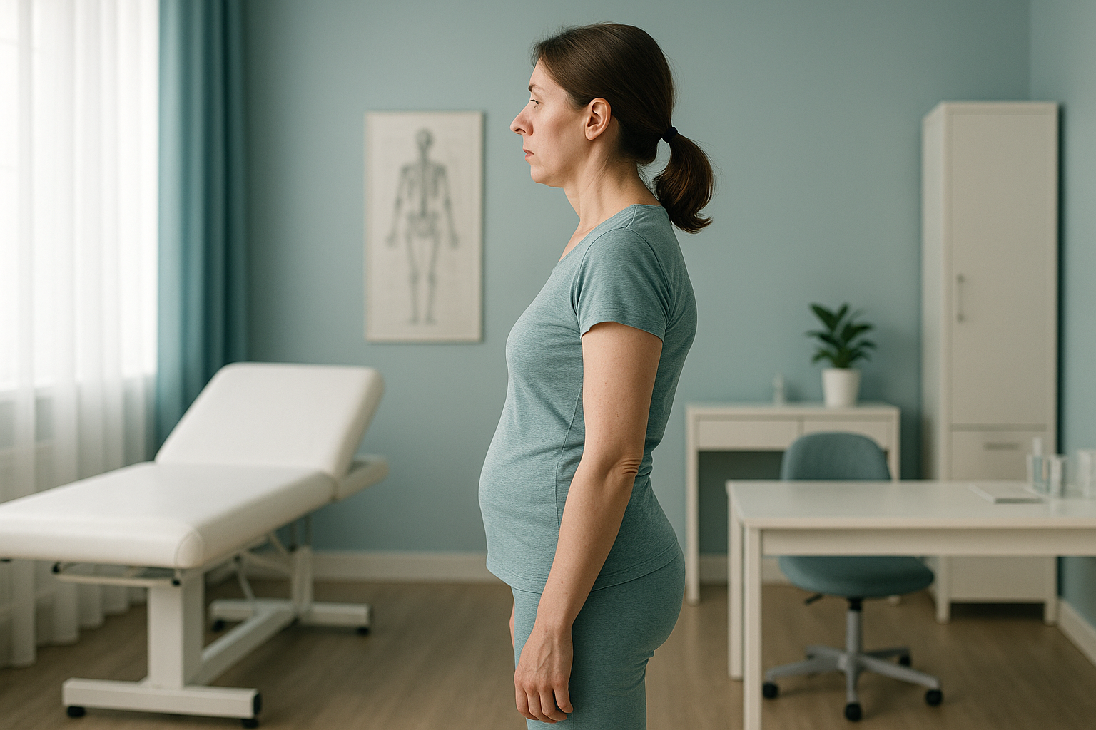
This pattern is different from an arched back posture (sometimes called hyperlordosis), where the lower back curves too much inward. With sway back, the lower curve actually flattens. The two are sometimes confused because both involve the pelvis, but the direction of the tilt and the shape of the spine are quite different.
How do you know if you have sway back posture?
You may notice some of these everyday signs. None of them is proof on its own, but a pattern of several together is a strong hint that your alignment needs a closer look.
If you also experience numbness, weakness in the legs, or loss of bladder or bowel control, please seek urgent medical attention from your GP, call 111, or go to A&E. These are red-flag symptoms that need immediate assessment.
What causes sway back posture?
Sway back posture develops when certain muscle groups weaken or tighten over time, pulling the pelvis out of its balanced position. It is rarely caused by a single event. The most common contributing factors are listed below.
Your body works as a connected chain from your feet to your head. When the pelvis shifts forward, the spine, knees and even neck adjust to keep you balanced. That is why sway back posture often produces pain far from where the real problem sits.
Why does sway back posture need a standing scan?
Most imaging, including standard X-rays, CT and MRI, is done while you are lying down. When you lie flat, gravity is no longer pulling on your skeleton, your muscles relax, and the misalignment that causes your symptoms can temporarily disappear. The scan may come back "normal" even though you are clearly in discomfort when upright.
A standing, weight-bearing scan captures your skeleton the way it actually works, under gravity, with your muscles engaged. EOS whole-body imaging does this in a single low-dose scan from head to toe. It uses about 90% less radiation than a standard X-ray, around ten times less. That means a clear, full-length picture of your alignment without unnecessary exposure.
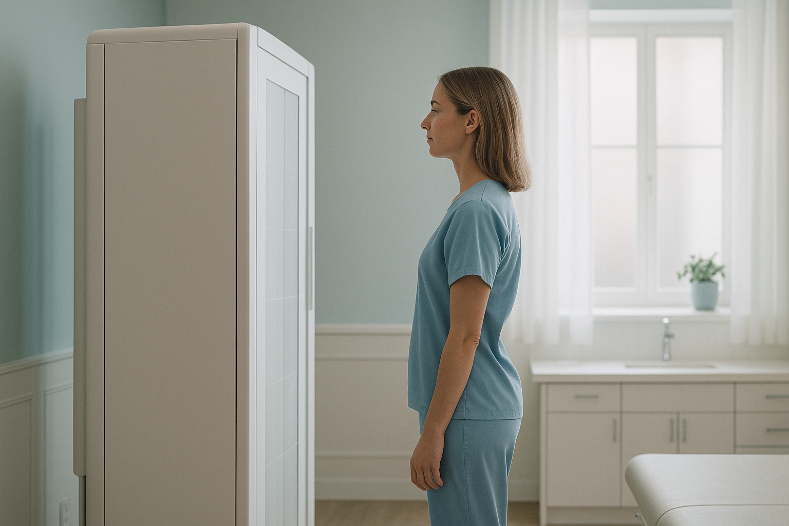
Because the EOS scan covers your entire skeleton in one image, the radiographer and your clinician can see exactly how far your pelvis has shifted, how your spine is compensating, and whether your knees or hips are bearing uneven load. This full-chain view is what single-area imaging misses.
Not sure if a standing scan is right for you? Talk to us first, at no cost.
Book a free video consultationWhat is the difference between sway back and flat back posture?
Both patterns involve a flattened lower back, but they are not the same thing. In flat back posture, the pelvis tucks under and the entire spine straightens, so you stand very upright but stiff. In sway back posture, the pelvis pushes forward while the upper body leans backward. The result is an S-shaped slouch rather than a straight line. The distinction matters because each pattern loads different joints and responds to different approaches.
How is sway back posture usually managed?
ScanAlign is a diagnostic imaging clinic, not a treatment provider. We provide the detailed alignment picture that helps your physiotherapist, osteopath or surgeon plan the right approach. Broadly, management of sway back posture may include targeted strengthening of the core and glutes, stretching of tight hip flexors, postural re-education, and sometimes manual therapy. The specific plan depends on what the imaging and clinical assessment show.
What matters most is getting an accurate, whole-body picture first. Without that, exercises risk addressing the symptom rather than the underlying alignment issue. A standing scan gives your treating clinician the information they need to tailor your programme properly.
When MRI is the better tool. EOS excels at showing bones, joints and whole-body alignment. If your clinician suspects a disc problem, nerve compression or soft-tissue injury, MRI is usually the better choice. The two scans answer different questions and often work well together.
Frequently asked questions about sway back posture
Yes. The forward shift of the pelvis flattens the natural curve of the lower spine, placing extra stress on the joints and ligaments. Over time this can produce chronic lower back stiffness and pain, especially after standing or walking.
No. An arched back (hyperlordosis) has an exaggerated inward curve in the lower spine. Sway back actually flattens that curve and pushes the hips forward. They are different patterns with different causes, even though both involve the pelvis.
It can. When the pelvis shifts forward, the legs angle differently to keep you balanced. This changes the load on the hip and knee joints and can contribute to pain in those areas over time.
An EOS scan captures your full skeleton while you stand under gravity. It shows exactly how the pelvis, spine, hips and knees relate to each other, revealing the compensations that lying-down imaging misses. Learn more about how EOS whole-body imaging works.
EOS uses about 90% less radiation than a standard X-ray, around ten times less. You can find more details on safety, cost and what to expect on our FAQs page.
No. ScanAlign is a private, self-pay clinic. You can refer yourself directly. We recommend starting with a free video consultation so we can check that a standing scan is the right next step for you.
Pricing details are on our FAQs page. The free video consultation is a good first step to discuss what scan package suits your situation.
Many people see improvement with targeted strengthening and postural work, guided by an accurate picture of their alignment. ScanAlign provides the diagnostic imaging. Your physiotherapist or specialist then uses the results to plan your management.
Standard X-rays usually focus on one area (for example, the lower back) and are often taken lying down. When you lie flat, gravity is removed and the postural shift disappears. A standing, full-body image captures what is actually happening when you are upright.
ScanAlign is at 19 Harley Street, London, inside The Harley Street Hospital. You can book a free video consultation first, so you know exactly what to expect before you visit.
Related alignment topics
Ready to see the full picture?
A free video consultation lets you discuss your symptoms with our team and find out whether a standing alignment scan is the right next step.
Book a free video consultationSources
- Roussouly P, Pinheiro-Franco JL. Biomechanical analysis of the spino-pelvic organisation and adaptation in pathology. European Spine Journal. 2011;20(Suppl 5):609-618.
- NHS. Back pain: causes, symptoms and treatments. nhs.uk/conditions/back-pain.
- Deschênes S, Charron G, Beaudoin G, et al. Diagnostic imaging of spinal deformities: reducing patients' radiation dose with a new slot-scanning X-ray imager. Spine. 2010;35(9):989-994.
Written by Abbas Dhami (Specialist Diagnostic Radiographer)
This page is general information and not a diagnosis.
ScanAlign is the trading name of CoreMed Solutions Ltd and operates under The Harley Street Hospital's CQC licence.